Radiopaedia

Last updated
Radiopaedia
Radiopaedia org logo small.jpg
Type of site
Wiki
Available inEnglish
OwnerRadiopaedia Australia Pty Ltd
EditorAssociate Professor Frank Gaillard
URL http://radiopaedia.org/
CommercialYes
RegistrationOptional (required to edit)
LaunchedDecember 2005
Current status16,719 articles with 56,720 cases (as of December 2023)

Radiopaedia is a wiki-based international collaborative educational web resource containing a radiology encyclopedia and imaging case repository. [1] It is currently the largest freely available radiology related resource in the world with more than 50,000 patient cases and over 16,000 reference articles on radiology-related topics. The open edit nature of articles allows radiologists, radiology trainees, radiographers, sonographers, and other healthcare professionals interested in medical imaging to refine most content through time. An editorial board peer reviews all contributions. [2]

Contents

Background

Radiopaedia was started as a past-time project to store radiology notes and cases online by the Australian neuroradiologist Associate Professor Frank Gaillard in December 2005, while he was a radiology resident. He later became passionate in building the website and decided to release it on the web, advocating free dissemination of knowledge. [3] [4]

The domain name for radiopaedia.org was registered on 11 January 2007. [5]

The Radiopaedia.org platform and text content are owned by Radiopaedia Australia Pty Ltd, a privately held company for which Gaillard is the chief executive officer. [6] One of its investors is Investling and its revenue derives from ads, courses, and paid supporters. [7] [8] For image content, contributors reserve some rights and license the content to Radiopaedia and its users under a Creative Commons license. [6]

The site was initially programmed using MediaWiki, the same program platform as Wikipedia, but now runs on a bespoke code written by TrikeApps. [9]

In 2010, almost all of the article and image collection from radswiki (a similar wiki-based radiology educational site) was donated to Radiopaedia. [10]

Its article content is currently limited to English.

Purpose

Radiopaedia’s mission is "to create the best radiology reference the world has ever seen and to make it available for free, for ever, for all." [11] Its intention is to benefit the radiology community and wider society and it relies on benevolent collaborations from radiologists and others with an interest in medical imaging.

Similarly to Wikipedia, registered users of the site are allowed to freely add and edit the majority of the content. This allows content to be progressively upgraded over years and for radiologists and society, in general, to continuously refine article content through time. The site also allows registered users to maintain their own personal case library of teaching cases. [12] Rather than individually publishing articles, users are encouraged to integrate content with links to cases and journal articles and collaboratively refine content. [13] In an attempt to reduce vandalism and to peer-review content, an editorial board moderates changes to ensure that the presented material is as accurate and relevant as possible. [14] As with similar open edit sites, unreliability of content has been a concern;[ citation needed ] however, despite its open edit nature, it is ranked relatively high among user reviews. [15] [16]

A survey done in 2020 shows that 90% of on-call radiology trainees in the United States are using Radiopedia and StatDx as the first and second line options to help them during their work. [17] Educational benefit was also demonstrated when integrating Radiopedia-based training in medical curriculum. [18]

Sub sites

Radiopaedia also maintains several other educational subsites which include

Editorial team

The editorial team, develop as well as help users to maintain the high-quality content of the website. [14]

The current editorial board (2021) is composed of individuals from a variety of countries and includes: [19]

Editor in chief

Academic director

Community director

Editorial director

Managing editors

iOS apps

In 2009, the first Radiopaedia iOS app series was released in two volumes. These apps package cases and articles for users to review and have sample questions and answers. [20] [21] More volumes were released subsequently. [22]

All the iOS apps were discontinued after March 2018. Instead, cases in the iOS apps exist in the form of "iOS case packs" playlists on the website. [23]

In 2019, nine out of 163 medical students surveyed in a single institution in Canada stated that they would use a radiology app for mobile learning such as "Figure 1", "Night in the ED" and "Radiopaedia" and will be willing to pay for a radiology app. [24]

Most of the content is shared under a Creative Commons non-commercial license. [1]

Related Research Articles

<span class="mw-page-title-main">Radiology</span> Branch of medicine

Radiology is the medical specialty that uses medical imaging to diagnose diseases and guide their treatment, within the bodies of humans and other animals. It began with radiography, but today it includes all imaging modalities, including those that use no ionizing electromagnetic radiation, as well as others that do, such as computed tomography (CT), fluoroscopy, and nuclear medicine including positron emission tomography (PET). Interventional radiology is the performance of usually minimally invasive medical procedures with the guidance of imaging technologies such as those mentioned above.

<span class="mw-page-title-main">Gestational sac</span> Cavity of fluid surrounding an embryo

The gestational sac is the large cavity of fluid surrounding the embryo. During early embryogenesis, it consists of the extraembryonic coelom, also called the chorionic cavity. The gestational sac is normally contained within the uterus. It is the only available structure that can be used to determine if an intrauterine pregnancy exists until the embryo can be identified.

The Royal Australian and New Zealand College of Radiologists (RANZCR) is the leading professional organisation for the promotion of the science and practice of the medical specialties of clinical radiology and radiation oncology in Australia and New Zealand. The college has members throughout the world. RANZCR provides the educational curricula for medical graduates training to enter the specialties.

<span class="mw-page-title-main">Radiological Society of North America</span> Organization of medical imaging professionals

The Radiological Society of North America (RSNA) is a non-profit organization and an international society of radiologists, medical physicists and other medical imaging professionals representing 31 radiologic subspecialties from 145 countries around the world. Based in Oak Brook, Illinois, it was established in 1915.

<span class="mw-page-title-main">Agger nasi</span>

The agger nasi is a small ridge on the lateral side of the nasal cavity. It is located midway at the anterior edge of the middle nasal concha, directly above the atrium of the middle meatus. It is formed by a mucous membrane that is covering the ethmoidal crest of the maxilla.

<span class="mw-page-title-main">Colic flexures</span> Two abrupt bends in the large intestine

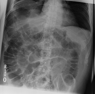
In the anatomy of the human digestive tract, there are two colic flexures, or curvatures in the transverse colon. The right colic flexure is also known as the hepatic flexure, and the left colic flexure is also known as the splenic flexure. Note that "right" refers to the patient's anatomical right, which may be depicted on the left of a diagram.

<span class="mw-page-title-main">Royal College of Radiologists</span> Professional association of clinical oncologist and radiologist

The Royal College of Radiologists (RCR) is the professional body responsible for the specialties of clinical oncology and clinical radiology throughout the United Kingdom. Its role is to advance the science and practice of radiology and oncology, further public education, and set appropriate professional standards of practice. The college sets and monitors the educational curriculum for those training to enter the profession, and administers the Fellowship of the Royal College of Radiologists exams. It is a registered charity in the United Kingdom (no. 211540).

<span class="mw-page-title-main">Radiologic sign</span> Medical sign from radiology

A radiologic sign is an objective indication of some medical fact that is detected by a physician during radiologic examination with medical imaging.

<span class="mw-page-title-main">Omental cake</span> Medical condition

Omental cake is a radiologic sign indicative of an abnormally thickened greater omentum. It refers to infiltration of the normal omental structure by other types of soft-tissue or chronic inflammation resulting in a thickened, or cake-like appearance.

<span class="mw-page-title-main">European Society of Radiology</span>

The European Society of Radiology (ESR) is an international medical society based in Vienna, Austria dedicated to the promotion and coordination of scientific, philanthropic, intellectual and professional activities of radiology in Europe. In addition to various other activities, the ESR serves as an umbrella organisation for European radiologists, organises the annual European Congress of Radiology (ECR) and coordinates the publication of European Radiology, a monthly peer-reviewed medical journal. Additionally, the ESR pilots the harmonisation of teaching programmes throughout Europe with various activities and initiatives.

WikiNodes is an app for the Apple iPad built by IDEA.org. WikiNodes was the first tablet app for browsing Wikipedia using a radial tree approach to visualize how articles and subsections of articles are interrelated. The app displays related items, which spread on the screen, as a spiderweb of icons.

<span class="mw-page-title-main">Tornwaldt cyst</span> Medical condition

A Tornwaldt cyst also spelt as Thornwaldt or Thornwald cyst is a benign cyst located in the upper posterior nasopharynx. It was first described by Gustav Ludwig Tornwaldt. It can be seen on computed tomography (CT) or magnetic resonance imaging (MRI) of the head as a well-circumscribed round mass lying in the midline. In most cases, treatment is not necessary. Indications for treatment include symptomatic lesions, large lesions (>1 cm), or lesions adjacent to the eustachian tube orifice.

<span class="mw-page-title-main">World of Knowledge</span> Educational project developed by WOKcraft

World of Knowledge is a global Wikipedia inspired commercial project to enhance learning developed by company WOKcraft based on a community of users creating multiple choice questions for free public use. The abbreviation for the project is WOK and used to market quiz game mobile apps in a social network called the Knowledge Network.

<span class="mw-page-title-main">MRI pulse sequence</span>

An MRI pulse sequence in magnetic resonance imaging (MRI) is a particular setting of pulse sequences and pulsed field gradients, resulting in a particular image appearance.

Fogging phenomenon in computerized tomography (CT) scanning of the head is vanishing signs of an infarct on the serial CT imaging in a patient with a recent stroke. It is a reversal of the hypodensity on the CT after an acute ischemic stroke. This happens as a result of re-nourishment of the infarcted area in subacute phase about one to three weeks after the stroke. In fact, resolution of the edema, which was caused by the accident, leads to increased attenuation of infarcted area that may regain near-normal density and mask the stroke. However, in the third week, parenchymal volume loss commonly appears as a hypoattenuation with a negative mass effect (shrinkage).

COVID-19 datasets are public databases for sharing case data and medical information related to the COVID-19 pandemic.

The empty delta sign is a radiologic sign seen on brain imaging which is associated with cerebral venous sinus thrombosis. It is usually seen on magnetic resonance imaging (MRI) or computed tomography (CT) scans with contrast. It is seen as dural wall enhancement in the absence of intra-sinus enhancement. This is due to the presence of a blood clot in the dural venous sinuses. The dural venous sinuses drain blood from the brain to the internal jugular veins, which in turn drains blood to the heart. It has been proposed that the empty delta sign occurs in dural venous thromboses due to contrast material filling the dural venous collateral circulation immediately surrounding the dura whilst being unable to fill the intra-dural sinus space due to the presence of a blood clot. The superior sagittal sinus is most commonly affected, but the radiologic sign may also be seen in the transverse sinuses.

Fat suppression is an MRI technique in which fat signal from adipose tissue is suppressed to better visualize uptake of contrast material by bodily tissues, reduce chemical shift artifact, and to characterize certain types of lesions such as adrenal gland tumors, bone marrow infiltration, fatty tumors, and steatosis by determining the fat content of the tissues. Due to short relaxation times, fat exhibits a strong signal in magnetic resonance imaging (MRI), easily discernible on scans.

References

  1. 1 2 "Radiopaedia.org, the wiki-based collaborative Radiology resource". Radiopaedia.org. Retrieved 26 April 2022.
  2. "General overview of Radiopaedia.org". Radiopaedia.org. Retrieved 2021-01-01.
  3. "Frank Gaillard". Radiopedia.org. Archived from the original on 22 January 2022. Retrieved 2 August 2022.
  4. "Leaders in Medical Education - Dr. Frank Gaillard, Radiopaedia". Osmosis.org. Archived from the original on 2 August 2022. Retrieved 2 August 2022.
  5. "Radiopedia.org domain name information". whois. 22 July 2022. Archived from the original on 12 March 2012. Retrieved 22 July 2022.
  6. 1 2 "Terms of Use". Radiopaedia.org. Retrieved 2021-01-01.
  7. "Our Businesses". Investling.com. Retrieved 2018-04-05.
  8. "Become a Supporter". Radiopaedia.org.
  9. Jones, Jeremy. "Editor-in-chief | Radiology Reference Article | Radiopaedia.org". Radiopaedia.org. Retrieved 26 April 2022.
  10. "Radswiki.net". Radiopaedia.org. Retrieved 26 April 2022.
  11. "About Radiopaedia.org". Radiopaedia.org. Retrieved 26 April 2022.
  12. Jones, Jeremy. "Cases | Radiology Reference Article | Radiopaedia.org". Radiopaedia.org. Retrieved 26 April 2022.
  13. Gaillard, Frank. "Articles | Radiology Reference Article | Radiopaedia.org". Radiopaedia.org. Retrieved 26 April 2022.
  14. 1 2 Jones, Jeremy. "Editorial team | Radiology Reference Article | Radiopaedia.org". Radiopaedia.org. Retrieved 26 April 2022.
  15. Talanow, Roland. "Learning Radiology :: Radiology learning resources in the Internet". Elearning-radiology.com. Archived from the original on 2010-12-08.
  16. "10 Best Radiology Learning Websites". gratirad.com.{{cite web}}: CS1 maint: url-status (link)
  17. Derakhshani A, Ding J, Vijayasarathi A (September 2021). "On-call radiology 2020: Where trainees look for help in a high stakes and time sensitive environment". Clinical Imaging. 77: 219–223. doi:10.1016/j.clinimag.2021.05.003. PMID   33992883.
  18. Röhrich, S; Heidinger, BH; Prayer, F (2 July 2022). "Impact of a content-based image retrieval system on the interpretation of chest CTs of patients with diffuse parenchymal lung disease". European Radiology. doi: 10.1007/s00330-022-08973-3 . PMC   9755072 . PMID   35779087.
  19. "Board of editors". Radiopedia. Archived from the original on 18 March 2022. Retrieved 22 July 2022.
  20. "Radiopaedia iPad app is great learning tool for radiologists and other imaging-based specialists". iMedicalApps. 22 September 2010. Archived from the original on 9 October 2010. Retrieved 1 May 2024.
  21. "Radiopaedia MSK iPhone app « A radiology geek's blog". Archived from the original on 2010-03-08.
  22. "App Guide - iPhone Apps matching radiopedia". MacWorld. Archived from the original on 10 June 2011. Retrieved 1 May 2024.
  23. "Goodbye iOS app". Radiopedia blog. Archived from the original on 3 June 2023. Retrieved 1 May 2024.
  24. Darras, Kathryn E.; van Merriënboer, Jeroen J. G.; Toom, Matthew; Roberson, Nathan D.; de Bruin, Anique B. H.; Nicolaou, Savvas; Forster, Bruce B. (August 2019). "Developing the Evidence Base for M-Learning in Undergraduate Radiology Education: Identifying Learner Preferences for Mobile Apps". Canadian Association of Radiologists Journal. 70 (3): 320–326. doi:10.1016/j.carj.2019.03.007. ISSN   0846-5371.