
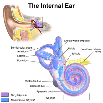
The inner ear is the innermost part of the vertebrate ear. In vertebrates, the inner ear is mainly responsible for sound detection and balance. In mammals, it consists of the bony labyrinth, a hollow cavity in the temporal bone of the skull with a system of passages comprising two main functional parts:

The sense of balance or equilibrioception is the perception of balance and spatial orientation. It helps prevent humans and nonhuman animals from falling over when standing or moving. Equilibrioception is the result of a number of sensory systems working together: the eyes, the inner ears, and the body's sense of where it is in space (proprioception) ideally need to be intact.

The vestibulocochlear nerve, known as the eighth cranial nerve, transmits sound and equilibrium (balance) information from the inner ear to the brain. Through olivocochlear fibers, it also transmits motor and modulatory information from the superior olivary complex in the brainstem to the cochlea.
The semicircular canals or semicircular ducts are three semicircular, interconnected tubes located in the innermost part of each ear, the inner ear. The three canals are the horizontal, superior and posterior semicircular canals.
The saccule is a bed of sensory cells in the inner ear. It translates head movements into neural impulses for the brain to interpret. The saccule detects linear accelerations and head tilts in the vertical plane. When the head moves vertically, the sensory cells of the saccule are disturbed and the neurons connected to them begin transmitting impulses to the brain. These impulses travel along the vestibular portion of the eighth cranial nerve to the vestibular nuclei in the brainstem.

The vestibular system, in vertebrates, is a sensory system that provides the leading contribution to the sense of balance and spatial orientation for the purpose of coordinating movement with balance. Together with the cochlea, a part of the auditory system, it constitutes the labyrinth of the inner ear in most mammals.
Spatial disorientation of an aviator is the inability to determine attitude, altitude or speed. It is most critical at night or in poor weather, when there is no visible horizon, since vision is the dominant sense for orientation. The auditory system, vestibular system, and proprioceptive system collectively work to co-ordinate movement with balance, and can also create illusory nonvisual sensations, resulting in spatial disorientation in the absence of strong visual cues.

An otolith, also called statoconium or otoconium or statolith, is a calcium carbonate structure in the saccule or utricle of the inner ear, specifically in the vestibular system of vertebrates. The saccule and utricle, in turn, together make the otolith organs. These organs are what allows an organism, including humans, to perceive linear acceleration, both horizontally and vertically (gravity). They have been identified in both extinct and extant vertebrates.

In the inner ear, stereocilia are the mechanosensing organelles of hair cells, which respond to fluid motion in numerous types of animals for various functions, including hearing and balance. They are about 10–50 micrometers in length and share some similar features of microvilli. The hair cells turn the fluid pressure and other mechanical stimuli into electric stimuli via the many microvilli that make up stereocilia rods. Stereocilia exist in the auditory and vestibular systems.

The vestibular nerve is one of the two branches of the vestibulocochlear nerve. In humans the vestibular nerve transmits sensory information transmitted by vestibular hair cells located in the two otolith organs and the three semicircular canals via the vestibular ganglion of Scarpa. Information from the otolith organs reflects gravity and linear accelerations of the head. Information from the semicircular canals reflects rotational movement of the head. Both are necessary for the sensation of body position and gaze stability in relation to a moving environment.

Human senses are not naturally geared for the inflight environment. Pilots may experience disorientation and loss of perspective, creating illusions that range from false horizons to sensory conflict with instrument readings or the misjudging of altitude over water.
The ampullary cupula, or cupula, is a structure in the vestibular system, providing the sense of spatial orientation.

The otolithic membrane is a fibrous structure located in the vestibular system of the inner ear. It plays a critical role in the brain's interpretation of equilibrium. The membrane serves to determine if the body or the head is tilted, in addition to the linear acceleration of the body. The linear acceleration could be in the horizontal direction as in a moving car or vertical acceleration such as that felt when an elevator moves up or down.
A kinocilium is a special type of cilium on the apex of hair cells located in the sensory epithelium of the vertebrate inner ear.
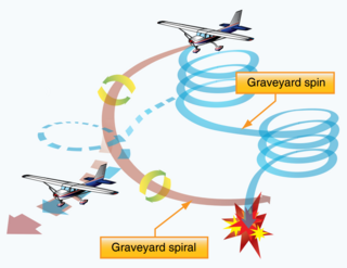
In aviation, a graveyard spiral is a type of dangerous spiral dive entered into accidentally by a pilot who is not trained or not proficient in flying in instrument meteorological conditions (IMC). Other names for this phenomenon include suicide spiral, deadly spiral, death spiral and vicious spiral.

The crista ampullaris is the sensory organ of rotation. They are found in the ampullae of each of the semicircular canals of the inner ear, meaning that there are 3 pairs in total. The function of the crista ampullaris is to sense angular acceleration and deceleration.
The saccule is the smaller sized vestibular sac ; it is globular in form, and lies in the recessus sphæricus near the opening of the scala vestibuli of the cochlea. Its anterior part exhibits an oval thickening, the macula of saccule, to which are distributed the saccular filaments of the acoustic nerve.
The vestibular evoked myogenic potential is a neurophysiological assessment technique used to determine the function of the otolithic organs of the inner ear. It complements the information provided by caloric testing and other forms of inner ear testing. There are two different types of VEMPs. One is the oVEMP and another is the cVEMP. The oVEMP measures integrity of the utricule and superior vestibular nerve and the cVemp measures the saccule and the inferior vestibular nerve.
The righting reflex, also known as the labyrinthine righting reflex, is a reflex that corrects the orientation of the body when it is taken out of its normal upright position. It is initiated by the vestibular system, which detects that the body is not erect and causes the head to move back into position as the rest of the body follows. The perception of head movement involves the body sensing linear acceleration or the force of gravity through the otoliths, and angular acceleration through the semicircular canals. The reflex uses a combination of visual system inputs, vestibular inputs, and somatosensory inputs to make postural adjustments when the body becomes displaced from its normal vertical position. These inputs are used to create what is called an efference copy. This means that the brain makes comparisons in the cerebellum between expected posture and perceived posture, and corrects for the difference. The reflex takes 6 or 7 weeks to perfect, but can be affected by various types of balance disorders.
The Ocular tilt reaction (OTR), comprises skew deviation, head tilt and ocular torsion involving structures of the inner ear responsible for maintenance of balance of the body i.e. the semi-circular canals (SCC), utricle and saccule.

