The ethmoid bone is an unpaired bone in the skull that separates the nasal cavity from the brain. It is located at the roof of the nose, between the two orbits. The cubical bone is lightweight due to a spongy construction. The ethmoid bone is one of the bones that make up the orbit of the eye.

The inferior nasal concha is one of the three paired nasal conchae in the nose. It extends horizontally along the lateral wall of the nasal cavity and consists of a lamina of spongy bone, curled upon itself like a scroll,. The inferior nasal conchae are considered a pair of facial bones. As the air passes through the turbinates, the air is churned against these mucosa-lined bones in order to receive warmth, moisture and cleansing. Superior to inferior nasal concha are the middle nasal concha and superior nasal concha which arise from the cranial portion of the skull. Hence, these two are considered as a part of the cranial bones.
Lamina is a general anatomical term meaning "plate" or "layer." It is used in both gross anatomy and microscopic anatomy to describe structures.

The spinocerebellar tract is a nerve tract originating in the spinal cord and terminating in the same side (ipsilateral) of the cerebellum.

The sphenoidal conchae are two thin, curved plates, situated at the anterior and lower part of the body of the sphenoid. An aperture of variable size exists in the anterior wall of each, and through this the sphenoidal sinus opens into the nasal cavity.
The pterygoid processes of the sphenoid, one on either side, descend perpendicularly from the regions where the body and the greater wings of the sphenoid bone unite.

The Labyrinth or Lateral Mass of the ethmoid bone consists of a number of thin-walled cellular cavities, the ethmoidal cells, arranged in three groups, anterior, middle, and posterior, and interposed between two vertical plates of bone; the lateral plate forms part of the orbit, the medial plate forms part of the nasal cavity. In the disarticulated bone many of these cells are opened into, but when the bones are articulated, they are closed in at every part, except where they open into the nasal cavity.
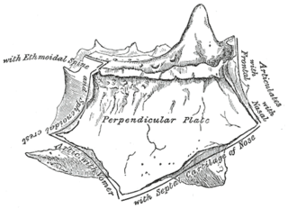
The perpendicular plate of the ethmoid bone is a thin, flattened lamina, polygonal in form, which descends from the under surface of the cribriform plate, and assists in forming the septum of the nose; it is generally deflected a little to one or other side. The anterior border articulates with the spine of the frontal bone and the crest of the nasal bones.

The median portion of the wall of the forebrain consists of a thin lamina, the lamina terminalis, which stretches from the Interventricular foramen to the recess at the base of the optic stalk and contains the vascular organ of the lamina terminalis, which regulates the osmotic concentration of the blood. The lamina terminalis is immediately anterior to the tuber cinereum; together they form the pituitary stalk.

The base of the cartilaginous portion of the auditory tube lies directly under the mucous membrane of the nasal part of the pharynx, where it forms an elevation, the torus tubarius, the torus of the auditory tube, or cushion, behind the pharyngeal orifice of the tube. The torus tubarius is very close to the tubal tonsil, which is sometimes also called the tonsil of (the) torus tubarius. Equating the torus with its tonsil however might be seen as incorrect or imprecise.

The petrous part of the temporal bone is pyramid-shaped and is wedged in at the base of the skull between the sphenoid and occipital bones. Directed medially, forward, and a little upward, it presents a base, an apex, three surfaces, and three angles, and houses in its interior, the components of the inner ear. The petrous portion is among the most basal elements of the skull and forms part of the endocranium. Petrous comes from the Latin word petrosus, meaning "stone-like, hard". It is one of the densest bones in the body.

The osseous spiral lamina is a bony shelf or ledge which projects from the modiolus into the interior of the canal, and, like the canal, takes two-and-three-quarter turns around the modiolus.
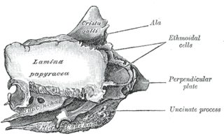
The orbital lamina of ethmoid bone, is a smooth, oblong bone plate which forms the lateral surface of the labyrinth of the ethmoid bone in the skull. The plate covers in the middle and posterior ethmoidal cells and forms a large part of the medial wall of the orbit.

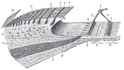
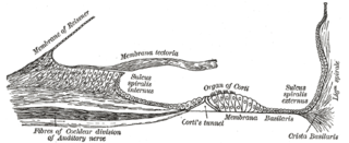
The basilar membrane stretches from the tympanic lip of the osseous spiral lamina to the basilar crest and consists of two parts, an inner and an outer. The inner is thin, and is named the inner tunnel : it supports the spiral organ of Corti.
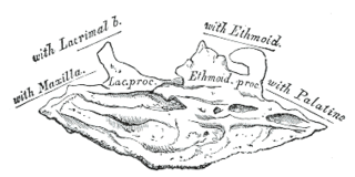
Behind the lacrimal process of the inferior nasal conchae lies a broad, thin plate, the ethmoidal process, which ascends to join the uncinate process of the ethmoid; from its lower border a thin lamina, the maxillary process, curves downward and lateralward; it articulates with the maxilla and forms a part of the medial wall of the maxillary sinus.
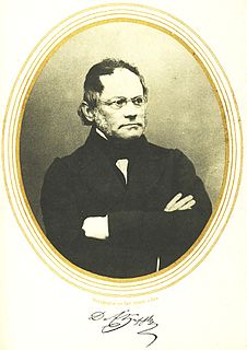
Emil Huschke was a German anatomist and embryologist who was a native of Weimar.

The reticular membrane is a thin, stiff lamina that extends from the outer hair cells to the Hensen's cells. The RM is composed of "minute-fiddle-shaped cuticular structures" called the phalangeal extensions of the outer hair cells, interspaced with extensions coming from the outer phalangeal cells. The RM separates endolymph in the cochlear duct from underlying corticolymph and perilymph of the scala tympani.

In the vertebrate spinal column, each vertebra is an irregular bone with a complex structure composed of bone and some hyaline cartilage, the proportions of which vary according to the segment of the backbone and the species of vertebrate.
A plate in animal anatomy may refer to several things: