Stapes

Last updated
Stapes
Gray918.png
Frontal view of stapes (A), and view from below (B).
Details
Pronunciation /ˈstpz/
Precursor Second branchial arch
Part of Middle ear
System Auditory system
Articulations Incudostapedial joint
Identifiers
Latin 'stapes'
MeSH D013199
TA98 A15.3.02.033
TA2 895
FMA 52751
Anatomical terms of bone

The stapes or stirrup is a bone in the middle ear of humans and other animals which is involved in the conduction of sound vibrations to the inner ear. This bone is connected to the oval window by its annular ligament, which allows the footplate (or base) to transmit sound energy through the oval window into the inner ear. The stapes is the smallest and lightest bone in the human body, and is so-called because of its resemblance to a stirrup (Latin : Stapes).

Contents

Structure

The size of the stapes, compared with a 10-cent euro coin. Stapes human ear.jpg
The size of the stapes, compared with a 10-cent euro coin.

The stapes is the third bone of the three ossicles in the middle ear and the smallest in the human body. It measures roughly 2 to 3 mm, greater along the head-base span. [1] It rests on the oval window, to which it is connected by an annular ligament and articulates with the incus , or anvil through the incudostapedial joint. [2] They are connected by anterior and posterior limbs (Latin : crura). [3] :862

Development

The stapes develops from the second pharyngeal arch during the sixth to eighth week of embryological life. The central cavity of the stapes, the obturator foramen, is due to the presence embryologically of the stapedial artery, which usually regresses in humans during normal development. [2] [4]

Animals

The stapes is one of three ossicles in mammals. In non-mammalian tetrapods, the bone homologous to the stapes is usually called the columella; however, in reptiles, either term may be used. In fish, the homologous bone is called the hyomandibular, and is part of the gill arch supporting either the spiracle or the jaw, depending on the species. The equivalent term in amphibians is the pars media plectra. [2] [5] :481–482

Variation

The stapes appears to be relatively constant in size in different ethnic groups. [6] In 0.01–0.02% of people, the stapedial artery does not regress, and persists in the central foramen. [7] In this case, a pulsatile sound may be heard in the affected ear, or there may be no symptoms at all. [8] Rarely, the stapes may be completely absent. [9] [10] :262

Function

Situated between the incus and the inner ear, the stapes transmits sound vibrations from the incus to the oval window, a membrane-covered opening to the inner ear. The stapes is also stabilized by the stapedius muscle, which is innervated by the facial nerve. [3] :861–863

Clinical relevance

Otosclerosis is a congenital or spontaneous-onset disease characterized by abnormal bone remodeling in the inner ear. Often this causes the stapes to adhere to the oval window, which impedes its ability to conduct sound, and is a cause of conductive hearing loss. Clinical otosclerosis is found in about 1% of people, although it is more common in forms that do not cause noticeable hearing loss. Otosclerosis is more likely in young age groups, and females. [11] Two common treatments are stapedectomy, the surgical removal of the stapes and replacement with an artificial prosthesis, and stapedotomy, the creation of a small hole in the base of the stapes followed by the insertion of an artificial prosthesis into that hole. [12] :661 Surgery may be complicated by a persistent stapedial artery, fibrosis-related damage to the base of the bone, or obliterative otosclerosis, resulting in obliteration of the base. [7] [10] :254–262

History

The stapes, as first described by Giovanni Filippo Ingrassia (Labeled M, bottom right). Ingrassia stapes noback.jpg
The stapes, as first described by Giovanni Filippo Ingrassia (Labeled M, bottom right).

The stapes is commonly described as having been discovered by the professor Giovanni Filippo Ingrassia in 1546 at the University of Naples, [13] although this remains the nature of some controversy, as Ingrassia's description was published posthumously in his 1603 anatomical commentary In Galeni librum de ossibus doctissima et expectatissima commentaria. Spanish anatomist Pedro Jimeno is first to have been credited with a published description, in Dialogus de re medica (1549). [14] The bone is so-named because of its resemblance to a stirrup (Latin : stapes), an example of a late Latin word, probably created in mediaeval times from "to stand" (Latin : stapia), as stirrups did not exist in the early Latin-speaking world. [15]

Related Research Articles

<span class="mw-page-title-main">Inner ear</span> Innermost part of the vertebrate ear

The inner ear is the innermost part of the vertebrate ear. In vertebrates, the inner ear is mainly responsible for sound detection and balance. In mammals, it consists of the bony labyrinth, a hollow cavity in the temporal bone of the skull with a system of passages comprising two main functional parts:

<span class="mw-page-title-main">Middle ear</span> Portion of the ear internal to the eardrum, and external to the oval window of the inner ear

The middle ear is the portion of the ear medial to the eardrum, and distal to the oval window of the cochlea.

<span class="mw-page-title-main">Cholesteatoma</span> Medical condition

Cholesteatoma is a destructive and expanding growth consisting of keratinizing squamous epithelium in the middle ear and/or mastoid process. Cholesteatomas are not cancerous as the name may suggest, but can cause significant problems because of their erosive and expansile properties. This can result in the destruction of the bones of the middle ear (ossicles), as well as growth through the base of the skull into the brain. They often become infected and can result in chronically draining ears. Treatment almost always consists of surgical removal.

The ossicles are three bones in either middle ear that are among the smallest bones in the human body. They serve to transmit sounds from the air to the fluid-filled labyrinth (cochlea). The absence of the auditory ossicles would constitute a moderate-to-severe hearing loss. The term "ossicle" literally means "tiny bone". Though the term may refer to any small bone throughout the body, it typically refers to the malleus, incus, and stapes of the middle ear.

<i>Incus</i> Bone of the middle ear

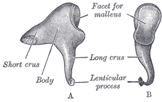
The incus or anvil in the ear is one of three small bones (ossicles) in the middle ear. The incus receives vibrations from the malleus, to which it is connected laterally, and transmits these to the stapes medially. The incus is named for its resemblance to an anvil.

<span class="mw-page-title-main">Cochlea</span> Snail-shaped part of inner ear involved in hearing

The cochlea is the part of the inner ear involved in hearing. It is a spiral-shaped cavity in the bony labyrinth, in humans making 2.75 turns around its axis, the modiolus. A core component of the cochlea is the organ of Corti, the sensory organ of hearing, which is distributed along the partition separating the fluid chambers in the coiled tapered tube of the cochlea.

<span class="mw-page-title-main">Otosclerosis</span> Condition characterized by an abnormal bone growth in the middle ear

Otosclerosis is a condition of the middle ear where portions of the dense enchondral layer of the bony labyrinth remodel into one or more lesions of irregularly-laid spongy bone. As the lesions reach the stapes the bone is resorbed, then hardened (sclerotized), which limits its movement and results in hearing loss, tinnitus, vertigo or a combination of these. The term otosclerosis is something of a misnomer: much of the clinical course is characterized by lucent rather than sclerotic bony changes, so the disease is also known as otospongiosis.

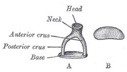
Stapedectomy is a surgical procedure in which the stapes bone is removed from the middle ear and replaced with a prosthesis.

<span class="mw-page-title-main">Ear</span> Organ of hearing and balance

An ear is the organ that enables hearing and body balance using the vestibular system. In mammals, the ear is usually described as having three parts: the outer ear, the middle ear and the inner ear. The outer ear consists of the pinna and the ear canal. Since the outer ear is the only visible portion of the ear in most animals, the word "ear" often refers to the external part alone. The middle ear includes the tympanic cavity and the three ossicles. The inner ear sits in the bony labyrinth, and contains structures which are key to several senses: the semicircular canals, which enable balance and eye tracking when moving; the utricle and saccule, which enable balance when stationary; and the cochlea, which enables hearing. The ear is a self cleaning organ through its relationship with earwax and the ear canals. The ears of vertebrates are placed somewhat symmetrically on either side of the head, an arrangement that aids sound localization.

<span class="mw-page-title-main">Acoustic reflex</span> Small muscle contraction in the middle ear in response to loud sound

The acoustic reflex is an involuntary muscle contraction that occurs in the middle ear in response to loud sound stimuli or when the person starts to vocalize.

<span class="mw-page-title-main">Stapedius muscle</span> Muscle in the human ear

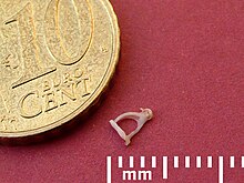
The stapedius is the smallest skeletal muscle in the human body. At just over one millimeter in length, its purpose is to stabilize the smallest bone in the body, the stapes or stirrup bone of the middle ear.

<span class="mw-page-title-main">Tensor tympani muscle</span> Muscle of the middle ear

The tensor tympani is a muscle within the middle ear, located in the bony canal above the bony part of the auditory tube, and connects to the malleus bone. Its role is to damp loud sounds, such as those produced from chewing, shouting, or thunder. Because its reaction time is not fast enough, the muscle cannot protect against hearing damage caused by sudden loud sounds, like explosions or gunshots.

<span class="mw-page-title-main">Evolution of mammalian auditory ossicles</span> Middle ear bones evolved from jaw bones

The evolution of mammalian auditory ossicles was an evolutionary process that resulted in the formation of the bones of the mammalian middle ear. These bones, or ossicles, are a defining characteristic of all mammals. The event is well-documented and important as a demonstration of transitional forms and exaptation, the re-purposing of existing structures during evolution.

<span class="mw-page-title-main">Columella (auditory system)</span> Bony structures in the skull that serve the purpose of transmitting sounds

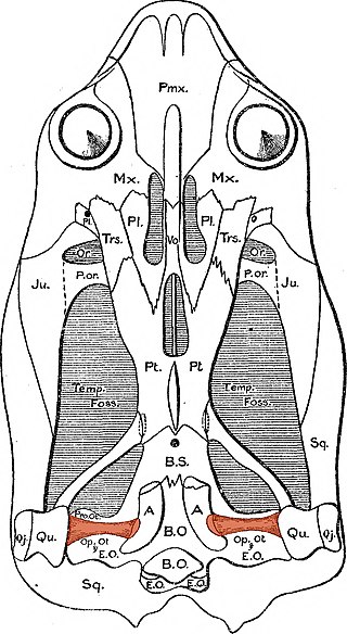
In the auditory system, the columella contributes to hearing in amphibians, reptiles and birds. The columella form thin, bony structures in the interior of the skull and serve the purpose of transmitting sounds from the eardrum. It is an evolutionary homolog of the stapes, one of the auditory ossicles in mammals.

<span class="mw-page-title-main">Persistent stapedial artery</span> Blood vessel anomaly

A persistent stapedial artery (PSA) is a rare anomaly in human anatomy where the stapedial branch of posterior auricular artery, or simply stapedial artery, remains within the ear of a fetus after the first ten weeks of pregnancy. Whilst not problematic for the majority of people with the anomaly, it can cause difficulties with hearing.

Endoscopic ear surgery (EES) is a minimally invasive alternative to traditional ear surgery and is defined as the use of the rigid endoscope, as opposed to a surgical microscope, to visualize the middle and inner ear during otologic surgery. During endoscopic ear surgery the surgeon holds the endoscope in one hand while working in the ear with the other. To allow this kind of single-handed surgery, different surgical instruments have to be used. Endoscopic visualization has improved due to high-definition video imaging and wide-field endoscopy, and being less invasive, EES is gaining importance as an adjunct to microscopic ear surgery.

<i>Malleus</i> Bone of the middle ear

The malleus, or hammer, is a hammer-shaped small bone or ossicle of the middle ear. It connects with the incus, and is attached to the inner surface of the eardrum. The word is Latin for 'hammer' or 'mallet'. It transmits the sound vibrations from the eardrum to the incus (anvil).

A middle ear implant is a hearing device that is surgically implanted into the middle ear. They help people with conductive, sensorineural or mixed hearing loss to hear. 

<span class="mw-page-title-main">Thickened earlobes-conductive deafness syndrome</span> Medical condition

Thickened earlobes-conductive deafness syndrome, also known as Escher-Hirt syndrome, or Schweitzer Kemink Graham syndrome, is a rare genetic disorder which is characterized by ear and jaw abnormalities associated with progressive hearing loss. Two families worldwide have been described with the disorder.

Chaoyangodens is an extinct genus of eutriconodont mammal from the Early Cretaceous of China. It includes a single species, Chaoyangodens lii, known from a single complete skeleton recovered from the Dawangzhangzi bed of the Yixian Formation, part of the fossiliferous Jehol biota. Chaoyangodens was a moderate-sized Mesozoic mammal. The generic name refers to Chaoyang Prefecture while the specific name honors the collector of the fossil, Hai-Jun Li. Chaoyangodens is intermediate in age between Liaoconodon and a diverse fauna of eutriconodonts from older beds of the Yixian Formation. Like Liaoconodon, it is not easily equated with other eutriconodonts, since it bears distinctive dental traits relative to recognized eutriconodont subgroups.

References

  1. Àwengen, D. F.; Nishihara, S.; Kurokawa, H.; Goode, R. L. (April 1995). "Measurements of the stapes superstructure". The Annals of Otology, Rhinology, and Laryngology. 104 (4 Pt 1): 311–6. doi:10.1177/000348949510400411. PMID   7717624. S2CID   43418740.
  2. 1 2 3 Chapman, S. C. (January 1, 2011). "Can you hear me now? Understanding vertebrate middle ear development". Frontiers in Bioscience. 16 (5): 1675–92. doi:10.2741/3813. PMC   3065862 . PMID   21196256.
  3. 1 2 Drake, Richard L.; Vogl, Wayne; Tibbitts, Adam W. M. Mitchell (2005). Gray's Anatomy for Students . Illustrations by Richard Tibbitts and Paul Richardson. Philadelphia: Elsevier/Churchill Livingstone. ISBN   978-0-8089-2306-0.
  4. Rodriguez-Vazquez, J. F. (August 2005). "Development of the stapes and associated structures in human embryos". Journal of Anatomy. 207 (2): 165–173. doi:10.1111/j.1469-7580.2005.00441.x. PMC   1571512 . PMID   16050903.
  5. Romer, Alfred Sherwood; Parsons, Thomas S (1977). The Vertebrate Body. Philadelphia, Pennsylvania: Holt-Saunders International. ISBN   978-0-03-910284-5.
  6. Arensburg, B.; Harell, M.; Nathan, H. (February 1981). "The human middle ear ossicles: Morphometry, and taxonomic implications". Journal of Human Evolution. 10 (2): 199–205. doi:10.1016/S0047-2484(81)80018-8.
  7. 1 2 Mutlu, C.; da Costa, S. S.; Paparella, MM; Schachern, Pennsylvania (1998). "Clinical-histopathological correlations of pitfalls in middle ear surgery". European Archives of Oto-Rhino-Laryngology. 255 (4): 189–194. doi:10.1007/s004050050041. PMID   9592676. S2CID   25682582.
  8. Silbergleit, R.; Quint, D. J.; Mehta, B. A.; Patel, S. C.; Metes, J. J.; Noujaim, S. E. (Mar 2000). "The persistent stapedial artery". American Journal of Neuroradiology. 21 (3): 572–577. PMC   8174972 . PMID   10730654.
  9. Reiber, M.; Schwaber, M. (February 1997). "Congenital absence of stapes and facial nerve dehiscence". Otolaryngology–Head and Neck Surgery. 116 (2): 278. doi:10.1016/S0194-5998(97)70343-7. PMID   9051082. S2CID   33351053.
  10. 1 2 Tympanoplasty, Mastoidectomy, and Stapes Surgery. Georg Thieme Verlag. 2008. ISBN   978-1-282-86537-2.
  11. Menger, D. J.; Tange, R. A. (April 2003). "The aetiology of otosclerosis: a review of the literature". Clinical Otolaryngology and Allied Sciences. 28 (2): 112–120. doi: 10.1046/j.1365-2273.2003.00675.x . PMID   12680829.
  12. Hall, John E.; Guyton, Arthur C. (2005). Textbook of medical physiology (11th ed.). Philadelphia: W. B. Saunders. ISBN   978-0-7216-0240-0.
  13. Dispenza, F.; Cappello, F.; Kulamarva, G.; De Stefano, A. (October 2013). "The discovery of stapes". Acta Otorhinolaryngologica Italica. 33 (5): 357–359. PMC   3825043 . PMID   24227905.
  14. Mudry, Albert (April 2013). "Disputes Surrounding the Discovery of the Stapes in the Mid 16th Century". Otology & Neurotology. 34 (3): 588–592. doi:10.1097/MAO.0b013e31827d8abc. PMID   23370557. S2CID   30466939.
  15. Harper, Douglas. "Stapes (n.)". Online Etymology Dictionary. Retrieved 27 December 2013.