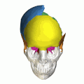
The skull is a bone protective cavity for the brain. The skull is composed of four types of bone i.e., cranial bones, facial bones, ear ossicles and hyoid bone, however two parts are more prominent: the cranium and the mandible. In humans, these two parts are the neurocranium (braincase) and the viscerocranium that includes the mandible as its largest bone. The skull forms the anterior-most portion of the skeleton and is a product of cephalisation—housing the brain, and several sensory structures such as the eyes, ears, nose, and mouth. In humans, these sensory structures are part of the facial skeleton.

The sphenoid bone is an unpaired bone of the neurocranium. It is situated in the middle of the skull towards the front, in front of the basilar part of the occipital bone. The sphenoid bone is one of the seven bones that articulate to form the orbit. Its shape somewhat resembles that of a butterfly or bat with its wings extended.

The occipital bone is a cranial dermal bone and the main bone of the occiput. It is trapezoidal in shape and curved on itself like a shallow dish. The occipital bone overlies the occipital lobes of the cerebrum. At the base of the skull in the occipital bone, there is a large oval opening called the foramen magnum, which allows the passage of the spinal cord.

The temporal bones are situated at the sides and base of the skull, and lateral to the temporal lobes of the cerebral cortex.

A snake skeleton consists primarily of the skull, vertebrae, and ribs, with only vestigial remnants of the limbs.

The basilar part of the occipital bone extends forward and upward from the foramen magnum, and presents in front an area more or less quadrilateral in outline.

Macroplata is an extinct genus of Early Jurassic rhomaleosaurid plesiosaur which grew up to 4.65 metres (15.3 ft) in length. Like other plesiosaurs, Macroplata probably lived on a diet of fish, using its sharp needle-like teeth to catch prey. Its shoulder bones were fairly large, indicating a powerful forward stroke for fast swimming. Macroplata also had a relatively long neck, twice the length of the skull, in contrast to pliosaurs.
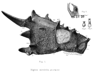
Elginia is an extinct genus of pareiasaurid known from the Late Permian of Scotland and China. It was named for the area around Elgin in Scotland, which has yielded many fossils referred to as the Elgin Reptiles.

Askeptosaurus is an extinct genus of askeptosauroid, a marine reptile from the extinct order Thalattosauria. Askeptosaurus is known from several well-preserved fossils found in Middle Triassic marine strata in what is now Italy and Switzerland.

Bauria is an extinct genus of the suborder Therocephalia that existed during the Early and Middle Triassic period, around 246-251 million years ago. It belonged to the family Bauriidae. Bauria was probably a carnivore or insectivore. It lived in South Africa, specifically in the Burgersdorp Formation in South Africa.

The endocranium in comparative anatomy is a part of the skull base in vertebrates and it represents the basal, inner part of the cranium. The term is also applied to the outer layer of the dura mater in human anatomy.

Leptopleuron is an extinct genus of procolophonid that lived in the dry lands during the late Triassic in Elgin of northern Scotland and was the first to be included in the clade of Procolophonidae. First described by English paleontologist and biologist Sir Richard Owen, Leptopleuron is derived from two Greek bases, leptos for "slender" and pleuron for "rib," describing it as having slender ribs. The fossil is also known by a second name, Telerpeton, which is derived from the Greek bases tele for "far off" and herpeton for "reptile." In Scotland, Leptopleuron was found specifically in the Lossiemouth Sandstone Formation. The yellow sandstone it was located in was poorly lithified with wind coming from the southwest. The environment is also described to consist of barchan dunes due to the winds, ranging up to 20 m tall that spread during dry phases into flood plains. Procolophonoids such as Leptopleuron were considered an essential addition to the terrestrial ecosystem during the Triassic.
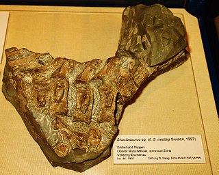
Phantomosaurus is an extinct genus of ichthyosaur that lived during the late Anisian stage of the Middle Triassic. Fossils have been found in southern Germany. It was discovered in 1965 and named in 1997 as a species of Shastasaurus by Sander in the rocks of the Upper Muschelkalk.

Vancleavea is a genus of extinct, armoured, non-archosaurian archosauriforms from the Late Triassic of western North America. The type and only known species is V. campi, named by Robert Long & Phillip A Murry in 1995. At that time, the genus was only known from fragmentary bones including osteoderms and vertebrae. However, since then many more fossils have been found, including a pair of nearly complete skeletons discovered in 2002. These finds have shown that members of the genus were bizarre semiaquatic reptiles. Vancleavea individuals had short snouts with large, fang-like teeth, and long bodies with small limbs. They were completely covered with bony plates known as osteoderms, which came in several different varieties distributed around the body. Phylogenetic analyses by professional paleontologists have shown that Vancleavea was an archosauriform, part of the lineage of reptiles that would lead to archosaurs such as dinosaurs and crocodilians. Vancleavea lacks certain traits which are present in most other archosauriforms, most notably the antorbital, mandibular and supratemporal fenestrae, which are weight-saving holes in the skulls of other taxa. However, other features clearly support its archosauriform identity, including a lack of intercentra, the presence of osteoderms, an ossified laterosphenoid, and several adaptations of the femur and ankle bones. In 2016, a new genus of archosauriform, Litorosuchus, was described. This genus resembled both Vancleavea and more typical archosauriforms in different respects, allowing Litorosuchus to act as a transitional fossil linking Vancleavea to less aberrant archosauriforms.

The skull roof or the roofing bones of the skull are a set of bones covering the brain, eyes and nostrils in bony fishes and all land-living vertebrates. The bones are derived from dermal bone and are part of the dermatocranium.

Athabascasaurus is an extinct genus of platypterygiine ophthalmosaurid ichthyosaur known from Alberta, Canada.
Huskerpeton is an extinct genus of recumbirostran from the Early Permian period. They belong to the order Microsauria, which was established in 1863 by Dawson, and was quickly expanded to include many different small taxa. They lived in what is now Nebraska and Kansas. The holotype of Huskerpeton was uncovered at the Eskridge formation in Nebraska, which is part of how it got its name.
This glossary explains technical terms commonly employed in the description of dinosaur body fossils. Besides dinosaur-specific terms, it covers terms with wider usage, when these are of central importance in the study of dinosaurs or when their discussion in the context of dinosaurs is beneficial. The glossary does not cover ichnological and bone histological terms, nor does it cover measurements.

Khunnuchelys was a genus of trionychine turtle from the Late Cretaceous of Asia. Three species are known, K. erinhotensis, the type species, K. kizylkumensis, and K. lophorhothon. K. erinhotensis is known from the Iren Dabasu Formation in China from the late Turonian until the middle Campanian. K. kizylkumensis is known from the late Turonian Bissekty Formation of Uzbekistan. The third species, described in 2013 by Danilov et al., is known from the early to middle Campanian aged Bostobe Formation of Kazakhstan.
Lohuecotitan is an extinct genus of titanosaurian sauropod dinosaur which lived during the Late Cretaceous in Spain. The only species known in the genus is Lohuecotitan pandafilandi, described and named in 2016.