Brow ridge

Last updated
Brow ridge
Gray134.png
Frontal bone. Outer surface. Brow ridge labelled as "superciliary arch" at center right).
Identifiers
TA98 A02.1.03.005
TA2 524
FMA 52850
Anatomical terms of bone
The brow ridges are often not well expressed in human females, as pictured above in a female skull, and are most easily seen in profile. Hallonflickans kranium English Supraorbital Ridge Labeled.png
The brow ridges are often not well expressed in human females, as pictured above in a female skull, and are most easily seen in profile.

The brow ridge, or supraorbital ridge known as superciliary arch in medicine, is a bony ridge located above the eye sockets of all primates and some other animals. In humans, the eyebrows are located on their lower margin.

Contents

Structure

The brow ridge is a nodule or crest of bone situated on the frontal bone of the skull. It forms the separation between the forehead portion itself (the squama frontalis) and the roof of the eye sockets (the pars orbitalis). Normally, in humans, the ridges arch over each eye, offering mechanical protection. In other primates, the ridge is usually continuous and often straight rather than arched. The ridges are separated from the frontal eminences by a shallow groove. The ridges are most prominent medially, and are joined to one another by a smooth elevation named the glabella.

Typically, the arches are more prominent in men than in women, [1] and vary between different ethnic groups. Behind the ridges, deeper in the bone, are the frontal sinuses.

Terminology

The brow ridges, being a prominent part of the face in some ethnic groups and a trait linked to both atavism and sexual dimorphism, have a number of names in different disciplines. In vernacular English, the terms eyebrow bone or eyebrow ridge are common. The more technical terms frontal or supraorbital arch, ridge or torus (or tori to refer to the plural, as the ridge is usually seen as a pair) are often found in anthropological or archaeological studies. In medicine, the term arcus superciliaris (Latin) or the English translation superciliary arch. This feature is different from the supraorbital margin and the margin of the orbit.

Some paleoanthropologists distinguish between frontal torus and supraorbital ridge. [2] In anatomy, a torus is a projecting shelf of bone that unlike a ridge is rectilinear, unbroken and goes through glabella. [3] Some fossil hominins, in this use of the word, have the frontal torus, [4] but almost all modern humans only have the ridge. [5]

Development

Spatial model

The Spatial model proposes that supraorbital torus development can be best explained in terms of the disparity between the anterior position of the orbital component relative to the neurocranium.

Much of the groundwork for the spatial model was laid down by Schultz (1940). He was the first to document that at later stages of development (after age 4) the growth of the orbit would outpace that of the eye. Consequently, he proposed that facial size is the most influential factor in orbital development, with orbital growth being only secondarily affected by size and ocular position.

Weindenreich (1941) and Biegert (1957, 1963) argued that the supraorbital region can best be understood as a product of the orientation of its two components, the face and the neurocranium.

The most composed articulation of the spatial model was presented by Moss and Young (1960), who stated that "the presence… of supraorbital ridges is only the reflection of the spatial relationship between two functionally unrelated cephalic components, the orbit and the brain" (Moss and Young, 1960, p282). They proposed (as first articulated by Biegert in 1957) that during infancy the neurocranium extensively overlaps the orbit, a condition that prohibits brow ridge development. As the splanchnocranium grows, however, the orbits begin to advance, thus causing the anterior displacement of the face relative to the brain. Brow ridges then form as a result of this separation.

Bio-mechanical model

The bio-mechanical model predicts that morphological variation in torus size is the direct product of differential tension caused by mastication, as indicated by an increase in load/lever ratio and broad craniofacial angle. [6]

Research done on this model has largely been based on earlier work of Endo. By applying pressure similar to the type associated with chewing, he carried out an analysis of the structural function of the supraorbital region on dry human and gorilla skulls. His findings indicated that the face acts as a pillar that carries and disperses tension caused by the forces produced during mastication. [7] [8] [9] [10] Russell and Oyen et al. elaborated on this idea, suggesting that amplified facial projection necessitates the application of enhanced force to the anterior dentition in order to generate the same bite power that individuals with a dorsal deflection of the facial skull exert. In more prognathic individuals, this increased pressure triggers bone deposition to reinforce the brow ridges, until equilibrium is reached. [11] [12] [13]

Native American man with pronounced brow ridge and sloping forehead. Wolf Robe yo.jpg
Native American man with pronounced brow ridge and sloping forehead.

Oyen et al. conducted a cross-section study of Papio anubis in order to ascertain the relationship between palate length, incisor load and Masseter lever efficiency, relative to torus enlargement. Indications found of osteoblastic deposition in the glabella were used as evidence for supraorbital enlargement. Oyen et al.’s data suggested that more prognathic individuals experienced a decrease in load/lever efficiency. This transmits tension via the frontal process of the maxilla to the supraorbital region, resulting in a contemporary reinforcement of this structure. This was also correlated to periods of tooth eruption. [14]

In a later series of papers, Russell developed aspects of this mode further. Employing an adult Australian sample, she tested the association between brow ridge formation and anterior dental loading, via the craniofacial angle (prosthion-nasion-metopion), maxilla breadth, and discontinuities in food preparation such as those observed between different age groups. Finding strong support for the first two criteria, she concluded that the supraorbital complex is formed as a result of increased tension due to the widening of the maxilla, thought to be positively correlated with the size of the masseter muscle, as well as with the improper orientation of bone in the superior orbital region. [11] [15]

Function

Some researchers have suggested that brow ridges function to protect the eyes and orbital bones during hand-to-hand combat, given that they are an incredibly dimorphic trait. [16]

Image of a rugby player with a pronounced supraorbital ridge. Rugby player with a pronounced supraorbital ridge.jpg
Image of a rugby player with a pronounced supraorbital ridge.

Paleolithic humans

Pronounced brow ridges were a common feature among paleolithic humans. Early modern people such as those from the finds from Jebel Irhoud and Skhul and Qafzeh had thick, large brow ridges, but they differ from those of archaic humans like Neanderthals by having a supraorbital foramen or notch, forming a groove through the ridge above each eye, although there were exceptions, such as Skhul 2 in which the ridge was unbroken, unlike other members of her tribe. [17] [18] This splits the ridge into central parts and distal parts. In current humans, almost always only the central sections of the ridge are preserved (if preserved at all). This contrasts with many archaic and early modern humans, where the brow ridge is pronounced and unbroken. [19]

Other animals

Gorilla with a frontal torus Gorilla Cin Zoo 020.jpg
Gorilla with a frontal torus

The size of these ridges varies also between different species of primates, either living or fossil. The closest living relatives of humans, the great apes and especially gorillas or chimpanzees, have a very pronounced supraorbital ridge, which has also been called a frontal torus, [4] while in modern humans and orangutans, it is relatively reduced. The fossil record indicates that the supraorbital ridge in early hominins was reduced as the cranial vault grew; the frontal portion of the brain became positioned above rather than behind the eyes, giving a more vertical forehead.

Supraorbital ridges are also present in some other animals, such as wild rabbits, [20] eagles [21] and certain species of sharks. [22] The presence of a supraorbital ridge in the Korean field mouse has been used to distinguish it among related species. [23]

See also

Related Research Articles

<span class="mw-page-title-main">Skull</span> Bony structure that forms the head in vertebrates

The skull is a bone protective cavity for the brain. The skull is composed of four types of bone i.e., cranial bones, facial bones, ear ossicles and hyoid bone, however two parts are more prominent: the cranium and the mandible. In humans, these two parts are the neurocranium (braincase) and the viscerocranium that includes the mandible as its largest bone. The skull forms the anterior-most portion of the skeleton and is a product of cephalisation—housing the brain, and several sensory structures such as the eyes, ears, nose, and mouth. In humans, these sensory structures are part of the facial skeleton.

Facial feminization surgery (FFS) is a set of reconstructive surgical procedures that alter typically male facial features to bring them closer in shape and size to typical female facial features. FFS can include various bony and soft tissue procedures such as brow lift, rhinoplasty, cheek implantation, and lip augmentation.

<span class="mw-page-title-main">Zygomatic bone</span> Facial bone

In the human skull, the zygomatic bone, also called cheekbone or malar bone, is a paired irregular bone which articulates with the maxilla, the temporal bone, the sphenoid bone and the frontal bone. It is situated at the upper and lateral part of the face and forms the prominence of the cheek, part of the lateral wall and floor of the orbit, and parts of the temporal fossa and the infratemporal fossa. It presents a malar and a temporal surface; four processes, and four borders.

<span class="mw-page-title-main">Frontal bone</span> Bone in the human skull

The frontal bone is a bone in the human skull. The bone consists of two portions. These are the vertically oriented squamous part, and the horizontally oriented orbital part, making up the bony part of the forehead, part of the bony orbital cavity holding the eye, and part of the bony part of the nose respectively. The name comes from the Latin word frons.

<span class="mw-page-title-main">Forehead</span> Area of the head between the eyebrows and the hairline

In human anatomy, the forehead is an area of the head bounded by three features, two of the skull and one of the scalp. The top of the forehead is marked by the hairline, the edge of the area where hair on the scalp grows. The bottom of the forehead is marked by the supraorbital ridge, the bone feature of the skull above the eyes. The two sides of the forehead are marked by the temporal ridge, a bone feature that links the supraorbital ridge to the coronal suture line and beyond. However, the eyebrows do not form part of the forehead.

<span class="mw-page-title-main">Corrugator supercilii muscle</span> Muscle near the eye

The corrugator supercilii muscle is a small, narrow, pyramidal muscle of the face. It arises from the medial end of the superciliary arch; it inserts into the deep surface of the skin of the eyebrow.

<span class="mw-page-title-main">Skull fracture</span> Medical condition

A skull fracture is a break in one or more of the eight bones that form the cranial portion of the skull, usually occurring as a result of blunt force trauma. If the force of the impact is excessive, the bone may fracture at or near the site of the impact and cause damage to the underlying structures within the skull such as the membranes, blood vessels, and brain.

<span class="mw-page-title-main">Occipitofrontalis muscle</span> Facial muscle helping to create facial expressions

The occipitofrontalis muscle is a muscle which covers parts of the skull. It consists of two parts or bellies: the occipital belly, near the occipital bone, and the frontal belly, near the frontal bone. It is supplied by the supraorbital artery, the supratrochlear artery, and the occipital artery. It is innervated by the facial nerve. In humans, the occipitofrontalis helps to create facial expressions.

<span class="mw-page-title-main">Frontal sinus</span> Airspace below ridge of eyebrow in humam anatomy

The frontal sinuses are one of the four pairs of paranasal sinuses that are situated behind the brow ridges. Sinuses are mucosa-lined airspaces within the bones of the face and skull. Each opens into the anterior part of the corresponding middle nasal meatus of the nose through the frontonasal duct which traverses the anterior part of the labyrinth of the ethmoid. These structures then open into the semilunar hiatus in the middle meatus.

<span class="mw-page-title-main">Supraorbital foramen</span> Opening above the eye socket, below the forehead

The supraorbital foramen, is a bony elongated opening located above the orbit and under the forehead. It is part of the frontal bone of the skull. The supraorbital foramen lies directly under the eyebrow. In some people this foramen is incomplete and is then known as the supraorbital notch.

<span class="mw-page-title-main">Squamous part of the frontal bone</span> Upper two-thirds of the forward skull

The squamous part of the frontal bone is the superior portion when viewed in standard anatomical orientation. There are two surfaces of the squamous part of the frontal bone: the external surface, and the internal surface.

<span class="mw-page-title-main">Zygomatic process</span>

The zygomatic processes are three processes (protrusions) from other bones of the skull which each articulate with the zygomatic bone. The three processes are:

<span class="mw-page-title-main">Post-orbital constriction</span>

In physical anthropology, post-orbital constriction is the narrowing of the cranium (skull) just behind the eye sockets found in most non-human primates and early hominins. This constriction is very noticeable in non-human primates, slightly less so in Australopithecines, even less in Homo erectus and completely disappears in modern Homo sapiens. Post-orbital constriction index in non-human primates and hominin range in category from increased constriction, intermediate, reduced constriction and disappearance. The post-orbital constriction index is defined by either a ratio of minimum frontal breadth (MFB), behind the supraorbital torus, divided by the maximum upper facial breadth (BFM), bifrontomalare temporale, or as the maximum width behind the orbit of the skull.

The postorbital bar is a bony arched structure that connects the frontal bone of the skull to the zygomatic arch, which runs laterally around the eye socket. It is a trait that only occurs in mammalian taxa, such as most strepsirrhine primates and the hyrax, while haplorhine primates have evolved fully enclosed sockets. One theory for this evolutionary difference is the relative importance of vision to both orders. As haplorrhines tend to be diurnal, and rely heavily on visual input, many strepsirrhines are nocturnal and have a decreased reliance on visual input.

<span class="mw-page-title-main">Neurocranium</span> Part of the skull around the brain

In human anatomy, the neurocranium, also known as the braincase, brainpan, or brain-pan is the upper and back part of the skull, which forms a protective case around the brain. In the human skull, the neurocranium includes the calvaria or skullcap. The remainder of the skull is the facial skeleton.

<i>Lufengpithecus</i> Extinct genus of primates

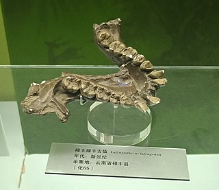
Lufengpithecus is an extinct genus of ape, known from the Late Miocene of East Asia. It is known from thousands of dental remains and a few skulls and probably weighed about 50 kg (110 lb). It contains three species: L. lufengensis, L. hudienensis and L. keiyuanensis. Lufengpithecus lufengensis is from the Late Miocene found in China, named after the Lufeng site and dated around 6.2 Ma. Lufengopithecus is either thought to be the sister group to Ponginae, or the sister to the clade containing Ponginae and Homininae.

Rooneyia viejaensis is a relatively small primate belonging to the extinct monotypic genus Rooneyia. Rooneyia viejaensis is known from the North American Eocene of the Sierra Vieja of West Texas; the species is only known from the type specimen. The lack of additional fossils at this time makes it difficult to hypothesize where and how Rooneyia may have evolved. The minimal wear upon the molar teeth of the specimen has led to the assumption that the type specimen is that of a young adult. Rooneyia does not consistently fall within any one group of fossil or extant primates.

<span class="mw-page-title-main">Chellean Man</span> Hominin fossil

Olduvai Hominid number 9, known as the Chellean Man, is a fossilized skull cap of an early hominin, found in LLK II, Olduvai Gorge by Louis S. B. Leakey in 1960. It is believed to be ca. 1.4 million years old. Its cranial capacity is estimated at than 1067 cm3, the largest value among all known African Homo erectus specimens. OH 9 is significant because the features it carried and its correlation to the species classification it resides in.

<i>Homo longi</i> Archaic human from China, 146,000 BP

Homo longi is an extinct species of archaic human identified from a nearly complete skull, nicknamed 'Dragon Man', from Harbin on the Northeast China Plain, dating to at minimum 146,000 years ago during the Middle Pleistocene. The skull was discovered in 1933 along the Songhua River while the Dongjiang Bridge was under construction for the Manchukuo National Railway. Due to a tumultuous wartime atmosphere, it was hidden and only brought to paleoanthropologists in 2018. The original describers postulated H. longi represents a member of the Denisovans, though this is unconfirmable without genetic testing. They also considered modern humans to be more closely related to H. longi than to the European Neanderthals, but DNA evidence suggests Denisovans are more closely related to Neanderthals than modern humans.

<span class="mw-page-title-main">Kocabaş cranium</span> Damaged Homo erectus calvarium.

The Kocabaş cranium is the damaged calvarium fossil of a young Homo erectus discovered near the village of Kocabaş, located in the Denizli Province of Turkey by quarry workers in 2002.

References

PD-icon.svgThis article incorporates text in the public domain from page 135 of the 20th edition of Gray's Anatomy (1918)

  1. Patton, Kevin T.; Thibodeau, Gary A. (2018). Anthony's Textbook of Anatomy & Physiology - E-Book. Elsevier Health Sciences. p. 276. ISBN   9780323709309.
  2. American Heritage Dictionary. supraorbital Archived 2007-12-14 at the Wayback Machine
  3. torus Archived 2007-12-15 at the Wayback Machine
  4. 1 2 Sollas, W.J. (1925). "The Taungs Skull". Nature. 115 (2902): 908–9. Bibcode:1925Natur.115..908S. doi:10.1038/115908a0. S2CID   4125405.
  5. Aloisi, Massimiliano (2000). The Origin of Humankind: Conference Proceedings of the International Symposium, Venice, 14-15 May 1998. IOS Press. ISBN   9781586030308.
  6. Oyen and Russell, 1984, p. 368-369
  7. Endo, B (1965). "Distribution of Stress and Strain Produced in the Human Facial Skeleton by the Masticatory Force". The Journal of Anthropological Society of Nippon. 73 (4): 123–136. doi: 10.1537/ase1911.73.123 .
  8. Endo, B (1970). "Analysis of Stresses around the Orbit Due to Masseter and Temporalis Muscles Respectively". The Journal of Anthropological Society of Nippon. 78 (4): 251–266. doi: 10.1537/ase1911.78.251 .
  9. Endo, B (1973). "Stress analysis of the gorilla face". Primates. 14: 37–45. doi:10.1007/bf01730514. S2CID   23751360.
  10. Endo B (July 1966). "A biomechanical study of the human facial skeleton by means of strain-sensitive lacquer". Okajimas Folia Anatomica Japonica. 42 (4): 205–17. doi: 10.2535/ofaj1936.42.4_205 . PMID   6013426. S2CID   16122254.
  11. 1 2 Russell, MD (1985). "The supraorbital torus: "A most remarkable peculiarity."". Current Anthropology. 26: 337. doi:10.1086/203279. S2CID   146857927.
  12. Russell MD (May 1982). "Tooth eruption and browridge formation" (PDF). Am J Phys Anthropol. 58 (1): 59–65. doi:10.1002/ajpa.1330580107. hdl: 2027.42/37614 . PMID   7124915.
  13. Oyen, OJ, Rice, RW, and Cannon, MS (1979). "Brow ridge structure and function in extant primates and Neanderthals". American Journal of Physical Anthropology. 51: 88–96. doi:10.1002/ajpa.1330510111.{{cite journal}}: CS1 maint: multiple names: authors list (link)
  14. Oyen OJ, Walker AC, Rice RW (September 1979). "Craniofacial growth in olive baboons (Papio cynocephalus anubis): browridge formation". Growth. 43 (3): 174–87. PMID   116911.
  15. Shea, Brian T.; Russell, Mary D. (July 1986). "On skull form and the supraorbital torus in primates". Current Anthropology. 27 (3): 257–260. doi:10.1086/203427. JSTOR   2742880. S2CID   145273372.
  16. Carrier, David; Morgan, Michael H. (2015). "Protective buttressing of the hominin face" (PDF). Biological Reviews. 90 (1): 330–346. doi:10.1111/brv.12112. PMID   24909544. S2CID   14777701.
  17. "Homo sapiens - H. sapiens (Anatomically Modern Humans - AMH) are the species we belong to". Archived from the original on 2011-09-08. Retrieved 2019-01-29.
  18. Bhupendra, P. "Forehead Anatomy". Medscape references. Medscape . Retrieved 11 December 2013.
  19. "How to ID a modern human?". News, 2012. Natural History Museum, London . Retrieved 11 December 2013.
  20. H. H. Kolb (June 1992). "The supraorbital ridge as an indicator of age in wild rabbits (Oryctolagus cuniculus)". Journal of Zoology. 227 (2): 334–338. doi:10.1111/j.1469-7998.1992.tb04830.x . Retrieved 16 February 2023.
  21. "Learn About Eagles from A to Z". National Eagle Center . Retrieved 16 February 2023.
  22. J. Douglas Ogilby (1893). "Description of a new shark from the Tasmanian coast". Records of the Australian Museum. Retrieved 16 February 2023.
  23. "First Report of the Herb Field Mouse, Apodemus uralensis (Pallas, 1811) from Mongolia". Mongolian Journal of Biological Sciences: 36. ISSN   2225-4994 . Retrieved 16 February 2023.

Further reading