The skull is a bone protective cavity for the brain. The skull is composed of four types of bone i.e., cranial bones, facial bones, ear ossicles and hyoid bone, however two parts are more prominent: the cranium and the mandible. In humans, these two parts are the neurocranium (braincase) and the viscerocranium that includes the mandible as its largest bone. The skull forms the anterior-most portion of the skeleton and is a product of cephalisation—housing the brain, and several sensory structures such as the eyes, ears, nose, and mouth. In humans, these sensory structures are part of the facial skeleton.

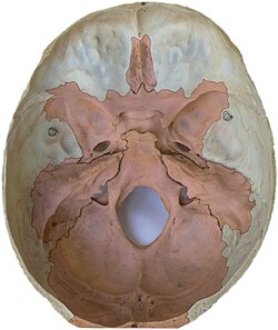
The foramen magnum is a large, oval-shaped opening in the occipital bone of the skull. It is one of the several oval or circular openings (foramina) in the base of the skull. The spinal cord, an extension of the medulla oblongata, passes through the foramen magnum as it exits the cranial cavity. Apart from the transmission of the medulla oblongata and its membranes, the foramen magnum transmits the vertebral arteries, the anterior and posterior spinal arteries, the tectorial membranes and alar ligaments. It also transmits the accessory nerve into the skull.

The sphenoid bone is an unpaired bone of the neurocranium. It is situated in the middle of the skull towards the front, in front of the basilar part of the occipital bone. The sphenoid bone is one of the seven bones that articulate to form the orbit. Its shape somewhat resembles that of a butterfly or bat with its wings extended.

The occipital bone is a cranial dermal bone and the main bone of the occiput. It is trapezoidal in shape and curved on itself like a shallow dish. The occipital bone overlies the occipital lobes of the cerebrum. At the base of the skull in the occipital bone, there is a large oval opening called the foramen magnum, which allows the passage of the spinal cord.
The parietal bones are two bones in the skull which, when joined at a fibrous joint, form the sides and roof of the cranium. In humans, each bone is roughly quadrilateral in form, and has two surfaces, four borders, and four angles. It is named from the Latin paries (-ietis), wall.

The temporal bones are situated at the sides and base of the skull, and lateral to the temporal lobes of the cerebral cortex.

The internal carotid artery is an artery in the neck which supplies the anterior circulation of the brain.

The posterior cranial fossa is the part of the cranial cavity located between the foramen magnum, and tentorium cerebelli. It is formed by the sphenoid bones, temporal bones, and occipital bone. It lodges the cerebellum, and parts of the brainstem.

The cranial cavity, also known as intracranial space, is the space within the skull that accommodates the brain. The skull minus the mandible is called the cranium. The cavity is formed by eight cranial bones known as the neurocranium that in humans includes the skull cap and forms the protective case around the brain. The remainder of the skull is called the facial skeleton. Meninges are protective membranes that surround the brain to minimize damage to the brain in the case of head trauma. Meningitis is the inflammation of meninges caused by bacterial or viral infections.

The greater wing of the sphenoid bone, or alisphenoid, is a bony process of the sphenoid bone, positioned in the skull behind each eye. There is one on each side, extending from the side of the body of the sphenoid and curving upward, laterally, and backward.

The carotid canal is a passage in the petrous part of the temporal bone of the skull through which the internal carotid artery and its internal carotid (nervous) plexus pass from the neck into the cranial cavity.

The petrous part of the temporal bone is pyramid-shaped and is wedged in at the base of the skull between the sphenoid and occipital bones. Directed medially, forward, and a little upward, it presents a base, an apex, three surfaces, and three angles, and houses in its interior, the components of the inner ear. The petrous portion is among the most basal elements of the skull and forms part of the endocranium. Petrous comes from the Latin word petrosus, meaning "stone-like, hard". It is one of the densest bones in the body. In other mammals, it is a separate bone, the petrosal bone.

The lateral parts of the occipital bone are situated at the sides of the foramen magnum; on their under surfaces are the condyles for articulation with the superior facets of the atlas.

The middle cranial fossa is formed by the sphenoid bones, and the temporal bones. It lodges the temporal lobes, and the pituitary gland. It is deeper than the anterior cranial fossa, is narrow medially and widens laterally to the sides of the skull. It is separated from the posterior cranial fossa by the clivus and the petrous crest.

This article describes the anatomy of the head and neck of the human body, including the brain, bones, muscles, blood vessels, nerves, glands, nose, mouth, teeth, tongue, and throat.

The base of skull, also known as the cranial base or the cranial floor, is the most inferior area of the skull. It is composed of the endocranium and the lower parts of the calvaria.

In human anatomy, the neurocranium, also known as the braincase, brainpan, or brain-pan, is the upper and back part of the skull, which forms a protective case around the brain. In the human skull, the neurocranium includes the calvaria or skullcap. The remainder of the skull is the facial skeleton.

The skull roof or the roofing bones of the skull are a set of bones covering the brain, eyes and nostrils in bony fishes and all land-living vertebrates. The bones are derived from dermal bone and are part of the dermatocranium.
The chondrocranium is the primitive cartilaginous skeletal structure of the fetal skull that grows to envelop the rapidly growing embryonic brain.
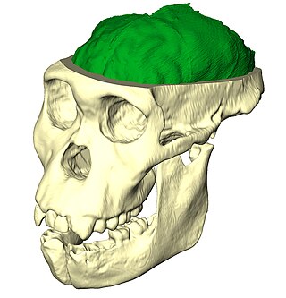
Paleoneurobiology is the study of brain evolution by analysis of brain endocasts to determine endocranial traits and volumes. Considered a subdivision of neuroscience, paleoneurobiology combines techniques from other fields of study including paleontology and archaeology. It reveals specific insight concerning human evolution. The cranium is unique in that it grows in response to the growth of brain tissue rather than genetic guidance, as is the case with bones that support movement. Fossil skulls and their endocasts can be compared to each other, to the skulls and fossils of recently deceased individuals, and even compared to those of other species to make inferences about functional anatomy, physiology and phylogeny. Paleoneurobiology is in large part influenced by developments in neuroscience as a whole; without substantial knowledge about current functionality, it would be impossible to make inferences about the functionality of ancient brains.