Related Research Articles
Cranial nerves are the nerves that emerge directly from the brain, of which there are conventionally considered twelve pairs. Cranial nerves relay information between the brain and parts of the body, primarily to and from regions of the head and neck, including the special senses of vision, taste, smell, and hearing.

In all bilaterian animals, the mesoderm is one of the three primary germ layers in the very early embryo. The other two layers are the ectoderm and endoderm, with the mesoderm as the middle layer between them.

The parasympathetic nervous system (PSNS) is one of the two divisions, the other being the sympathetic, that together are called the autonomic nervous system, which is a division of another system called the peripheral nervous system (PNS)). The autonomic nervous system is responsible for regulating the body's unconscious actions. The parasympathetic system is responsible for stimulation of "rest-and-digest" or "feed and breed" activities that occur when the body is at rest, especially after eating, including sexual arousal, salivation, lacrimation (tears), urination, digestion and defecation. Its action is described as being complementary to that of the sympathetic nervous system, which is responsible for stimulating activities associated with the fight-or-flight response.

The facial nerve is the seventh cranial nerve, or simply CN VII. It emerges from the pons of the brainstem, controls the muscles of facial expression, and functions in the conveyance of taste sensations from the anterior two-thirds of the tongue. The nerves typically travels from the pons through the facial canal in the temporal bone and exits the skull at the stylomastoid foramen. It arises from the brainstem from an area posterior to the cranial nerve VI and anterior to cranial nerve VIII.
Articles related to anatomy include:

In anatomy, the orbit is the cavity or socket of the skull in which the eye and its appendages are situated. "Orbit" can refer to the bony socket, or it can also be used to imply the contents. In the adult human, the volume of the orbit is 30 millilitres, of which the eye occupies 6.5 ml. The orbital contents comprise the eye, the orbital and retrobulbar fascia, extraocular muscles, cranial nerves II, III, IV, V, and VI, blood vessels, fat, the lacrimal gland with its sac and duct, the eyelids, medial and lateral palpebral ligaments, check ligaments, the suspensory ligament, septum, ciliary ganglion and short ciliary nerves.
A germ layer is a primary layer of cells that forms during embryonic development. The three germ layers in vertebrates are particularly pronounced; however, all eumetazoans produce two or three primary germ layers. Some animals, like cnidarians, produce two germ layers making them diploblastic. Other animals such as chordates produce a third layer between these two layers, making them triploblastic. Germ layers eventually give rise to all of an animal’s tissues and organs through the process of organogenesis.

Neural crest cells are a temporary group of cells unique to vertebrates that arise from the embryonic ectoderm germ layer, and in turn give rise to a diverse cell lineage—including melanocytes, craniofacial cartilage and bone, smooth muscle, peripheral and enteric neurons and glia.

In embryology and prenatal development, the dental papilla is a condensation of ectomesenchymal cells called odontoblasts, seen in histologic sections of a developing tooth. It lies below a cellular aggregation known as the enamel organ. The dental papilla appears after 8–10 weeks intra uteral life. The dental papilla gives rise to the dentin and pulp of a tooth.

In vertebrates, an odontoblast is a cell of neural crest origin that is part of the outer surface of the dental pulp, and whose biological function is dentinogenesis, which is the formation of dentin, the substance beneath the tooth enamel on the crown and the cementum on the root.
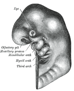
The pharyngeal arches, also known as visceral arches, are structures seen in the embryonic development of vertebrates that are recognisable precursors for many structures. In fish, the arches are known as the branchial arches, or gill arches.

This article describes the anatomy of the head and neck of the human body, including the brain, bones, muscles, blood vessels, nerves, glands, nose, mouth, teeth, tongue, and throat.

The facial muscles are a group of striated skeletal muscles supplied by the facial nerve that, among other things, control facial expression. These muscles are also called mimetic muscles.
Ectomesenchyme has properties similar to mesenchyme. The origin of the ectomesenchyme is disputed. It is either like the mesenchyme, arising from mesodermic cells, or conversely arising from neural crest cells. The neural crest is a critical group of cells that form in the cranial region during early vertebrate development. Ectomesenchyme plays a critical role in the formation of the hard and soft tissues of the head and neck such as bones, muscles, teeth and, notably, the pharyngeal arches.

The human nose is the most protruding part of the face. It bears the nostrils and is the first organ of the respiratory system. It is also the principal organ in the olfactory system. The shape of the nose is determined by the nasal bones and the nasal cartilages, including the nasal septum which separates the nostrils and divides the nasal cavity into two. On average the nose of a male is larger than that of a female.

The following outline is provided as an overview of and topical guide to human anatomy:
Neural crest cells are multipotent cells required for the development of cells, tissues and organ systems. A subpopulation of neural crest cells are the cardiac neural crest complex. This complex refers to the cells found amongst the midotic placode and somite 3 destined to undergo epithelial-mesenchymal transformation and migration to the heart via pharyngeal arches 3, 4 and 6.
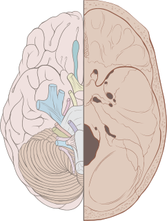
The face and neck development of the human embryo refers to the development of the structures from the third to eighth week that give rise to the future head and neck. They consist of three layers, the ectoderm, mesoderm and endoderm, which form the mesenchyme, neural crest and neural placodes. The paraxial mesoderm forms structures named somites and somitomeres that contribute to the development of the floor of the brain and voluntary muscles of the craniofacial region. The lateral plate mesoderm consists of the laryngeal cartilages. The three tissue layers give rise to the pharyngeal apparatus, formed by six pairs of pharyngeal arches, a set of pharyngeal pouches and pharyngeal grooves, which are the most typical feature in development of the head and neck. The formation of each region of the face and neck is due to the migration of the neural crest cells which come from the ectoderm. These cells determine the future structure to develop in each pharyngeal arch. Eventually, they also form the neurectoderm, which forms the forebrain, midbrain and hindbrain, cartilage, bone, dentin, tendon, dermis, pia mater and arachnoid mater, sensory neurons, and glandular stroma.

Trabecular cartilages are paired, rod-shaped cartilages, which develop in the head of the vertebrate embryo. They are the primordia of the anterior part of the cranial base, and are derived from the cranial neural crest cells.
References
- ↑ "The Neural Crest" . Retrieved 2009-05-31.
- ↑ Grenier J, Teillet MA, Grifone R, Kelly RG, Duprez D (2009). Callaerts P (ed.). "Relationship between Neural Crest Cells and Cranial Mesoderm during Head Muscle Development". PLOS ONE. 4 (2): e4381. doi:10.1371/journal.pone.0004381. PMC 2634972 . PMID 19198652.
- ↑ Jiang HB, Tian WD, Liu LK, Xu Y (June 2008). "In vitro odontoblast-like cell differentiation of cranial neural crest cells induced by fibroblast growth factor 8 and dentin non-collagen proteins". Cell Biol. Int. 32 (6): 671–8. doi:10.1016/j.cellbi.2008.01.293. PMID 18339562. S2CID 5586748.
- ↑ Nie X, Zhang YJ, Tian WD, et al. (January 2007). "Improvement of peripheral nerve regeneration by a tissue-engineered nerve filled with ectomesenchymal stem cells". Int J Oral Maxillofac Surg. 36 (1): 32–8. doi:10.1016/j.ijom.2006.06.005. PMID 17169530.
| This developmental biology article is a stub. You can help Wikipedia by expanding it. |