Neurula

Last updated
Cross section of a vertebrate embryo in the neurula stage Vetebrate Embryo.jpg
Cross section of a vertebrate embryo in the neurula stage

A neurula is a vertebrate embryo at the early stage of development in which neurulation occurs. The neurula stage is preceded by the gastrula stage; consequentially, neurulation is preceded by gastrulation. [1] Neurulation marks the beginning of the process of organogenesis. [2]

Contents

Mice, chicks, and frogs are common experimental models for studying the neurula. Depending on the species, embryos reach the neurula stage at different time points and spend a varying amount of time in this stage. [3] [4] For oviparous organisms, incubation temperature also affects the length of neurulation. [2] In addition to development of the neural tube, other processes occur in a neurula stage embryo depending on the species. For example, in reptiles, extra-embryonic membrane tissues become distinct from the embryo. [2]

The neurula embryo has five regions of mesoderm that surround the neural tube. [5] Anterior mesoderm develops into the head region, while posterior mesoderm develops into the trunk. [1] Various molecules, including proteoglycans in the extracellular matrix, and genes, including Pax transcription factors, are essential for the development and closure of the neural tube in the neurula stage embryo. [6] [7]

Neurulation

Neurulation is a process in vertebrate embryos at the neurula stage in which the neural tube is formed. [6] [8] There are two types of neurulation: primary and secondary neurulation. Primary neurulation refers to the formation and inward folding of the neural plate upon itself to form the neural tube. [6] [8] In secondary neurulation, the neural tube forms via the merging of cavities in the medullary cord. [6] [8] [9] In amphibians and reptiles, primary neurulation forms the whole neural tube, and the neural tube closes simultaneously along its length. [8] Contrarily, in fish, secondary neurulation forms the neural tube. [10] Both primary and secondary neurulation occur in birds and mammals, although with slight differences. Primary neurulation occurs in the cranial and upper spinal regions, which gives rise to the brain and upper regions of the spinal cord. Secondary neurulation occurs in the lower sacral and caudal regions, resulting in the formation of the lower regions of the spinal cord. [6] [10] In birds, the neural tube closes anterior to posterior, while in mammals, the middle closes first, followed by the closure of both ends. [8]

Developmental timing

The point at which the embryo reaches the neurula stage differs among species, while for oviparous organisms, the length of neurulation is additionally affected by incubation temperature. In general, the lower the temperature, the greater the length of neurulation. Chick embryos reach the neurula stage on day 2 post-fertilization, and they undergo neurulation up to day 5. Reptiles, including crocodiles, lizards, and turtles, tend to spend a longer time in the neurula stage. [2] A typical frog embryo, incubated at 18 °C, is an early stage neurula by 50 hours post-fertilization and a late stage neurula by 67 hours. [3] The mouse embryo begins neurulation on day 7.5 of gestation and remains in the neurula stage until day 9. [4]

Morphology

The mesoderm of a vertebrate embryo in the neurula stage can be divided into five regions. Ventral to the neural tube is the chordamesoderm. Lateral to either side of the neural tube is the paraxial mesoderm, while the intermediate lateral region to the neural tube is the intermediate mesoderm. The fourth region is the lateral plate mesoderm, and the last region is the head mesenchym. [5] Anterior portions of the mesoderm develop into rostral regions of an organism, such as the head, while posterior mesoderm develops into caudal regions, such as the trunk or tail. [1] The paraxial mesoderm, also termed somitic mesoderm, develops into somites, blocks of tissue that occur in a segmental pattern. Somites, in turn, give rise to vertebrae, ribs, skeletal muscle, cartilage, tendons, and skin. [8] [11]

Transition from the gastrula stage to the neurula stage Comparative embryology of the vertebrates; with 2057 drawings and photos. grouped as 380 illus (1953) (20047812224).jpg
Transition from the gastrula stage to the neurula stage

In Xenopus laevis, the transition from the gastrula to the neurula involves morphological changes in two regions surrounding the blastopore: the dorsal involuting marginal zone (IMZ) and the overlying non-involuting marginal zone (NIMZ) of the gastrula. Following involution at the mid-gastrula stage, the IMZ undergoes convergent extension, in which the lateral regions narrow and move towards the midline and the anterior end lengthens. This has the effect of narrowing the blastopore. The NIMZ, which does not involute, simultaneously extends in the opposite direction and at a greater rate to cover regions no longer occupied by the IMZ. The convergent extension of the IMZ and NIMZ begins in the second half of gastrulation and continues into the late neurula stage. Eventually, deep tissue of the IMZ forms the central notochord and the surrounding paraxial mesoderm. By the early neurula stage, the notochord is clearly distinguished. Notochordal cells become arranged in a formation representing a stack of coins in a process termed circumferential intercalation. The superficial layer of the IMZ develops into the roof of the archenteron, or the primitive gut, while the underlying endoderm forms the archenteron floor. The NIMZ develops into a structure resembling the early neural tube. The outer ectodermal layer of the neurula is formed by uniform expansion of the cells at the animal pole, known as the animal cap. The ectoderm then differentiates into neural and epidermal tissue. [12]

In reptilian embryos, beginning in the late-stage neurula and carrying over into the early stages of organogenesis, extra-embryonic membrane tissues comprising the yolk sac, chorion, and amnion become distinct from the tissues of the embryo. The mesoderm splits to create the extra-embryonic coelom, which consists of two layers. The vascularized mesoderm-endoderm inner layer, termed the splanchnopleure, develops into the yolk sac, while the nonvascularized ectoderm-mesoderm outer layer, termed the somatopleure, becomes the amnion and chorion. During organogenesis, these three extra-embryonic tissues become fully developed. Additionally, within the reptilian neurula, tissues of the brain begin to differentiate and the heart and blood vessels start to form. [2]

Chemical composition

Mouse neurula tissues divide rapidly, with an average cell cycle lasting 8–10 hours. Proteoglycans in the extracellular matrix (ECM) of neurula-stage cells play an important role in promoting functional cranial neurulation and neural fold elevation; hyaluronic acid (HA) is synthesized and becomes accumulated, while the cell maintains a low level of sulfated glycosaminoglycans (GAGs). HA is involved in creation of biconvex neural folds, while sulfated GAGs are critical in manipulating the neural groove into a V-shape, as well as in neural tube closure. The ECM does not play a major role in spinal neurulation due to the close-packed nature of the mesodermal cells in the spinal region, which allows little intercellular space. Additionally, actin-containing microfilaments are believed to be necessary in cranial neurulation. They may act as the mechanism for neural folding, or they may stabilize neural folds that have already formed; however, their exact role has not been determined. There is some evidence that growth factors, such as insulin or transferrin, also play a role in neurulation, but this link has not been well-studied. [6]

Gene activation

A variety of genes have been found to be expressed in the neurula stage embryo. Different genes are activated for different neurulation events, such as those occurring in separate regions of the developing neural tube. [6] These genes are necessary for proper neurulation and closure of the neural tube. Signaling molecules such as Wnts, FGFs, and BMFs along with the transcription factors that include Msx, Snail s, Sox8/9/10, and Pax3 /7 genes play key roles in neural crest formation. [6]

Pax transcriptional factors have an important role in early development, especially with regards to the CNS and neural crest. Pax3 and Pax7 are promoters of both neural crest cell survival along with promoting environmental stress resistance. [7] In mouse embryos Pax3 blocks the tumor suppressor gene p53, which is necessary for controlled proliferation and genomic stability, is expressed in all cells of the neurula. [6] [7] During early development Pax3 is expressed at the posterior and lateral area of the neural plate, the same region that the neural crest arises from. [7] Neural crest defects were found to occur in mouse and human Pax3 mutants, indicating an importance of functionality. [7] [13] Within chicks, frogs and fish Pax3/Pax7 are activated by Wnt and FGF signaling. [7] Pax3 and Pax7 are also required for neural crest induction after depletion of the two genes resulted in the lack of activation of the specific neural crest genes Snail2 and Foxd3, which didn't allow further development or emigration of neural crest. [7] Using knockouts has been helpful for understanding the role and functions of several genes found in the neurula. For example, Wnt-1 was found to have no role in the closing of the neural plate, despite being present at the tip of the neural folds when it is closing. Though mutants of Wnt-1 does lead to pattern defects within the brain. Notch1 is involved with formation of somites. HNF-3 is needed for development of the notochord and node. [14] The gene Apolipoprotein B, which is involved in transporting and metabolizing fat soluble molecules in the blood, is expressed in the yolk sac and fetal liver. [6] Within the neurula in Xenopus laevis, development genes Xwnt-3 and Xwnt-4 are present. [15]

Related Research Articles

<span class="mw-page-title-main">Ontogeny</span> Origination and development of an organism

Ontogeny is the origination and development of an organism, usually from the time of fertilization of the egg to adult. The term can also be used to refer to the study of the entirety of an organism's lifespan.

<span class="mw-page-title-main">Mesoderm</span> Middle germ layer of embryonic development

The mesoderm is the middle layer of the three germ layers that develops during gastrulation in the very early development of the embryo of most animals. The outer layer is the ectoderm, and the inner layer is the endoderm.

<span class="mw-page-title-main">Neural tube</span> Developmental precursor to the central nervous system

In the developing chordate, the neural tube is the embryonic precursor to the central nervous system, which is made up of the brain and spinal cord. The neural groove gradually deepens as the neural fold become elevated, and ultimately the folds meet and coalesce in the middle line and convert the groove into the closed neural tube. In humans, neural tube closure usually occurs by the fourth week of pregnancy.
<span class="mw-page-title-main">Gastrulation</span> Stage in embryonic development in which germ layers form

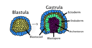
Gastrulation is the stage in the early embryonic development of most animals, during which the blastula, or in mammals the blastocyst is reorganized into a multilayered structure known as the gastrula. Before gastrulation, the embryo is a continuous epithelial sheet of cells; by the end of gastrulation, the embryo has begun differentiation to establish distinct cell lineages, set up the basic axes of the body, and internalized one or more cell types including the prospective gut.

<span class="mw-page-title-main">Ectoderm</span> Outer germ layer of embryonic development

The ectoderm is one of the three primary germ layers formed in early embryonic development. It is the outermost layer, and is superficial to the mesoderm and endoderm. It emerges and originates from the outer layer of germ cells. The word ectoderm comes from the Greek ektos meaning "outside", and derma meaning "skin".

<span class="mw-page-title-main">Neurulation</span> Embryological process forming the neural tube

Neurulation refers to the folding process in vertebrate embryos, which includes the transformation of the neural plate into the neural tube. The embryo at this stage is termed the neurula.

<span class="mw-page-title-main">Somite</span> Each of several blocks of mesoderm that flank the neural tube on either side in embryogenesis

The somites are a set of bilaterally paired blocks of paraxial mesoderm that form in the embryonic stage of somitogenesis, along the head-to-tail axis in segmented animals. In vertebrates, somites subdivide into the dermatomes, myotomes, sclerotomes and syndetomes that give rise to the vertebrae of the vertebral column, rib cage, part of the occipital bone, skeletal muscle, cartilage, tendons, and skin.

A germ layer is a primary layer of cells that forms during embryonic development. The three germ layers in vertebrates are particularly pronounced; however, all eumetazoans produce two or three primary germ layers. Some animals, like cnidarians, produce two germ layers making them diploblastic. Other animals such as bilaterians produce a third layer between these two layers, making them triploblastic. Germ layers eventually give rise to all of an animal's tissues and organs through the process of organogenesis.

Organogenesis is the phase of embryonic development that starts at the end of gastrulation and continues until birth. During organogenesis, the three germ layers formed from gastrulation form the internal organs of the organism.

<span class="mw-page-title-main">Animal embryonic development</span> Process by which the embryo forms and develops

In developmental biology, animal embryonic development, also known as animal embryogenesis, is the developmental stage of an animal embryo. Embryonic development starts with the fertilization of an egg cell (ovum) by a sperm cell, (spermatozoon). Once fertilized, the ovum becomes a single diploid cell known as a zygote. The zygote undergoes mitotic divisions with no significant growth and cellular differentiation, leading to development of a multicellular embryo after passing through an organizational checkpoint during mid-embryogenesis. In mammals, the term refers chiefly to the early stages of prenatal development, whereas the terms fetus and fetal development describe later stages.

<span class="mw-page-title-main">Neural plate</span>

The neural plate is a key developmental structure that serves as the basis for the nervous system. Cranial to the primitive node of the embryonic primitive streak, ectodermal tissue thickens and flattens to become the neural plate. The region anterior to the primitive node can be generally referred to as the neural plate. Cells take on a columnar appearance in the process as they continue to lengthen and narrow. The ends of the neural plate, known as the neural folds, push the ends of the plate up and together, folding into the neural tube, a structure critical to brain and spinal cord development. This process as a whole is termed primary neurulation.

<span class="mw-page-title-main">Neural crest</span> Pluripotent embyronic cell group giving rise to diverse cell lineages

Neural crest cells are a temporary group of cells that arise from the embryonic ectoderm germ layer, and in turn give rise to a diverse cell lineage—including melanocytes, craniofacial cartilage and bone, smooth muscle, peripheral and enteric neurons and glia.

<span class="mw-page-title-main">Paraxial mesoderm</span>

Paraxial mesoderm, also known as presomitic or somitic mesoderm, is the area of mesoderm in the neurulating embryo that flanks and forms simultaneously with the neural tube. The cells of this region give rise to somites, blocks of tissue running along both sides of the neural tube, which form muscle and the tissues of the back, including connective tissue and the dermis.

<span class="mw-page-title-main">Mesenchyme</span> Type of animal embryonic connective tissue

Mesenchyme is a type of loosely organized animal embryonic connective tissue of undifferentiated cells that give rise to most tissues, such as skin, blood or bone. The interactions between mesenchyme and epithelium help to form nearly every organ in the developing embryo.
<span class="mw-page-title-main">Eye development</span> Formation of the eye during embryonic development

Eye formation in the human embryo begins at approximately three weeks into embryonic development and continues through the tenth week. Cells from both the mesodermal and the ectodermal tissues contribute to the formation of the eye. Specifically, the eye is derived from the neuroepithelium, surface ectoderm, and the extracellular mesenchyme which consists of both the neural crest and mesoderm.

Convergent extension (CE), sometimes called convergence and extension (C&E), is the process by which the tissue of an embryo is restructured to converge (narrow) along one axis and extend (elongate) along a perpendicular axis by cellular movement.

<span class="mw-page-title-main">Human embryonic development</span> Development and formation of the human embryo

Human embryonic development, or human embryogenesis, is the development and formation of the human embryo. It is characterised by the processes of cell division and cellular differentiation of the embryo that occurs during the early stages of development. In biological terms, the development of the human body entails growth from a one-celled zygote to an adult human being. Fertilization occurs when the sperm cell successfully enters and fuses with an egg cell (ovum). The genetic material of the sperm and egg then combine to form the single cell zygote and the germinal stage of development commences. Embryonic development in the human, covers the first eight weeks of development; at the beginning of the ninth week the embryo is termed a fetus. The eight weeks has 23 stages.

<span class="mw-page-title-main">Homeobox protein goosecoid</span> Protein-coding gene in the species Homo sapiens

Homeobox protein goosecoid(GSC) is a homeobox protein that is encoded in humans by the GSC gene. Like other homeobox proteins, goosecoid functions as a transcription factor involved in morphogenesis. In Xenopus, GSC is thought to play a crucial role in the phenomenon of the Spemann-Mangold organizer. Through lineage tracing and timelapse microscopy, the effects of GSC on neighboring cell fates could be observed. In an experiment that injected cells with GSC and observed the effects of uninjected cells, GSC recruited neighboring uninjected cells in the dorsal blastopore lip of the Xenopus gastrula to form a twinned dorsal axis, suggesting that the goosecoid protein plays a role in the regulation and migration of cells during gastrulation.

<span class="mw-page-title-main">Ectoderm specification</span> Stage in embryonic development

In Xenopus laevis, the specification of the three germ layers occurs at the blastula stage. Great efforts have been made to determine the factors that specify the endoderm and mesoderm. On the other hand, only a few examples of genes that are required for ectoderm specification have been described in the last decade. The first molecule identified to be required for the specification of ectoderm was the ubiquitin ligase Ectodermin ; later, it was found that the deubiquitinating enzyme, FAM/USP9x, is able to overcome the effects of ubiquitination made by Ectodermin in Smad4. Two transcription factors have been proposed to control gene expression of ectodermal specific genes: POU91/Oct3/4 and FoxIe1/Xema. A new factor specific for the ectoderm, XFDL156, has shown to be essential for suppression of mesoderm differentiation from pluripotent cells.

This glossary of developmental biology is a list of definitions of terms and concepts commonly used in the study of developmental biology and related disciplines in biology, including embryology and reproductive biology, primarily as they pertain to vertebrate animals and particularly to humans and other mammals. The developmental biology of invertebrates, plants, fungi, and other organisms is treated in other articles; e.g. terms relating to the reproduction and development of insects are listed in Glossary of entomology, and those relating to plants are listed in Glossary of botany.

References

  1. 1 2 3 Stern, Claudio D. (February 2001). "Initial Patterning of the Central Nervous System: How Many Organizers?" (PDF). Nature Reviews Neuroscience. 2 (2): 92–98. doi:10.1038/35053563. PMID   11252999. S2CID   13652379.[ permanent dead link ]
  2. 1 2 3 4 5 Andrews, R. M. (2004). "Patterns of embryonic development" (PDF). Reptilian Incubation: Environment, Evolution and Behaviour: 75–102. S2CID   42246488. Archived from the original (PDF) on 2018-11-30.
  3. 1 2 Hill, M.A. (2018, November 30) EmbryologyFrog Development. Retrieved from https://embryology.med.unsw.edu.au/embryology/index.php/Frog_Development .
  4. 1 2 Hill, M.A. (2018, November 30) EmbryologyMouse Timeline Detailed. Retrieved from https://embryology.med.unsw.edu.au/embryology/index.php/Mouse_Timeline_Detailed .
  5. 1 2 Gilbert, Scott F. (2000). "Paraxial and intermediate mesoderm". Developmental Biology. 6th Edition.
  6. 1 2 3 4 5 6 7 8 9 10 Fleming, A; Gerrelli, D; Greene, N D; Copp, A J (2002-03-01). "Mechanisms of normal and abnormal neurulation: evidence from embryo culture studies". International Journal of Developmental Biology. 41 (2). ISSN   0214-6282.
  7. 1 2 3 4 5 6 7 Monsoro-Burq, Anne H. (2015-08-01). "PAX transcription factors in neural crest development". Seminars in Cell & Developmental Biology. 44: 87–96. doi:10.1016/j.semcdb.2015.09.015. ISSN   1084-9521. PMID   26410165.
  8. 1 2 3 4 5 6 Slonczewski, Joan. "Chapter 14. Gastrulation and Neurulation". biology.kenyon.edu. Retrieved 2018-11-16.
  9. Shimokita, Eisuke; Takahashi, Yoshiko (April 2011). "Secondary neurulation: Fate-mapping and gene manipulation of the neural tube in tail bud". Development, Growth & Differentiation. 53 (3): 401–410. doi:10.1111/j.1440-169x.2011.01260.x. ISSN   0012-1592. PMID   21492152. S2CID   31342085.
  10. 1 2 Schoenwolf, Gary C.; Smith, Jodi L. (2000), "Mechanisms of Neurulation" , Developmental Biology Protocols, Humana Press, vol. 136, pp.  125–134, doi:10.1385/1-59259-065-9:125, ISBN   978-1592590650, PMID   10840705
  11. Larsen, William J. (2001). Human embryology (3. ed.). Philadelphia, Pa.: Churchill Livingstone. pp. 53–86. ISBN   0-443-06583-7.
  12. Keller, R. E., Danilchik, M., Gimlich, R., & Shih, J. (1985). "The function and mechanism of convergent extension during gastrulation of Xenopus laevis" (PDF). Development, 89(Supplement), 185-209.
  13. Tassabehji, Mayada; Read, Andrew P.; Newton, Valerie E.; Harris, Rodney; Balling, Rudi; Gruss, Peter; Strachan, Tom (1992-02-13). "Waardenburg's syndrome patients have mutations in the human homologue of the Pax-3 paired box gene". Nature. 355 (6361): 635–636. Bibcode:1992Natur.355..635T. doi:10.1038/355635a0. ISSN   0028-0836. PMID   1347148. S2CID   4330804.
  14. Lenkiewicz, E.; Ferencowa, A.; Szewczykowa, E. (2012-03-17). "Subconjunctival auto-hemotherapy of eye burns in our cases". Klinika Oczna. 94 (4): 113–4. doi:10.4016/46352.01. PMID   1405409.
  15. Christian, Jan L.; Gavin, Brian J.; McMahon, Andrew P.; Moon, Randall T. (1991-02-01). "Isolation of cDNAs partially encoding four XenopusWnt-1int-1-related proteins and characterization of their transient expression during embryonic development". Developmental Biology. 143 (2): 230–234. doi:10.1016/0012-1606(91)90073-C. ISSN   0012-1606. PMID   1991549.