Cranial cavity

Last updated
Cranial cavity
Scheme body cavities-en.svg
Body cavities
Gray194.png
Cranial cavity
Details
FunctionContains and protects the brain
Identifiers
Latin cavitas cranii
TA98 A01.1.00.048
A02.1.00.012
TA2 100, 413
FMA 9644
Anatomical terminology

The cranial cavity, also known as intracranial space, is the space within the skull that accommodates the brain. The skull minus the mandible is called the cranium. The cavity is formed by eight cranial bones known as the neurocranium that in humans includes the skull cap and forms the protective case around the brain. The remainder of the skull is called the facial skeleton. Meninges are protective membranes that surround the brain to minimize damage to the brain in the case of head trauma. Meningitis is the inflammation of meninges caused by bacterial or viral infections.

Contents

Structure

The capacity of an adult human cranial cavity is 1,200–1,700 cm3. [1]

The spaces between meninges and the brain are filled with a clear cerebrospinal fluid, increasing the protection of the brain. Facial bones of the skull are not included in the cranial cavity. There are only eight cranial bones: The occipital, sphenoid, frontal, ethmoid, two parietal, and two temporal bones are fused together by the ossification of fixed fibrous sutures. The frontal and sphenoid bones are towards the front middle of the skull and in front of the temporal bone. The ethmoid bone is the bone at the roof of the nose that separates the naval cavity from the brain. It is a part of the dorsal cavity the cranial cavity and the spinal cord. The occipital bone is at the back of the skull. The dorsal cavity is lined by the three meninges. The three meninges are the three membranes that envelop the brain and spinal cord, in which the central nervous system developed, which are the pia mater, the arachnoid mater, and the dura mater. The latter is the thickest and outermost of the three membrane layers; it contains the most collagen, and it is derived from the mesoderm - the middle germ layer or the primary layer of the cells formed in embryogenic development via epigenetic effects induced by developmental cues, in the early embryo. Also there are the two parietal bones and the two temporal bones, which are a part of the dorsal cavity located on the posterior of the body. The occipital bone found in the rear of the skull is thicker to limit fractures caused by blows to the back of the head. The eight bones are blended together to form the cranial cavity. The pituitary gland is also found in the make up of the cranial cavity. It plays a major role in the body, creating and secreting many bodily hormones. The gland secretes different fluids that are important for the body to function. The body's temperature, physical, and sexual functions are regulated by this gland. One of the major glands are controlled through this cavity.

The cerebrum is the most anterior part of the brain, located in the top half of the skull, consisting of two hemispheres separated by a fissure and connected by the corpus callosum. It is responsible for integrating complex sensory and neural functions, and subsequently initiating and coordinating voluntary activity in the body.

The cortex is the outer layer of the cerebrum, composed of folded grey matter. Its neuron cell bodies, dendrites, synapses, axons, and axon terminals play a crucial role in consciousness. The two hemispheres are divided into four lobes, distinct sections of the organ: the frontal lobe, parietal lobe, temporal lobe, and occipital lobe. Our understanding of the specific functions of the cerebral cortex are based on the theories of localisation and lateralisation. Localisation is the theory that specific areas of the brain are associated with/responsible for particular physical and psychological functions. Lateralisation is the theory that one hemisphere is dominant over the other/responsible for particular physical and psychological functions.

The meninges are the three membranes that line the skull and vertebral canal, and enclose the brain and spinal cord. The Cerebrospinal Fluid serves a vital function in the cerebral autoregulation of cerebral blood flow. Cerebrospinal Fluid occupies the subarachnoid space and the ventricular system around and inside the brain and spinal cord.

Function

There are twelve cranial nerves that are responsible for controlling the cranial cavity. These nerves are responsible for providing necessary sensory information for things such as smell, taste, hearing, and sight. The ability to sleep and chew is also a part of one of the things the cranial cavities in charge of. In order for the cranial cavity to be able to do all these functions it has to have the organs to be able to control those systems. The cranial cavity houses the Brain, Meninges, and the Cerebrospinal Fluid. The primary function of the brain is supplying information to the rest of the body and to help it function as whole. It helps supply some of the cranial nerves from the face to the feet and also to help get the body performing critical bodily functions.

The cranial cavity includes eight cranial bones and they are collectively combined to form this area. A gland that is found in this cavity is called the pituitary gland which secretes different fluids that are necessary for different parts of the body to function. There are many things that are regulated by this gland such as body temperature, physical growth, and sexual functions. The pituitary gland also controls the thyroid gland.

The cranial cavity has a variety of spinal and cranial nerves residing in it. The cranial nerves are responsible for storing sensory information needed for taste, smell, sight and the ability to hear. The spinal nerves allow for the sensory and motor signals to be received, which provide a normal feeling and function for the arms and legs.

Clinical significance

In the event that the brain strikes the inside of the skull, there is a risk of a minor traumatic brain injury (concussion). This type of injury can alter brain function, cause memory loss, headaches and/or nausea. A minor traumatic brain injury can be followed by post concussion syndrome. As there are a number of structures in the cranial cavity, a traumatic injury to the head can potentially cause major damage to the brain. This is why it is recommended to get medical attention following a head injury.

The cranial cavity houses the meninges, as well as other necessary organs, and these membranes surround the central nervous system. If an infection forms, it can lead to a disease called meningitis which can be potentially fatal. [2]

See also

Related Research Articles

<span class="mw-page-title-main">Central nervous system</span> Brain and spinal cord

The central nervous system (CNS) is the part of the nervous system consisting primarily of the brain and spinal cord. The CNS is so named because the brain integrates the received information and coordinates and influences the activity of all parts of the bodies of bilaterally symmetric and triploblastic animals—that is, all multicellular animals except sponges and diploblasts. It is a structure composed of nervous tissue positioned along the rostral to caudal axis of the body and may have an enlarged section at the rostral end which is a brain. Only arthropods, cephalopods and vertebrates have a true brain.

<span class="mw-page-title-main">Body cavity</span> Internal space within a multicellular organism

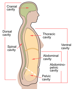
A body cavity is any space or compartment, or potential space, in an animal body. Cavities accommodate organs and other structures; cavities as potential spaces contain fluid.

<span class="mw-page-title-main">Skull</span> Bony structure that forms the head in vertebrates

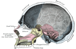
The skull is a bone protective cavity for the brain. The skull is composed of four types of bone i.e., cranial bones, facial bones, ear ossicles and hyoid bone. However two parts are more prominent: the cranium and the mandible. In humans, these two parts are the neurocranium and the viscerocranium that includes the mandible as its largest bone. The skull forms the anterior-most portion of the skeleton and is a product of cephalisation—housing the brain, and several sensory structures such as the eyes, ears, nose, and mouth. In humans these sensory structures are part of the facial skeleton.

<span class="mw-page-title-main">Fontanelle</span> Anatomical feature of the infant human skull

A fontanelle is an anatomical feature of the infant human skull comprising soft membranous gaps (sutures) between the cranial bones that make up the calvaria of a fetus or an infant. Fontanelles allow for stretching and deformation of the neurocranium both during birth and later as the brain expands faster than the surrounding bone can grow. Premature complete ossification of the sutures is called craniosynostosis.

Articles related to anatomy include:

<span class="mw-page-title-main">Trigeminal nerve</span> Cranial nerve responsible for the faces senses and motor functions

In neuroanatomy, the trigeminal nerve (lit. triplet nerve), also known as the fifth cranial nerve, cranial nerve V, or simply CN V, is a cranial nerve responsible for sensation in the face and motor functions such as biting and chewing; it is the most complex of the cranial nerves. Its name (trigeminal, from Latin tri- 'three', and -geminus 'twin') derives from each of the two nerves (one on each side of the pons) having three major branches: the ophthalmic nerve (V1), the maxillary nerve (V2), and the mandibular nerve (V3). The ophthalmic and maxillary nerves are purely sensory, whereas the mandibular nerve supplies motor as well as sensory (or "cutaneous") functions. Adding to the complexity of this nerve is that autonomic nerve fibers as well as special sensory fibers (taste) are contained within it.

<span class="mw-page-title-main">Sphenoid bone</span> Bone at the front of the skull

The sphenoid bone is an unpaired bone of the neurocranium. It is situated in the middle of the skull towards the front, in front of the basilar part of the occipital bone. The sphenoid bone is one of the seven bones that articulate to form the orbit. Its shape somewhat resembles that of a butterfly or bat with its wings extended.

<span class="mw-page-title-main">Occipital bone</span> Saucer-shaped membrane bone situated at the back and lower part of the cranium

The occipital bone is a cranial dermal bone and the main bone of the occiput. It is trapezoidal in shape and curved on itself like a shallow dish. The occipital bone overlies the occipital lobes of the cerebrum. At the base of the skull in the occipital bone, there is a large oval opening called the foramen magnum, which allows the passage of the spinal cord.

<span class="mw-page-title-main">Temporal bone</span> Sides and base of skull, connecting to the jaw and occipital bone

The temporal bones are situated at the sides and base of the skull, and lateral to the temporal lobes of the cerebral cortex.

<span class="mw-page-title-main">Meninges</span> Three membranes that envelop the brain and spinal cord

In anatomy, the meninges are the four membranes that envelop the brain and spinal cord. In mammals, the meninges are the dura mater, the arachnoid mater, the SLYM and the pia mater. Cerebrospinal fluid is located in the subarachnoid space between the arachnoid mater and the pia mater. The primary function of the meninges is to protect the central nervous system.

<span class="mw-page-title-main">Pia mater</span> Delicate innermost layer of the meninges, the membranes surrounding the brain and spinal cord

Pia mater, often referred to as simply the pia, is the delicate innermost layer of the meninges, the membranes surrounding the brain and spinal cord. Pia mater is medieval Latin meaning "tender mother". The other two meningeal membranes are the dura mater and the arachnoid mater. Both the pia and arachnoid mater are derivatives of the neural crest while the dura is derived from embryonic mesoderm. The pia mater is a thin fibrous tissue that is permeable to water and small solutes. The pia mater allows blood vessels to pass through and nourish the brain. The perivascular space between blood vessels and pia mater is proposed to be part of a pseudolymphatic system for the brain. When the pia mater becomes irritated and inflamed the result is meningitis.

<span class="mw-page-title-main">Human brain</span> Central organ of the human nervous system

The human brain is the central organ of the human nervous system, and with the spinal cord makes up the central nervous system. The brain consists of the cerebrum, the brainstem and the cerebellum. It controls most of the activities of the body, processing, integrating, and coordinating the information it receives from the sense organs, and making decisions as to the instructions sent to the rest of the body. The brain is contained in, and protected by, the skull bones of the head.

<span class="mw-page-title-main">Dura mater</span> Outermost layer of the protective tissues around the central nervous system (meninges)

In neuroanatomy, dura mater is a thick membrane made of dense irregular connective tissue that surrounds the brain and spinal cord. It is the outermost of the three layers of membrane called the meninges that protect the central nervous system. The other two meningeal layers are the arachnoid mater and the pia mater. It envelops the arachnoid mater, which is responsible for keeping in the cerebrospinal fluid. It is derived primarily from the neural crest cell population, with postnatal contributions of the paraxial mesoderm.

<span class="mw-page-title-main">Crown (anatomy)</span> Top of the head

The crown is the top portion of the head behind the vertex. The anatomy of the crown varies between different organisms. The human crown is made of three layers of the scalp above the skull. The crown also covers a range of bone sutures, and contains blood vessels and branches of the trigeminal nerve.

<span class="mw-page-title-main">Skull fracture</span> Medical condition

A skull fracture is a break in one or more of the eight bones that form the cranial portion of the skull, usually occurring as a result of blunt force trauma. If the force of the impact is excessive, the bone may fracture at or near the site of the impact and cause damage to the underlying structures within the skull such as the membranes, blood vessels, and brain.

<span class="mw-page-title-main">Basilar skull fracture</span> Medical condition

A basilar skull fracture is a break of a bone in the base of the skull. Symptoms may include bruising behind the ears, bruising around the eyes, or blood behind the ear drum. A cerebrospinal fluid (CSF) leak occurs in about 20% of cases and may result in fluid leaking from the nose or ear. Meningitis occurs in about 14% of cases. Other complications include injuries to the cranial nerves or blood vessels.

<span class="mw-page-title-main">Superior sagittal sinus</span> Anatomical structure of the brain

The superior sagittal sinus, within the human head, is an unpaired area along the attached margin of the falx cerebri. It allows blood to drain from the lateral aspects of anterior cerebral hemispheres to the confluence of sinuses. Cerebrospinal fluid drains through arachnoid granulations into the superior sagittal sinus and is returned to venous circulation.

<span class="mw-page-title-main">Middle cranial fossa</span>

The middle cranial fossa is formed by the sphenoid bones, and the temporal bones. It lodges the temporal lobes, and the pituitary gland. It is deeper than the anterior cranial fossa, is narrow medially and widens laterally to the sides of the skull. It is separated from the posterior fossa by the clivus and the petrous crest.

<span class="mw-page-title-main">Head and neck anatomy</span>

This article describes the anatomy of the head and neck of the human body, including the brain, bones, muscles, blood vessels, nerves, glands, nose, mouth, teeth, tongue, and throat.

Cerebrospinal fluid rhinorrhoea refers to the drainage of cerebrospinal fluid through the nose (rhinorrhoea). It is typically caused by a basilar skull fracture, which presents complications such as infection. It may be diagnosed using brain scans, and by testing to see if discharge from the nose is cerebrospinal fluid. Treatment may be conservative, but usually involves neurosurgery.

References

  1. Turchin, Valentin. "The Phenomenon of Science".
  2. Martini R, Ober W, Garrison C, Welch K, and Hutchings RT. 2001. Fundamentals of Anatomy and Physiology, 5th ed. Prentice Hall, New Jersey. p. 195.