Auditory system

Last updated
Auditory system
Anatomical terminology
How sounds make their way from the source to the brain

The auditory system is the sensory system for the sense of hearing. It includes both the sensory organs (the ears) and the auditory parts of the sensory system. [1]

Contents

System overview

The outer ear funnels sound vibrations to the eardrum, increasing the sound pressure in the middle frequency range. The middle-ear ossicles further amplify the vibration pressure roughly 20 times. The base of the stapes couples vibrations into the cochlea via the oval window, which vibrates the perilymph liquid (present throughout the inner ear) and causes the round window to bulb out as the oval window bulges in.[ citation needed ]

Vestibular and tympanic ducts are filled with perilymph, and the smaller cochlear duct between them is filled with endolymph, a fluid with a very different ion concentration and voltage. [2] [3] [4] Vestibular duct perilymph vibrations bend organ of Corti outer cells (4 lines) causing prestin to be released in cell tips. This causes the cells to be chemically elongated and shrunk (somatic motor), and hair bundles to shift which, in turn, electrically affects the basilar membrane's movement (hair-bundle motor). These motors (outer hair cells) amplify the traveling wave amplitudes over 40-fold. [5] The outer hair cells (OHC) are minimally innervated by spiral ganglion in slow (unmyelinated) reciprocal communicative bundles (30+ hairs per nerve fiber); this contrasts inner hair cells (IHC) that have only afferent innervation (30+ nerve fibers per one hair) but are heavily connected. There are three to four times as many OHCs as IHCs. The basilar membrane (BM) is a barrier between scalae, along the edge of which the IHCs and OHCs sit. Basilar membrane width and stiffness vary to control the frequencies best sensed by the IHC. At the cochlear base the BM is at its narrowest and most stiff (high-frequencies), while at the cochlear apex it is at its widest and least stiff (low-frequencies). The tectorial membrane (TM) helps facilitate cochlear amplification by stimulating OHC (direct) and IHC (via endolymph vibrations). TM width and stiffness parallels BM's and similarly aids in frequency differentiation. [6] [7] [8] [9] [10] [11] [12] [13] [14] [ excessive citations ]

The superior olivary complex (SOC), in the pons, is the first convergence of the left and right cochlear pulses. SOC has 14 described nuclei; their abbreviation are used here (see Superior olivary complex for their full names). MSO determines the angle the sound came from by measuring time differences in left and right info. LSO normalizes sound levels between the ears; it uses the sound intensities to help determine sound angle. LSO innervates the IHC. VNTB innervate OHC. MNTB inhibit LSO via glycine. LNTB are glycine-immune, used for fast signalling. DPO are high-frequency and tonotopical. DLPO are low-frequency and tonotopical. VLPO have the same function as DPO, but act in a different area. PVO, CPO, RPO, VMPO, ALPO and SPON (inhibited by glycine) are various signalling and inhibiting nuclei. [15] [16] [17] [18]

The trapezoid body is where most of the cochlear nucleus (CN) fibers decussate (cross left to right and vice versa); this cross aids in sound localization. [19] The CN breaks into ventral (VCN) and dorsal (DCN) regions. The VCN has three nuclei.[ clarification needed ] Bushy cells transmit timing info, their shape averages timing jitters. Stellate (chopper) cells encode sound spectra (peaks and valleys) by spatial neural firing rates based on auditory input strength (rather than frequency). Octopus cells have close to the best temporal precision while firing, they decode the auditory timing code. The DCN has 2 nuclei. DCN also receives info from VCN. Fusiform cells integrate information to determine spectral cues to locations (for example, whether a sound originated from in front or behind). Cochlear nerve fibers (30,000+) each have a most sensitive frequency and respond over a wide range of levels. [20] [21]

Simplified, nerve fibers' signals are transported by bushy cells to the binaural areas in the olivary complex, while signal peaks and valleys are noted by stellate cells, and signal timing is extracted by octopus cells. The lateral lemniscus has three nuclei: dorsal nuclei respond best to bilateral input and have complexity tuned responses; intermediate nuclei have broad tuning responses; and ventral nuclei have broad and moderately complex tuning curves. Ventral nuclei of lateral lemniscus help the inferior colliculus (IC) decode amplitude modulated sounds by giving both phasic and tonic responses (short and long notes, respectively). IC receives inputs not shown, including visual (pretectal area: moves eyes to sound. superior colliculus: orientation and behavior toward objects, as well as eye movements (saccade)) areas, pons (superior cerebellar peduncle: thalamus to cerebellum connection/hear sound and learn behavioral response), spinal cord (periaqueductal grey: hear sound and instinctually move), and thalamus. The above are what implicate IC in the 'startle response' and ocular reflexes. Beyond multi-sensory integration IC responds to specific amplitude modulation frequencies, allowing for the detection of pitch. IC also determines time differences in binaural hearing. [22] The medial geniculate nucleus divides into ventral (relay and relay-inhibitory cells: frequency, intensity, and binaural info topographically relayed), dorsal (broad and complex tuned nuclei: connection to somatosensory info), and medial (broad, complex, and narrow tuned nuclei: relay intensity and sound duration). The auditory cortex (AC) brings sound into awareness/perception. AC identifies sounds (sound-name recognition) and also identifies the sound's origin location. AC is a topographical frequency map with bundles reacting to different harmonies, timing and pitch. Right-hand-side AC is more sensitive to tonality, left-hand-side AC is more sensitive to minute sequential differences in sound. [23] [24] Rostromedial and ventrolateral prefrontal cortices are involved in activation during tonal space and storing short-term memories, respectively. [25] The Heschl's gyrus/transverse temporal gyrus includes Wernicke's area and functionality, it is heavily involved in emotion-sound, emotion-facial-expression, and sound-memory processes. The entorhinal cortex is the part of the 'hippocampus system' that aids and stores visual and auditory memories. [26] [27] The supramarginal gyrus (SMG) aids in language comprehension and is responsible for compassionate responses. SMG links sounds to words with the angular gyrus and aids in word choice. SMG integrates tactile, visual, and auditory info. [28] [29]

Structure

Anatomy of the human ear (The length of the auditory canal is exaggerated in this image.).
.mw-parser-output .legend{page-break-inside:avoid;break-inside:avoid-column}.mw-parser-output .legend-color{display:inline-block;min-width:1.25em;height:1.25em;line-height:1.25;margin:1px 0;text-align:center;border:1px solid black;background-color:transparent;color:black}.mw-parser-output .legend-text{}
Brown is outer ear.
Red is middle ear.
Purple is inner ear. Anatomy of the Human Ear en.svg
Anatomy of the human ear (The length of the auditory canal is exaggerated in this image.).
  Brown is outer ear.
  Red is middle ear.
  Purple is inner ear.

Outer ear

The folds of cartilage surrounding the ear canal are called the auricle. Sound waves are reflected and attenuated when they hit the auricle, and these changes provide additional information that will help the brain determine the sound direction.

The sound waves enter the auditory canal, a deceptively simple tube. The ear canal amplifies sounds that are between 3 and 12 kHz.[ citation needed ] The tympanic membrane, at the far end of the ear canal marks the beginning of the middle ear.

Middle ear

Auditory ossicles from a deep dissection of the tympanic cavity Slide1ghe.JPG
Auditory ossicles from a deep dissection of the tympanic cavity

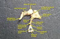
Sound waves travel through the ear canal and hit the tympanic membrane, or eardrum. This wave information travels across the air-filled middle ear cavity via a series of delicate bones: the malleus (hammer), incus (anvil) and stapes (stirrup). These ossicles act as a lever, converting the lower-pressure eardrum sound vibrations into higher-pressure sound vibrations at another, smaller membrane called the oval window or vestibular window. The manubrium (handle) of the malleus articulates with the tympanic membrane, while the footplate (base) of the stapes articulates with the oval window. Higher pressure is necessary at the oval window than at the tympanic membrane because the inner ear beyond the oval window contains liquid rather than air. The stapedius reflex of the middle ear muscles helps protect the inner ear from damage by reducing the transmission of sound energy when the stapedius muscle is activated in response to sound. The middle ear still contains the sound information in wave form; it is converted to nerve impulses in the cochlea.

Inner ear

Cochlea
Gray928.png
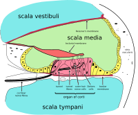
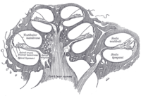
Diagrammatic longitudinal section of the cochlea. The cochlear duct, or scala media, is labeled as ductus cochlearis at right.
Anatomical terminology

The inner ear consists of the cochlea and several non-auditory structures. The cochlea has three fluid-filled sections (i.e. the scala media, scala tympani and scala vestibuli), and supports a fluid wave driven by pressure across the basilar membrane separating two of the sections. Strikingly, one section, called the cochlear duct or scala media, contains endolymph. The organ of Corti is located in this duct on the basilar membrane, and transforms mechanical waves to electric signals in neurons. The other two sections are known as the scala tympani and the scala vestibuli. These are located within the bony labyrinth, which is filled with fluid called perilymph, similar in composition to cerebrospinal fluid. The chemical difference between the fluids endolymph and perilymph fluids is important for the function of the inner ear due to electrical potential differences between potassium and calcium ions.[ citation needed ]

The plan view of the human cochlea (typical of all mammalian and most vertebrates) shows where specific frequencies occur along its length. The frequency is an approximately exponential function of the length of the cochlea within the Organ of Corti. In some species, such as bats and dolphins, the relationship is expanded in specific areas to support their active sonar capability.

Organ of Corti

The organ of Corti located at the scala media Cochlea-crosssection.svg
The organ of Corti located at the scala media

The organ of Corti forms a ribbon of sensory epithelium which runs lengthwise down the cochlea's entire scala media. Its hair cells transform the fluid waves into nerve signals. The journey of countless nerves begins with this first step; from here, further processing leads to a panoply of auditory reactions and sensations.

Hair cell

Hair cells are columnar cells, each with a "hair bundle" of 100–200 specialized stereocilia at the top, for which they are named. There are two types of hair cells specific to the auditory system; inner and outerhaircells. Inner hair cells are the mechanoreceptors for hearing: they transduce the vibration of sound into electrical activity in nerve fibers, which is transmitted to the brain. Outer hair cells are a motor structure. Sound energy causes changes in the shape of these cells, which serves to amplify sound vibrations in a frequency specific manner. Lightly resting atop the longest cilia of the inner hair cells is the tectorial membrane, which moves back and forth with each cycle of sound, tilting the cilia, which is what elicits the hair cells' electrical responses.

Inner hair cells, like the photoreceptor cells of the eye, show a graded response, instead of the spikes typical of other neurons. These graded potentials are not bound by the "all or none" properties of an action potential.

At this point, one may ask how such a wiggle of a hair bundle triggers a difference in membrane potential. The current model is that cilia are attached to one another by "tip links", structures which link the tips of one cilium to another. Stretching and compressing, the tip links may open an ion channel and produce the receptor potential in the hair cell. Recently it has been shown that cadherin-23 CDH23 and protocadherin-15 PCDH15 are the adhesion molecules associated with these tip links. [30] It is thought that a calcium driven motor causes a shortening of these links to regenerate tensions. This regeneration of tension allows for apprehension of prolonged auditory stimulation. [31]

Neurons

Afferent neurons innervate cochlear inner hair cells, at synapses where the neurotransmitter glutamate communicates signals from the hair cells to the dendrites of the primary auditory neurons.

There are far fewer inner hair cells in the cochlea than afferent nerve fibers – many auditory nerve fibers innervate each hair cell. The neural dendrites belong to neurons of the auditory nerve, which in turn joins the vestibular nerve to form the vestibulocochlear nerve, or cranial nerve number VIII. [32] The region of the basilar membrane supplying the inputs to a particular afferent nerve fibre can be considered to be its receptive field.

Efferent projections from the brain to the cochlea also play a role in the perception of sound, although this is not well understood. Efferent synapses occur on outer hair cells and on afferent (towards the brain) dendrites under inner hair cells

Neuronal structure

Cochlear nucleus

The cochlear nucleus is the first site of the neuronal processing of the newly converted "digital" data from the inner ear (see also binaural fusion). In mammals, this region is anatomically and physiologically split into two regions, the dorsal cochlear nucleus (DCN), and ventral cochlear nucleus (VCN). The VCN is further divided by the nerve root into the posteroventral cochlear nucleus (PVCN) and the anteroventral cochlear nucleus (AVCN). [33]

Trapezoid body

The trapezoid body is a bundle of decussating fibers in the ventral pons that carry information used for binaural computations in the brainstem. Some of these axons come from the cochlear nucleus and cross over to the other side before traveling on to the superior olivary nucleus. This is believed to help with localization of sound. [34]

Superior olivary complex

The superior olivary complex is located in the pons, and receives projections predominantly from the ventral cochlear nucleus, although the dorsal cochlear nucleus projects there as well, via the ventral acoustic stria. Within the superior olivary complex lies the lateral superior olive (LSO) and the medial superior olive (MSO). The former is important in detecting interaural level differences while the latter is important in distinguishing interaural time difference. [17]

Lateral lemniscus in red, as it connects the cochlear nucleus, superior olivary nucleus and the inferior colliculus, seen from behind Lateral lemniscus.PNG
Lateral lemniscus in red, as it connects the cochlear nucleus, superior olivary nucleus and the inferior colliculus, seen from behind

Lateral lemniscus

The lateral lemniscus is a tract of axons in the brainstem that carries information about sound from the cochlear nucleus to various brainstem nuclei and ultimately the contralateral inferior colliculus of the midbrain.

Inferior colliculi

The inferior colliculi (IC) are located just below the visual processing centers known as the superior colliculi. The central nucleus of the IC is a nearly obligatory relay in the ascending auditory system, and most likely acts to integrate information (specifically regarding sound source localization from the superior olivary complex [16] and dorsal cochlear nucleus) before sending it to the thalamus and cortex. [1] The inferior colliculus also receives descending inputs from the auditory cortex and auditory thalamus (or medial geniculate nucleus). [35]

Medial geniculate nucleus

The medial geniculate nucleus is part of the thalamic relay system.

Primary auditory cortex

The primary auditory cortex is the first region of cerebral cortex to receive auditory input.

Perception of sound is associated with the left posterior superior temporal gyrus (STG). The superior temporal gyrus contains several important structures of the brain, including Brodmann areas 41 and 42, marking the location of the primary auditory cortex, the cortical region responsible for the sensation of basic characteristics of sound such as pitch and rhythm. We know from research in nonhuman primates that the primary auditory cortex can probably be divided further into functionally differentiable subregions. [36] [37] [38] [39] [40] [41] [42] The neurons of the primary auditory cortex can be considered to have receptive fields covering a range of auditory frequencies and have selective responses to harmonic pitches. [43] Neurons integrating information from the two ears have receptive fields covering a particular region of auditory space.

The primary auditory cortex is surrounded by secondary auditory cortex, and interconnects with it. These secondary areas interconnect with further processing areas in the superior temporal gyrus, in the dorsal bank of the superior temporal sulcus, and in the frontal lobe. In humans, connections of these regions with the middle temporal gyrus are probably important for speech perception. The frontotemporal system underlying auditory perception allows us to distinguish sounds as speech, music, or noise.

The auditory ventral and dorsal streams

Dual stream connectivity between the auditory cortex and frontal lobe of monkeys and humans. Top: The auditory cortex of the monkey (left) and human (right) is schematically depicted on the supratemporal plane and observed from above (with the parieto- frontal operculi removed). Bottom: The brain of the monkey (left) and human (right) is schematically depicted and displayed from the side. Orange frames mark the region of the auditory cortex, which is displayed in the top sub-figures. Top and Bottom: Blue colors mark regions affiliated with the ADS, and red colors mark regions affiliated with the AVS (dark red and blue regions mark the primary auditory fields). Abbreviations: AMYG-amygdala, HG-Heschl's gyrus, FEF-frontal eye field, IFG-inferior frontal gyrus, INS-insula, IPS-intra parietal sulcus, MTG-middle temporal gyrus, PC-pitch center, PMd-dorsal premotor cortex, PP-planum polare, PT-planum temporale, TP-temporal pole, Spt-sylvian parietal-temporal, pSTG/mSTG/aSTG-posterior/middle/anterior superior temporal gyrus, CL/ ML/AL/RTL-caudo-/middle-/antero-/rostrotemporal-lateral belt area, CPB/RPB-caudal/rostral parabelt fields. Used with permission from Poliva O. From where to what: a neuroanatomically based evolutionary model of the emergence of speech in humans. Material was copied from this source, which is available under a Creative Commons Attribution 4.0 International License. Neurolinguistics.png
Dual stream connectivity between the auditory cortex and frontal lobe of monkeys and humans. Top: The auditory cortex of the monkey (left) and human (right) is schematically depicted on the supratemporal plane and observed from above (with the parieto- frontal operculi removed). Bottom: The brain of the monkey (left) and human (right) is schematically depicted and displayed from the side. Orange frames mark the region of the auditory cortex, which is displayed in the top sub-figures. Top and Bottom: Blue colors mark regions affiliated with the ADS, and red colors mark regions affiliated with the AVS (dark red and blue regions mark the primary auditory fields). Abbreviations: AMYG-amygdala, HG-Heschl's gyrus, FEF-frontal eye field, IFG-inferior frontal gyrus, INS-insula, IPS-intra parietal sulcus, MTG-middle temporal gyrus, PC-pitch center, PMd-dorsal premotor cortex, PP-planum polare, PT-planum temporale, TP-temporal pole, Spt-sylvian parietal-temporal, pSTG/mSTG/aSTG-posterior/middle/anterior superior temporal gyrus, CL/ ML/AL/RTL-caudo-/middle-/antero-/rostrotemporal-lateral belt area, CPB/RPB-caudal/rostral parabelt fields. Used with permission from Poliva O. From where to what: a neuroanatomically based evolutionary model of the emergence of speech in humans. CC-BY icon.svg Material was copied from this source, which is available under a Creative Commons Attribution 4.0 International License.

From the primary auditory cortex emerge two separate pathways: the auditory ventral stream and auditory dorsal stream. [44] The auditory ventral stream includes the anterior superior temporal gyrus, anterior superior temporal sulcus, middle temporal gyrus and temporal pole. Neurons in these areas are responsible for sound recognition, and extraction of meaning from sentences. The auditory dorsal stream includes the posterior superior temporal gyrus and sulcus, inferior parietal lobule and intra-parietal sulcus. Both pathways project in humans to the inferior frontal gyrus. The most established role of the auditory dorsal stream in primates is sound localization. In humans, the auditory dorsal stream in the left hemisphere is also responsible for speech repetition and articulation, phonological long-term encoding of word names, and verbal working memory.

Clinical significance

Proper function of the auditory system is required to able to sense, process, and understand sound from the surroundings. Difficulty in sensing, processing and understanding sound input has the potential to adversely impact an individual's ability to communicate, learn and effectively complete routine tasks on a daily basis. [45]

In children, early diagnosis and treatment of impaired auditory system function is an important factor in ensuring that key social, academic and speech/language developmental milestones are met. [46]

Impairment of the auditory system can include any of the following:

See also

Related Research Articles

<span class="mw-page-title-main">Inner ear</span> Innermost part of the vertebrate ear

The inner ear is the innermost part of the vertebrate ear. In vertebrates, the inner ear is mainly responsible for sound detection and balance. In mammals, it consists of the bony labyrinth, a hollow cavity in the temporal bone of the skull with a system of passages comprising two main functional parts:

<span class="mw-page-title-main">Cochlea</span> Snail-shaped part of inner ear involved in hearing

The cochlea is the part of the inner ear involved in hearing. It is a spiral-shaped cavity in the bony labyrinth, in humans making 2.75 turns around its axis, the modiolus. A core component of the cochlea is the organ of Corti, the sensory organ of hearing, which is distributed along the partition separating the fluid chambers in the coiled tapered tube of the cochlea.

Articles related to anatomy include:

<span class="mw-page-title-main">Vestibulocochlear nerve</span> Cranial nerve VIII, for hearing and balance

The vestibulocochlear nerve or auditory vestibular nerve, also known as the eighth cranial nerve, cranial nerve VIII, or simply CN VIII, is a cranial nerve that transmits sound and equilibrium (balance) information from the inner ear to the brain. Through olivocochlear fibers, it also transmits motor and modulatory information from the superior olivary complex in the brainstem to the cochlea.

<span class="mw-page-title-main">Basilar membrane</span> Stiff structural element within the cochlea of the inner ear which separates two liquid-filled tubes

The basilar membrane is a stiff structural element within the cochlea of the inner ear which separates two liquid-filled tubes that run along the coil of the cochlea, the scala media and the scala tympani. The basilar membrane moves up and down in response to incoming sound waves, which are converted to traveling waves on the basilar membrane.

<span class="mw-page-title-main">Organ of Corti</span> Receptor organ for hearing

The organ of Corti, or spiral organ, is the receptor organ for hearing and is located in the mammalian cochlea. This highly varied strip of epithelial cells allows for transduction of auditory signals into nerve impulses' action potential. Transduction occurs through vibrations of structures in the inner ear causing displacement of cochlear fluid and movement of hair cells at the organ of Corti to produce electrochemical signals.

<span class="mw-page-title-main">Inferior colliculus</span> Midbrain structure involved in the auditory pathway

The inferior colliculus (IC) is the principal midbrain nucleus of the auditory pathway and receives input from several peripheral brainstem nuclei in the auditory pathway, as well as inputs from the auditory cortex. The inferior colliculus has three subdivisions: the central nucleus, a dorsal cortex by which it is surrounded, and an external cortex which is located laterally. Its bimodal neurons are implicated in auditory-somatosensory interaction, receiving projections from somatosensory nuclei. This multisensory integration may underlie a filtering of self-effected sounds from vocalization, chewing, or respiration activities.

In physiology, tonotopy is the spatial arrangement of where sounds of different frequency are processed in the brain. Tones close to each other in terms of frequency are represented in topologically neighbouring regions in the brain. Tonotopic maps are a particular case of topographic organization, similar to retinotopy in the visual system.

<span class="mw-page-title-main">Pontine tegmentum</span>

The pontine tegmentum, or dorsal pons, is located within the brainstem, and is one of two parts of the pons, the other being the ventral pons or basilar part of the pons. The pontine tegmentum can be defined in contrast to the basilar pons: basilar pons contains the corticospinal tract running craniocaudally and can be considered the rostral extension of the ventral medulla oblongata; however, basilar pons is distinguished from ventral medulla oblongata in that it contains additional transverse pontine fibres that continue laterally to become the middle cerebellar peduncle. The pontine tegmentum is all the material dorsal from the basilar pons to the fourth ventricle. Along with the dorsal surface of the medulla, it forms part of the rhomboid fossa – the floor of the fourth ventricle.

<span class="mw-page-title-main">Cochlear nucleus</span> Two cranial nerve nuclei of the human brainstem

The cochlear nuclear (CN) complex comprises two cranial nerve nuclei in the human brainstem, the ventral cochlear nucleus (VCN) and the dorsal cochlear nucleus (DCN). The ventral cochlear nucleus is unlayered whereas the dorsal cochlear nucleus is layered. Auditory nerve fibers, fibers that travel through the auditory nerve carry information from the inner ear, the cochlea, on the same side of the head, to the nerve root in the ventral cochlear nucleus. At the nerve root the fibers branch to innervate the ventral cochlear nucleus and the deep layer of the dorsal cochlear nucleus. All acoustic information thus enters the brain through the cochlear nuclei, where the processing of acoustic information begins. The outputs from the cochlear nuclei are received in higher regions of the auditory brainstem.

<span class="mw-page-title-main">Dorsal cochlear nucleus</span>

The dorsal cochlear nucleus is a cortex-like structure on the dorso-lateral surface of the brainstem. Along with the ventral cochlear nucleus (VCN), it forms the cochlear nucleus (CN), where all auditory nerve fibers from the cochlea form their first synapses.

<span class="mw-page-title-main">Superior olivary complex</span> Collection of brainstem nuclei related to hearing

The superior olivary complex (SOC) or superior olive is a collection of brainstem nuclei that is located in pons, functions in multiple aspects of hearing and is an important component of the ascending and descending auditory pathways of the auditory system. The SOC is intimately related to the trapezoid body: most of the cell groups of the SOC are dorsal to this axon bundle while a number of cell groups are embedded in the trapezoid body. Overall, the SOC displays a significant interspecies variation, being largest in bats and rodents and smaller in primates.

<span class="mw-page-title-main">Interaural time difference</span> Difference in time that it takes a sound to travel between two ears

The interaural time difference when concerning humans or animals, is the difference in arrival time of a sound between two ears. It is important in the localization of sounds, as it provides a cue to the direction or angle of the sound source from the head. If a signal arrives at the head from one side, the signal has further to travel to reach the far ear than the near ear. This pathlength difference results in a time difference between the sound's arrivals at the ears, which is detected and aids the process of identifying the direction of sound source.

Binaural fusion or binaural integration is a cognitive process that involves the combination of different auditory information presented binaurally, or to each ear. In humans, this process is essential in understanding speech as one ear may pick up more information about the speech stimuli than the other.

<span class="mw-page-title-main">Ventral cochlear nucleus</span>

In the ventral cochlear nucleus (VCN), auditory nerve fibers enter the brain via the nerve root in the VCN. The ventral cochlear nucleus is divided into the anterior ventral (anteroventral) cochlear nucleus (AVCN) and the posterior ventral (posteroventral) cochlear nucleus (PVCN). In the VCN, auditory nerve fibers bifurcate, the ascending branch innervates the AVCN and the descending branch innervates the PVCN and then continue to the dorsal cochlear nucleus. The orderly innervation by auditory nerve fibers gives the AVCN a tonotopic organization along the dorsoventral axis. Fibers that carry information from the apex of the cochlea that are tuned to low frequencies contact neurons in the ventral part of the AVCN; those that carry information from the base of the cochlea that are tuned to high frequencies contact neurons in the dorsal part of the AVCN. Several populations of neurons populate the AVCN. Bushy cells receive input from auditory nerve fibers through particularly large endings called end bulbs of Held. They contact stellate cells through more conventional boutons.

<span class="mw-page-title-main">Hearing</span> Sensory perception of sound by living organisms

Hearing, or auditory perception, is the ability to perceive sounds through an organ, such as an ear, by detecting vibrations as periodic changes in the pressure of a surrounding medium. The academic field concerned with hearing is auditory science.

The neural encoding of sound is the representation of auditory sensation and perception in the nervous system. The complexities of contemporary neuroscience are continually redefined. Thus what is known of the auditory system has been continually changing. The encoding of sounds includes the transduction of sound waves into electrical impulses along auditory nerve fibers, and further processing in the brain.

Electrocochleography is a technique of recording electrical potentials generated in the inner ear and auditory nerve in response to sound stimulation, using an electrode placed in the ear canal or tympanic membrane. The test is performed by an otologist or audiologist with specialized training, and is used for detection of elevated inner ear pressure or for the testing and monitoring of inner ear and auditory nerve function during surgery.

<span class="mw-page-title-main">Sign language in the brain</span>

Sign language refers to any natural language which uses visual gestures produced by the hands and body language to express meaning. The brain's left side is the dominant side utilized for producing and understanding sign language, just as it is for speech. In 1861, Paul Broca studied patients with the ability to understand spoken languages but the inability to produce them. The damaged area was named Broca's area, and located in the left hemisphere’s inferior frontal gyrus. Soon after, in 1874, Carl Wernicke studied patients with the reverse deficits: patients could produce spoken language, but could not comprehend it. The damaged area was named Wernicke's area, and is located in the left hemisphere’s posterior superior temporal gyrus.

<span class="mw-page-title-main">Sound localization in owls</span> Ability of owls to locate sounds in 3D space

Most owls are nocturnal or crepuscular birds of prey. Because they hunt at night, they must rely on non-visual senses. Experiments by Roger Payne have shown that owls are sensitive to the sounds made by their prey, not the heat or the smell. In fact, the sound cues are both necessary and sufficient for localization of mice from a distant location where they are perched. For this to work, the owls must be able to accurately localize both the azimuth and the elevation of the sound source.

References

  1. 1 2 Demanez JP, Demanez L (2003). "Anatomophysiology of the central auditory nervous system: basic concepts". Acta Oto-Rhino-Laryngologica Belgica. 57 (4): 227–36. PMID   14714940.
  2. Tillotson JK, McCann S (2013). Kaplan medical anatomy flashcards. Kaplan Publishing. ISBN   978-1-60714-984-2.
  3. Ashwell K (2016). Barron's anatomy flash cards. Barron's Educational Series. ISBN   978-1-4380-7717-8.
  4. "How Does My Hearing Work?". NZ Audiological Society. Archived from the original on 23 August 2019. Retrieved 27 March 2016.
  5. Zheng J, Shen W, He DZ, Long KB, Madison LD, Dallos P (May 2000). "Prestin is the motor protein of cochlear outer hair cells". Nature. 405 (6783): 149–55. Bibcode:2000Natur.405..149Z. doi:10.1038/35012009. PMID   10821263. S2CID   4409772.
  6. Zwislocki JJ, Cefaratti LK (November 1989). "Tectorial membrane. II: Stiffness measurements in vivo". Hearing Research. 42 (2–3): 211–27. doi:10.1016/0378-5955(89)90146-9. PMID   2606804. S2CID   4706357.
  7. Richter CP, Emadi G, Getnick G, Quesnel A, Dallos P (September 2007). "Tectorial membrane stiffness gradients". Biophysical Journal. 93 (6): 2265–76. Bibcode:2007BpJ....93.2265R. doi:10.1529/biophysj.106.094474. PMC   1959565 . PMID   17496047.
  8. Meaud J, Grosh K (March 2010). "The effect of tectorial membrane and basilar membrane longitudinal coupling in cochlear mechanics". The Journal of the Acoustical Society of America. 127 (3): 1411–21. Bibcode:2010ASAJ..127.1411M. doi:10.1121/1.3290995. PMC   2856508 . PMID   20329841.
  9. Gueta R, Barlam D, Shneck RZ, Rousso I (October 2006). "Measurement of the mechanical properties of isolated tectorial membrane using atomic force microscopy". Proceedings of the National Academy of Sciences of the United States of America. 103 (40): 14790–5. Bibcode:2006PNAS..10314790G. doi: 10.1073/pnas.0603429103 . PMC   1595430 . PMID   17001011.
  10. Freeman DM, Abnet CC, Hemmert W, Tsai BS, Weiss TF (June 2003). "Dynamic material properties of the tectorial membrane: a summary". Hearing Research. 180 (1–2): 1–10. doi:10.1016/S0378-5955(03)00073-X. PMID   12782348. S2CID   24187159.
  11. Legan PK, Lukashkina VA, Goodyear RJ, Kössi M, Russell IJ, Richardson GP (October 2000). "A targeted deletion in alpha-tectorin reveals that the tectorial membrane is required for the gain and timing of cochlear feedback". Neuron. 28 (1): 273–85. doi: 10.1016/S0896-6273(00)00102-1 . PMID   11087000.
  12. Canlon B (1988). "The effect of acoustic trauma on the tectorial membrane, stereocilia, and hearing sensitivity: possible mechanisms underlying damage, recovery, and protection". Scandinavian Audiology. Supplementum. 27: 1–45. PMID   3043645.
  13. Zwislocki JJ (1979). "Tectorial membrane: a possible sharpening effect on the frequency analysis in the cochlea". Acta Oto-Laryngologica. 87 (3–4): 267–9. doi:10.3109/00016487909126419. PMID   443008.
  14. Teudt IU, Richter CP (October 2014). "Basilar membrane and tectorial membrane stiffness in the CBA/CaJ mouse". Journal of the Association for Research in Otolaryngology. 15 (5): 675–94. doi:10.1007/s10162-014-0463-y. PMC   4164692 . PMID   24865766.
  15. Thompson AM, Schofield BR (November 2000). "Afferent projections of the superior olivary complex". Microscopy Research and Technique. 51 (4): 330–54. doi:10.1002/1097-0029(20001115)51:4<330::AID-JEMT4>3.0.CO;2-X. PMID   11071718. S2CID   27179535.
  16. 1 2 Oliver DL (November 2000). "Ascending efferent projections of the superior olivary complex". Microscopy Research and Technique. 51 (4): 355–63. doi:10.1002/1097-0029(20001115)51:4<355::AID-JEMT5>3.0.CO;2-J. PMID   11071719. S2CID   36633546.
  17. 1 2 Moore JK (November 2000). "Organization of the human superior olivary complex". Microscopy Research and Technique. 51 (4): 403–12. doi: 10.1002/1097-0029(20001115)51:4<403::AID-JEMT8>3.0.CO;2-Q . PMID   11071722. S2CID   10151612.
  18. Yang L, Monsivais P, Rubel EW (March 1999). "The superior olivary nucleus and its influence on nucleus laminaris: a source of inhibitory feedback for coincidence detection in the avian auditory brainstem". The Journal of Neuroscience. 19 (6): 2313–25. doi:10.1523/JNEUROSCI.19-06-02313.1999. PMC   6782562 . PMID   10066281.
  19. Paolini AG, FitzGerald JV, Burkitt AN, Clark GM (September 2001). "Temporal processing from the auditory nerve to the medial nucleus of the trapezoid body in the rat". Hearing Research. 159 (1–2): 101–16. doi:10.1016/S0378-5955(01)00327-6. PMID   11520638. S2CID   25279502.
  20. Bajo VM, Merchán MA, Malmierca MS, Nodal FR, Bjaalie JG (May 1999). "Topographic organization of the dorsal nucleus of the lateral lemniscus in the cat". The Journal of Comparative Neurology. 407 (3): 349–66. doi:10.1002/(SICI)1096-9861(19990510)407:3<349::AID-CNE4>3.0.CO;2-5. PMID   10320216. S2CID   25724084.
  21. Young ED, Davis KA (2002). "Circuitry and Function of the Dorsal Cochlear Nucleus". In Oertel D, Fay RR, Popper AN (eds.). Integrative Functions in the Mammalian Auditory Pathway. Springer Handbook of Auditory Research. Vol. 15. New York, NY: Springer. pp. 160–206. doi:10.1007/978-1-4757-3654-0_5. ISBN   978-1-4757-3654-0.
  22. Oliver DL (2005). "Neuronal Organization in the Inferior Colliculus". In Winer JA, Schreiner CE (eds.). The Inferior Colliculus. New York, NY: Springer. pp. 69–114. doi:10.1007/0-387-27083-3_2. ISBN   978-0-387-27083-8.
  23. Janata P, Birk JL, Van Horn JD, Leman M, Tillmann B, Bharucha JJ (December 2002). "The cortical topography of tonal structures underlying Western music". Science. 298 (5601): 2167–70. Bibcode:2002Sci...298.2167J. doi:10.1126/science.1076262. PMID   12481131. S2CID   3031759.
  24. Morosan P, Rademacher J, Schleicher A, Amunts K, Schormann T, Zilles K (April 2001). "Human primary auditory cortex: cytoarchitectonic subdivisions and mapping into a spatial reference system". NeuroImage. 13 (4): 684–701. CiteSeerX   10.1.1.420.7633 . doi:10.1006/nimg.2000.0715. PMID   11305897. S2CID   16472551.
  25. Romanski LM, Tian B, Fritz J, Mishkin M, Goldman-Rakic PS, Rauschecker JP (December 1999). "Dual streams of auditory afferents target multiple domains in the primate prefrontal cortex". Nature Neuroscience. 2 (12): 1131–6. doi:10.1038/16056. PMC   2778291 . PMID   10570492.
  26. Badre D, Wagner AD (October 2007). "Left ventrolateral prefrontal cortex and the cognitive control of memory". Neuropsychologia. 45 (13): 2883–901. doi:10.1016/j.neuropsychologia.2007.06.015. PMID   17675110. S2CID   16062085.
  27. Amunts K, Kedo O, Kindler M, Pieperhoff P, Mohlberg H, Shah NJ, Habel U, Schneider F, Zilles K (December 2005). "Cytoarchitectonic mapping of the human amygdala, hippocampal region and entorhinal cortex: intersubject variability and probability maps". Anatomy and Embryology. 210 (5–6): 343–52. doi:10.1007/s00429-005-0025-5. PMID   16208455. S2CID   6984617.
  28. Penniello MJ, Lambert J, Eustache F, Petit-Taboué MC, Barré L, Viader F, Morin P, Lechevalier B, Baron JC (June 1995). "A PET study of the functional neuroanatomy of writing impairment in Alzheimer's disease. The role of the left supramarginal and left angular gyri". Brain: A Journal of Neurology. 118 ( Pt 3) (3): 697–706. doi:10.1093/brain/118.3.697. PMID   7600087.
  29. Stoeckel C, Gough PM, Watkins KE, Devlin JT (October 2009). "Supramarginal gyrus involvement in visual word recognition". Cortex; A Journal Devoted to the Study of the Nervous System and Behavior. 45 (9): 1091–6. doi:10.1016/j.cortex.2008.12.004. PMC   2726132 . PMID   19232583.
  30. Lelli A, Kazmierczak P, Kawashima Y, Müller U, Holt JR (August 2010). "Development and regeneration of sensory transduction in auditory hair cells requires functional interaction between cadherin-23 and protocadherin-15". The Journal of Neuroscience. 30 (34): 11259–69. doi:10.1523/JNEUROSCI.1949-10.2010. PMC   2949085 . PMID   20739546.
  31. Peng AW, Salles FT, Pan B, Ricci AJ (November 2011). "Integrating the biophysical and molecular mechanisms of auditory hair cell mechanotransduction". Nature Communications. 2: 523. Bibcode:2011NatCo...2..523P. doi:10.1038/ncomms1533. PMC   3418221 . PMID   22045002.
  32. "Meddean – CN VIII. Vestibulocochlear Nerve". Archived from the original on 2012-10-06. Retrieved 2007-10-27.
  33. Middlebrooks JC (2009). "Auditory System: Central Pathways". In Squire LF (ed.). Encyclopedia of Neuroscience. Academic Press. pp. 745–752, here: p. 745 f. ISBN   978-0-08-044617-2.
  34. Mendoza JE (2011). "Trapezoid Body". In Kreutzer JS, DeLuca J, Caplan B (eds.). Encyclopedia of Clinical Neuropsychology. New York: Springer. p. 2549. doi:10.1007/978-0-387-79948-3_807. ISBN   978-0-387-79947-6.
  35. Schreiner, Christoph (2005). Winer, Jeffery A; Schreiner, Christoph E (eds.). The Inferior Colliculus. Springer Science+ Business Media, Incorporated. doi:10.1007/b138578. ISBN   0-387-22038-0. Archived from the original on 2022-05-17. Retrieved 2022-05-17.
  36. Pandya DN (1995). "Anatomy of the auditory cortex". Revue Neurologique. 151 (8–9): 486–94. PMID   8578069.
  37. Kaas JH, Hackett TA (1998). "Subdivisions of auditory cortex and levels of processing in primates". Audiology & Neuro-Otology. 3 (2–3): 73–85. doi:10.1159/000013783. PMID   9575378. S2CID   46858666.
  38. Kaas JH, Hackett TA, Tramo MJ (April 1999). "Auditory processing in primate cerebral cortex". Current Opinion in Neurobiology. 9 (2): 164–70. doi:10.1016/S0959-4388(99)80022-1. PMID   10322185. S2CID   22984374.
  39. Kaas JH, Hackett TA (October 2000). "Subdivisions of auditory cortex and processing streams in primates". Proceedings of the National Academy of Sciences of the United States of America. 97 (22): 11793–9. Bibcode:2000PNAS...9711793K. doi: 10.1073/pnas.97.22.11793 . PMC   34351 . PMID   11050211.
  40. Hackett TA, Preuss TM, Kaas JH (December 2001). "Architectonic identification of the core region in auditory cortex of macaques, chimpanzees, and humans". The Journal of Comparative Neurology. 441 (3): 197–222. doi:10.1002/cne.1407. PMID   11745645. S2CID   21776552.
  41. Scott SK, Johnsrude IS (February 2003). "The neuroanatomical and functional organization of speech perception". Trends in Neurosciences. 26 (2): 100–7. CiteSeerX   10.1.1.323.8534 . doi:10.1016/S0166-2236(02)00037-1. PMID   12536133. S2CID   10390565.
  42. Tian B, Reser D, Durham A, Kustov A, Rauschecker JP (April 2001). "Functional specialization in rhesus monkey auditory cortex". Science. 292 (5515): 290–3. Bibcode:2001Sci...292..290T. doi:10.1126/science.1058911. PMID   11303104. S2CID   32846215.
  43. Wang X (December 2013). "The harmonic organization of auditory cortex". Frontiers in Systems Neuroscience. 7: 114. doi: 10.3389/fnsys.2013.00114 . PMC   3865599 . PMID   24381544.
  44. Hickok G, Poeppel D (May 2007). "The cortical organization of speech processing". Nature Reviews. Neuroscience. 8 (5): 393–402. doi:10.1038/nrn2113. PMID   17431404. S2CID   6199399.
  45. "Hearing Loss" (PDF). HearingLoss.org. National Academy on an Aging Society. Archived from the original (PDF) on 1 May 2013. Retrieved 28 January 2018.
  46. Ciorba A, Corazzi V, Negossi L, Tazzari R, Bianchini C, Aimoni C (December 2017). "Moderate-Severe Hearing Loss in Children: A Diagnostic and Rehabilitative Challenge". The Journal of International Advanced Otology. 13 (3): 407–413. doi: 10.5152/iao.2017.4162 . PMID   29360094.

Further reading