Two-streams hypothesis

Last updated

The two-streams hypothesis is a model of the neural processing of vision as well as hearing. [1] The hypothesis, given its initial characterisation in a paper by David Milner and Melvyn A. Goodale in 1992, argues that humans possess two distinct visual systems. [2] Recently there seems to be evidence of two distinct auditory systems as well. As visual information exits the occipital lobe, and as sound leaves the phonological network, it follows two main pathways, or "streams". The ventral stream (also known as the "what pathway") leads to the temporal lobe, which is involved with object and visual identification and recognition. The dorsal stream (or, "how pathway") leads to the parietal lobe, which is involved with processing the object's spatial location relative to the viewer and with speech repetition.

Contents

History

Ventral-dorsal streams.svg
How
What
Ventral-dorsal streams.svg
The dorsal stream (green) and ventral stream (purple) are shown. They originate from a common source in the visual cortex

Several researchers had proposed similar ideas previously. The authors themselves credit the inspiration of work on blindsight by Weiskrantz, and previous neuroscientific vision research. Schneider first proposed the existence of two visual systems for localisation and identification in 1969. [3] Ingle described two independent visual systems in frogs in 1973. [4] Ettlinger reviewed the existing neuropsychological evidence of a distinction in 1990. [5] Moreover, Trevarthen had offered an account of two separate mechanisms of vision in monkeys back in 1968. [6]

In 1982, Ungerleider and Mishkin distinguished the dorsal and ventral streams, as processing spatial and visual features respectively, from their lesion studies of monkeys – proposing the original where vs what distinction. [7] Though this framework was superseded by that of Milner & Goodale, it remains influential. [8]

One hugely influential source of information that has informed the model has been experimental work exploring the extant abilities of visual agnosic patient D.F. The first, and most influential report, came from Goodale and colleagues in 1991 [9] and work is still being published on her two decades later. [10] This has been the focus of some criticism of the model due to the perceived over-reliance on findings from a single case.

Two visual systems

Goodale and Milner [2] amassed an array of anatomical, neuropsychological, electrophysiological, and behavioural evidence for their model. According to their data, the ventral 'perceptual' stream computes a detailed map of the world from visual input, which can then be used for cognitive operations, and the dorsal 'action' stream transforms incoming visual information to the requisite egocentric (head-centered) coordinate system for skilled motor planning. The model also posits that visual perception encodes spatial properties of objects, such as size and location, relative to other objects in the visual field; in other words, it utilizes relative metrics and scene-based frames of reference. Visual action planning and coordination, on the other hand, uses absolute metrics determined via egocentric frames of reference, computing the actual properties of objects relative to the observer. Thus, grasping movements directed towards objects embedded in size-contrast-ambiguous scenes have been shown to escape the effects of these illusions, as different frames of references and metrics are involved in the perception of the illusion versus the execution of the grasping act. [11]

Norman [12] proposed a similar dual-process model of vision, and described eight main differences between the two systems consistent with other two-system models.

FactorVentral system (what)Dorsal system (how)
FunctionRecognition/identificationVisually guided behaviour
SensitivityHigh spatial frequencies - detailsHigh temporal frequencies - motion
MemoryLong-term stored representationsOnly very short-term storage
SpeedRelatively slowRelatively fast
ConsciousnessTypically highTypically low
Frame of referenceAllocentric or object-centeredEgocentric or viewer-centered
Visual inputMainly foveal or parafovealAcross retina
Monocular visionGenerally reasonably small effectsOften large effects e.g. motion parallax

Dorsal stream

The dorsal stream is proposed to be involved in the guidance of actions and recognizing where objects are in space. The dorsal stream projects from the primary visual cortex to the posterior parietal cortex. It was initially termed the "where" pathway since it was thought that the dorsal stream processes information regarding the spatial properties of an object. [13] However, later research conducted on a famous neuropsychological patient, Patient D.F., revealed that the dorsal stream is responsible for processing the visual information needed to construct the representations of objects one wishes to manipulate. Those findings led the nickname of the dorsal stream to be updated to the "how" pathway. [14] [15] The dorsal stream is interconnected with the parallel ventral stream (the "what" stream) which runs downward from V1 into the temporal lobe.

General features

The dorsal stream is involved in spatial awareness and guidance of actions (e.g., reaching). In this it has two distinct functional characteristics—it contains a detailed map of the visual field, and is also good at detecting and analyzing movements.

The dorsal stream commences with purely visual functions in the occipital lobe before gradually transferring to spatial awareness at its termination in the parietal lobe.

The posterior parietal cortex is essential for "the perception and interpretation of spatial relationships, accurate body image, and the learning of tasks involving coordination of the body in space". [16]

It contains individually functioning lobules. The lateral intraparietal sulcus (LIP) contains neurons that produce enhanced activation when attention is moved onto the stimulus or the animal saccades towards a visual stimulus, and the ventral intraparietal sulcus (VIP) where visual and somatosensory information are integrated.

Effects of damage or lesions

Damage to the posterior parietal cortex causes a number of spatial disorders including:

  • Simultanagnosia: where the patient can only describe single objects without the ability to perceive it as a component of a set of details or objects in a context (as in a scenario, e.g. the forest for the trees).
  • Optic ataxia: where the patient cannot use visuospatial information to guide arm movements.
  • Hemispatial neglect: where the patient is unaware of the contralesional half of space (that is, they are unaware of things in their left field of view and focus only on objects in the right field of view; or appear unaware of things in one field of view when they perceive them in the other). For example, a person with this disorder may draw a clock, and then label all twelve of the numbers on one side of the face and consider the drawing complete.
  • Akinetopsia: inability to perceive motion.
  • Apraxia: inability to produce discretionary or volitional movement in the absence of muscular disorders.

Ventral stream

The ventral stream is associated with object recognition and form representation. Also described as the "what" stream, it has strong connections to the medial temporal lobe (which is associated with long-term memories), the limbic system (which controls emotions), and the dorsal stream (which deals with object locations and motion).

The ventral stream gets its main input from the parvocellular (as opposed to magnocellular) layer of the lateral geniculate nucleus of the thalamus. These neurons project to V1 sublayers 4Cβ, 4A, 3B and 2/3a [17] successively. From there, the ventral pathway goes through V2 and V4 to areas of the inferior temporal lobe: PIT (posterior inferotemporal), CIT (central inferotemporal), and AIT (anterior inferotemporal). Each visual area contains a full representation of visual space. That is, it contains neurons whose receptive fields together represent the entire visual field. Visual information enters the ventral stream through the primary visual cortex and travels through the rest of the areas in sequence.

Moving along the stream from V1 to AIT, receptive fields increase their size, latency, and the complexity of their tuning. For example, recent studies have shown that the V4 area is responsible for color perception in humans, and the V8 (VO1) area is responsible for shape perception, while the VO2 area, which is located between these regions and the parahippocampal cortex, integrates information about the color and shape of stimuli into a holistic image. [18]

All the areas in the ventral stream are influenced by extraretinal factors in addition to the nature of the stimulus in their receptive field. These factors include attention, working memory, and stimulus salience. Thus the ventral stream does not merely provide a description of the elements in the visual world—it also plays a crucial role in judging the significance of these elements.

Damage to the ventral stream can cause inability to recognize faces or interpret facial expression. [19]

Two auditory systems

Ventral stream

comprehension of the phrase 'my cat' in the extended version of Hickok and Poeppel's dual pathway model T16-VentralComprehension.png
comprehension of the phrase 'my cat' in the extended version of Hickok and Poeppel's dual pathway model

Along with the visual ventral pathway being important for visual processing, there is also a ventral auditory pathway emerging from the primary auditory cortex. [20] In this pathway, phonemes are processed posteriorly to syllables and environmental sounds. [21] The information then joins the visual ventral stream at the middle temporal gyrus and temporal pole. Here the auditory objects are converted into audio-visual concepts. [22]

Dorsal stream

The function of the auditory dorsal pathway is to map the auditory sensory representations onto articulatory motor representations. Hickok & Poeppel claim that the auditory dorsal pathway is necessary because, "learning to speak is essentially a motor learning task. The primary input to this is sensory, speech in particular. So, there must be a neural mechanism that both codes and maintains instances of speech sounds, and can use these sensory traces to guide the tuning of speech gestures so that the sounds are accurately reproduced." [23]

repetition of the phrase 'what is your name?' in the extended version of Hickok and Poeppel's dual pathway model T16-DorsalRepetition.png
repetition of the phrase 'what is your name?' in the extended version of Hickok and Poeppel's dual pathway model

In contrast to the ventral stream's auditory processing, information enters from the primary auditory cortex into the posterior superior temporal gyrus and posterior superior temporal sulcus. From there the information moves to the beginning of the dorsal pathway, which is located at the boundary of the temporal and parietal lobes near the Sylvian fissure. The first step of the dorsal pathway begins in the sensorimotor interface, located in the left Sylvian parietal temporal (Spt) (within the Sylvian fissure at the parietal-temporal boundary). The spt is important for perceiving and reproducing sounds. This is evident because its ability to acquire new vocabulary, be disrupted by lesions and auditory feedback on speech production, articulatory decline in late-onset deafness and the non-phonological residue of Wernicke's aphasia; deficient self-monitoring. It is also important for the basic neuronal mechanisms for phonological short-term memory. Without the Spt, language acquisition is impaired. The information then moves onto the articulatory network, which is divided into two separate parts. The articulatory network 1, which processes motor syllable programs, is located in the left posterior inferior temporal gyrus and Brodmann's area 44 (pIFG-BA44). The articulatory network 2 is for motor phoneme programs and is located in the left M1-vBA6. [24]

Conduction aphasia affects a subject's ability to reproduce speech (typically by repetition), though it has no influence on the subject's ability to comprehend spoken language. This shows that conduction aphasia must reflect not an impairment of the auditory ventral pathway but instead of the auditory dorsal pathway. Buchsbaum et al [25] found that conduction aphasia can be the result of damage, particularly lesions, to the Spt (Sylvian parietal temporal). This is shown by the Spt's involvement in acquiring new vocabulary, for while experiments have shown that most conduction aphasiacs can repeat high-frequency, simple words, their ability to repeat low-frequency, complex words is impaired. The Spt is responsible for connecting the motor and auditory systems by making auditory code accessible to the motor cortex. It appears that the motor cortex recreates high-frequency, simple words (like cup) in order to more quickly and efficiently access them, while low-frequency, complex words (like Sylvian parietal temporal) require more active, online regulation by the Spt. This explains why conduction aphasiacs have particular difficulty with low-frequency words which requires a more hands-on process for speech production. "Functionally, conduction aphasia has been characterized as a deficit in the ability to encode phonological information for production," namely because of a disruption in the motor-auditory interface. [26] Conduction aphasia has been more specifically related to damage of the arcuate fasciculus, which is vital for both speech and language comprehension, as the arcuate fasiculus makes up the connection between Broca and Wernicke's areas. [26]

Criticisms

Goodale & Milner's innovation was to shift the perspective from an emphasis on input distinctions, such as object location versus properties, to an emphasis on the functional relevance of vision to behaviour, for perception or for action. Contemporary perspectives however, informed by empirical work over the past two decades, offer a more complex account than a simple separation of function into two-streams. [27] Recent experimental work for instance has challenged these findings, and has suggested that the apparent dissociation between the effects of illusions on perception and action is due to differences in attention, task demands, and other confounds. [28] [29] There are other empirical findings, however, that cannot be so easily dismissed which provide strong support for the idea that skilled actions such as grasping are not affected by pictorial illusions. [30] [31] [32] [33]

Moreover, recent neuropsychological research has questioned the validity of the dissociation of the two streams that has provided the cornerstone of evidence for the model. The dissociation between visual agnosia and optic ataxia has been challenged by several researchers as not as strong as originally portrayed; Hesse and colleagues demonstrated dorsal stream impairments in patient DF; [34] Himmelbach and colleagues reassessed DF's abilities and applied more rigorous statistical analysis demonstrating that the dissociation was not as strong as first thought. [10]

A 2009 review of the accumulated evidence for the model concluded that whilst the spirit of the model has been vindicated the independence of the two streams has been overemphasised. [35] Goodale & Milner themselves have proposed the analogy of tele-assistance, one of the most efficient schemes devised for the remote control of robots working in hostile environments. In this account, the dorsal stream is viewed as a semi-autonomous function that operates under guidance of executive functions which themselves are informed by ventral stream processing. [36]

Thus the emerging perspective within neuropsychology and neurophysiology is that, whilst a two-systems framework was a necessary advance to stimulate study of the highly complex and differentiated functions of the two neural pathways; the reality is more likely to involve considerable interaction between vision-for-action and vision-for-perception. Robert McIntosh and Thomas Schenk summarize this position as follows:

We should view the model not as a formal hypothesis, but as a set of heuristics to guide experiment and theory. The differing informational requirements of visual recognition and action guidance still offer a compelling explanation for the broad relative specializations of dorsal and ventral streams. However, to progress the field, we may need to abandon the idea that these streams work largely independently of one other, and to address the dynamic details of how the many visual brain areas arrange themselves from task to task into novel functional networks. [35] :62[ verification needed ]

See also

Related Research Articles

<span class="mw-page-title-main">Visual cortex</span> Region of the brain that processes visual information

The visual cortex of the brain is the area of the cerebral cortex that processes visual information. It is located in the occipital lobe. Sensory input originating from the eyes travels through the lateral geniculate nucleus in the thalamus and then reaches the visual cortex. The area of the visual cortex that receives the sensory input from the lateral geniculate nucleus is the primary visual cortex, also known as visual area 1 (V1), Brodmann area 17, or the striate cortex. The extrastriate areas consist of visual areas 2, 3, 4, and 5.

<span class="mw-page-title-main">Parietal lobe</span> Part of the brain responsible for sensory input and some language processing

The parietal lobe is one of the four major lobes of the cerebral cortex in the brain of mammals. The parietal lobe is positioned above the temporal lobe and behind the frontal lobe and central sulcus.

<span class="mw-page-title-main">Temporal lobe</span> One of the four lobes of the mammalian brain

The temporal lobe is one of the four major lobes of the cerebral cortex in the brain of mammals. The temporal lobe is located beneath the lateral fissure on both cerebral hemispheres of the mammalian brain.

<span class="mw-page-title-main">Wernicke's area</span> Speech comprehension region in the dominant hemisphere of the hominid brain

Wernicke's area, also called Wernicke's speech area, is one of the two parts of the cerebral cortex that are linked to speech, the other being Broca's area. It is involved in the comprehension of written and spoken language, in contrast to Broca's area, which is primarily involved in the production of language. It is traditionally thought to reside in Brodmann area 22, which is located in the superior temporal gyrus in the dominant cerebral hemisphere, which is the left hemisphere in about 95% of right-handed individuals and 70% of left-handed individuals.

<span class="mw-page-title-main">Visual memory</span> Ability to process visual and spatial information

Visual memory describes the relationship between perceptual processing and the encoding, storage and retrieval of the resulting neural representations. Visual memory occurs over a broad time range spanning from eye movements to years in order to visually navigate to a previously visited location. Visual memory is a form of memory which preserves some characteristics of our senses pertaining to visual experience. We are able to place in memory visual information which resembles objects, places, animals or people in a mental image. The experience of visual memory is also referred to as the mind's eye through which we can retrieve from our memory a mental image of original objects, places, animals or people. Visual memory is one of several cognitive systems, which are all interconnected parts that combine to form the human memory. Types of palinopsia, the persistence or recurrence of a visual image after the stimulus has been removed, is a dysfunction of visual memory.

Simultanagnosia is a rare neurological disorder characterized by the inability of an individual to perceive more than a single object at a time. This type of visual attention problem is one of three major components of Bálint's syndrome, an uncommon and incompletely understood variety of severe neuropsychological impairments involving space representation. The term "simultanagnosia" was first coined in 1924 by Wolpert to describe a condition where the affected individual could see individual details of a complex scene but failed to grasp the overall meaning of the image.

<span class="mw-page-title-main">Language processing in the brain</span> How humans use words to communicate

In psycholinguistics, language processing refers to the way humans use words to communicate ideas and feelings, and how such communications are processed and understood. Language processing is considered to be a uniquely human ability that is not produced with the same grammatical understanding or systematicity in even human's closest primate relatives.

Visual agnosia is an impairment in recognition of visually presented objects. It is not due to a deficit in vision, language, memory, or intellect. While cortical blindness results from lesions to primary visual cortex, visual agnosia is often due to damage to more anterior cortex such as the posterior occipital and/or temporal lobe(s) in the brain.[2] There are two types of visual agnosia: apperceptive agnosia and associative agnosia.

<span class="mw-page-title-main">Inferior temporal gyrus</span> One of three gyri of the temporal lobe of the brain

The inferior temporal gyrus is one of three gyri of the temporal lobe and is located below the middle temporal gyrus, connected behind with the inferior occipital gyrus; it also extends around the infero-lateral border on to the inferior surface of the temporal lobe, where it is limited by the inferior sulcus. This region is one of the higher levels of the ventral stream of visual processing, associated with the representation of objects, places, faces, and colors. It may also be involved in face perception, and in the recognition of numbers and words.

The perirhinal cortex is a cortical region in the medial temporal lobe that is made up of Brodmann areas 35 and 36. It receives highly processed sensory information from all sensory regions, and is generally accepted to be an important region for memory. It is bordered caudally by postrhinal cortex or parahippocampal cortex and ventrally and medially by entorhinal cortex.

Vision for perception and vision for action in neuroscience literature refers to two types of visual processing in the brain: visual processing to obtain information about the features of objects such as color, size, shape versus processing needed to guide movements such as catching a baseball. An idea is currently debated that these types of processing are done by anatomically different brain networks. Ventral visual stream subserves vision for perception, whereas dorsal visual stream subserves vision for action. This idea finds support in clinical research and animal experiments.

Auditory agnosia is a form of agnosia that manifests itself primarily in the inability to recognize or differentiate between sounds. It is not a defect of the ear or "hearing", but rather a neurological inability of the brain to process sound meaning. While auditory agnosia impairs the understanding of sounds, other abilities such as reading, writing, and speaking are not hindered. It is caused by bilateral damage to the anterior superior temporal gyrus, which is part of the auditory pathway responsible for sound recognition, the auditory "what" pathway.

The neuroanatomy of memory encompasses a wide variety of anatomical structures in the brain.

Recognition memory, a subcategory of explicit memory, is the ability to recognize previously encountered events, objects, or people. When the previously experienced event is reexperienced, this environmental content is matched to stored memory representations, eliciting matching signals. As first established by psychology experiments in the 1970s, recognition memory for pictures is quite remarkable: humans can remember thousands of images at high accuracy after seeing each only once and only for a few seconds.

<span class="mw-page-title-main">Superior temporal sulcus</span> Part of the brains temporal lobe

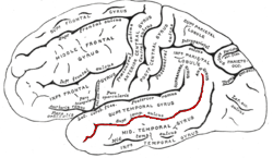
In the human brain, the superior temporal sulcus (STS) is the sulcus separating the superior temporal gyrus from the middle temporal gyrus in the temporal lobe of the brain. A sulcus is a deep groove that curves into the largest part of the brain, the cerebrum, and a gyrus is a ridge that curves outward of the cerebrum.

Visual object recognition refers to the ability to identify the objects in view based on visual input. One important signature of visual object recognition is "object invariance", or the ability to identify objects across changes in the detailed context in which objects are viewed, including changes in illumination, object pose, and background context.

<span class="mw-page-title-main">Parasol cell</span>

A parasol cell, sometimes called an M cell or M ganglion cell, is one type of retinal ganglion cell (RGC) located in the ganglion cell layer of the retina. These cells project to magnocellular cells in the lateral geniculate nucleus (LGN) as part of the magnocellular pathway in the visual system. They have large cell bodies as well as extensive branching dendrite networks and as such have large receptive fields. Relative to other RGCs, they have fast conduction velocities. While they do show clear center-surround antagonism, they receive no information about color. Parasol ganglion cells contribute information about the motion and depth of objects to the visual system.

Phonagnosia is a type of agnosia, or loss of knowledge, that involves a disturbance in the recognition of familiar voices and the impairment of voice discrimination abilities in which the affected individual does not suffer from comprehension deficits. Phonagnosia is an auditory agnosia, an acquired auditory processing disorder resulting from brain damage, other auditory agnosias include cortical deafness and auditory verbal agnosia also known as pure word deafness.

Constructional apraxia is a neurological disorder in which people are unable to perform tasks or movements even though they understand the task, are willing to complete it, and have the physical ability to perform the movements. It is characterized by an inability or difficulty to build, assemble, or draw objects. Constructional apraxia may be caused by lesions in the parietal lobe following stroke or it may serve as an indicator for Alzheimer's disease.

<span class="mw-page-title-main">Melvyn A. Goodale</span>

Melvyn Alan Goodale FRSC, FRS is a Canadian neuroscientist. He was the founding Director of the Brain and Mind Institute at the University of Western Ontario where he holds the Canada Research Chair in Visual Neuroscience. He holds appointments in the Departments of Psychology, Physiology & Pharmacology, and Ophthalmology at Western. Goodale's research focuses on the neural substrates of visual perception and visuomotor control.

References

  1. Eyesenck MW, Keane MT (2010). Cognitive Psychology: A Student's Handbook . Hove, UK: Psychology Press.
  2. 1 2 Goodale MA, Milner AD (1992). "Separate visual pathways for perception and action". Trends Neurosci. 15 (1): 20–5. doi:10.1016/0166-2236(92)90344-8. PMID   1374953. S2CID   793980.
  3. Schneider, GE. (Feb 1969). "Two visual systems". Science. 163 (3870): 895–902. Bibcode:1969Sci...163..895S. doi:10.1126/science.163.3870.895. PMID   5763873.
  4. Ingle, D. (Sep 1973). "Two visual systems in the frog". Science. 181 (4104): 1053–5. Bibcode:1973Sci...181.1053I. doi:10.1126/science.181.4104.1053. PMID   4542178. S2CID   42609212.
  5. Ettlinger G. (1990). ""Object vision" and "spatial vision": the neuropsychological evidence for the distinction". Cortex. 26 (3): 319–41. doi: 10.1016/s0010-9452(13)80084-6 . PMID   2123426. S2CID   4488069.
  6. Trevarthen, CB. (1968). "Two mechanisms of vision in primates". Psychol Forsch. 31 (4): 299–348. doi:10.1007/bf00422717. PMID   4973634. S2CID   27943693.
  7. Mishkin M, Ungerleider LG (1982). "Contribution of striate inputs to the visuospatial functions of parieto-preoccipital cortex in monkeys". Behav Brain Res. 6 (1): 57–77. doi:10.1016/0166-4328(82)90081-X. PMID   7126325. S2CID   33359587.
  8. Schenk, Thomas; McIntosh, Robert D. (2010). "Do we have independent visual streams for perception and action?". Cognitive Neuroscience. 1 (1): 52–62. doi:10.1080/17588920903388950. ISSN   1758-8928. PMID   24168245. S2CID   3560697.
  9. Goodale, MA.; Milner, AD.; Jakobson, LS.; Carey, DP. (Jan 1991). "A neurological dissociation between perceiving objects and grasping them". Nature. 349 (6305): 154–6. Bibcode:1991Natur.349..154G. doi:10.1038/349154a0. PMID   1986306. S2CID   4238254.
  10. 1 2 Himmelbach, M.; Boehme, R.; Karnath, HO. (Jan 2012). "20 years later: a second look on DF's motor behaviour". Neuropsychologia. 50 (1): 139–44. doi:10.1016/j.neuropsychologia.2011.11.011. PMID   22154499. S2CID   7513099.
  11. Aglioti S, DeSouza JF, Goodale MA (1995). "Size-contrast illusions deceive the eye but not the hand". Curr. Biol. 5 (6): 679–85. doi: 10.1016/S0960-9822(95)00133-3 . PMID   7552179. S2CID   206111613.
  12. Norman J. (2002). "Two visual systems and two theories of perception: An attempt to reconcile the constructivist and ecological approaches". Behav Brain Sci. 25 (1): 73–144. doi:10.1017/s0140525x0200002x. PMID   12625088.
  13. Mishkin, Mortimer; Ungerleider, Leslie G.; Macko, Kathleen A. (1983-01-01). "Object vision and spatial vision: two cortical pathways". Trends in Neurosciences. 6: 414–417. doi:10.1016/0166-2236(83)90190-X. ISSN   0166-2236. S2CID   15565609.
  14. Goodale, M. A.; Milner, A. D.; Jakobson, L. S.; Carey, D. P. (January 1991). "A neurological dissociation between perceiving objects and grasping them". Nature. 349 (6305): 154–156. Bibcode:1991Natur.349..154G. doi:10.1038/349154a0. ISSN   1476-4687. PMID   1986306. S2CID   4238254.
  15. Milner, A.D.; Goodale, M.A. (2006). The Visual Brain in Action (2nd ed.). OUP Oxford. ISBN   978-0198524724.
  16. Mark F Bear; Barry Connors; Michael Paradiso (2007). Neuroscience: Exploring the Brain . Hagerstown, MD: Lippincott Williams & Wilkins. ISBN   978-0-7817-6003-4.
  17. Lamme, Victor AF; Supèr, Hans; Spekreijse, Henk (1998). "Feedforward, horizontal, and feedback processing in the visual cortex". Current Opinion in Neurobiology. 8 (4): 529–535. doi:10.1016/S0959-4388(98)80042-1. PMID   9751656. S2CID   1499090.
  18. Kozlovskiy, Stanislav; Rogachev, Anton (2021). "How Areas of Ventral Visual Stream Interact When We Memorize Color and Shape Information". Advances in Intelligent Systems and Computing. 1358 (95–100). Springer-Nature: 95–100. doi:10.1007/978-3-030-71637-0_10. ISBN   978-3-030-71636-3. ISSN   2194-5357. S2CID   234902744.
  19. "Impairment of vision due to damage to the brain". www.ssc.education.ed.ac.uk.
  20. Hickok, Gregory; Poeppel, David (2007-04-13). "The cortical organization of speech processing". Nature Reviews Neuroscience. 8 (5): 393–402. doi:10.1038/nrn2113. ISSN   1471-003X. PMID   17431404. S2CID   6199399.
  21. DeWitt, I.; Rauschecker, J. P. (2012-02-01). "Phoneme and word recognition in the auditory ventral stream". Proceedings of the National Academy of Sciences. 109 (8): E505–E514. Bibcode:2012PNAS..109E.505D. doi: 10.1073/pnas.1113427109 . ISSN   0027-8424. PMC   3286918 . PMID   22308358.
  22. Gow, David W. (June 2012). "The cortical organization of lexical knowledge: A dual lexicon model of spoken language processing". Brain and Language. 121 (3): 273–288. doi:10.1016/j.bandl.2012.03.005. ISSN   0093-934X. PMC   3348354 . PMID   22498237.
  23. Hickok, Gregory (2012). "The cortical organization of speech processing: Feedback control and predictive coding the context of a dual-stream model". Journal of Communication Disorders. 45 (6): 393–402. doi:10.1016/j.jcomdis.2012.06.004. PMC   3468690 . PMID   22766458.
  24. Howard, Harry. "The dorsal stream". Brain and Language. Archived from the original on 2017-05-10. Retrieved 5 December 2015.
  25. Buchsbaum, Bradley R.; Baldo, Juliana; Okada, Kayoko; Berman, Karen F.; Dronkers, Nina; D'Esposito, Mark; Hickok, Gregory (December 2011). "Conduction aphasia, sensory-motor integration, and phonological short-term memory – An aggregate analysis of lesion and fMRI data". Brain and Language. 119 (3): 119–128. doi:10.1016/j.bandl.2010.12.001. ISSN   0093-934X. PMC   3090694 . PMID   21256582.
  26. 1 2 Howard, Harry. "The sensorimotor interface". Brain and Language.
  27. Milner, AD.; Goodale, MA. (February 2008). "Two visual systems re-viewed". Neuropsychologia. 46 (3): 774–85. doi:10.1016/j.neuropsychologia.2007.10.005. PMID   18037456. S2CID   1475566.
  28. Franz VH, Gegenfurtner KR, Bülthoff HH, Fahle M (2000). "Grasping visual illusions: no evidence for a dissociation between perception and action". Psychol. Sci. 11 (1): 20–5. doi:10.1111/1467-9280.00209. PMID   11228838. S2CID   6461170.
  29. Franz VH, Scharnowski F, Gegenfurtner KR (2005). "Illusion effects on grasping are temporally constant not dynamic". J Exp Psychol Hum Percept Perform. 31 (6): 1359–78. doi:10.1037/0096-1523.31.6.1359. PMID   16366795.
  30. Ganel T, Goodale MA (2003). "Visual control of action but not perception requires analytical processing of object shape". Nature. 426 (6967): 664–7. Bibcode:2003Natur.426..664G. doi:10.1038/nature02156. PMID   14668865. S2CID   4314969.
  31. Ganel T, Tanzer M, Goodale MA (2008). "A double dissociation between action and perception in the context of visual illusions: opposite effects of real and illusory size". Psychol. Sci. 19 (3): 221–5. doi:10.1111/j.1467-9280.2008.02071.x. PMID   18315792. S2CID   15679825.
  32. Cardoso-Leite, Pedro; Gorea, Andrei (2010). "On the Perceptual/Motor Dissociation: A Review of Concepts, Theory, Experimental Paradigms and Data Interpretations". Seeing and Perceiving. 23 (2): 89–151. doi:10.1163/187847510X503588. PMID   20550823.
  33. Goodale MA. (2011). "Transforming vision into action". Vision Res. 51 (14): 1567–87. doi: 10.1016/j.visres.2010.07.027 . PMID   20691202.
  34. Hesse, C.; Ball, K.; Schenk, T. (Jan 2012). "Visuomotor performance based on peripheral vision is impaired in the visual form agnostic patient DF". Neuropsychologia. 50 (1): 90–7. doi:10.1016/j.neuropsychologia.2011.11.002. PMID   22085864. S2CID   3092864.
  35. 1 2 McIntosh, RD.; Schenk, T. (May 2009). "Two visual streams for perception and action: current trends". Neuropsychologia. 47 (6): 1391–6. doi:10.1016/j.neuropsychologia.2009.02.009. PMID   19428404. S2CID   32937236.
  36. Milner, A.D.; Goodale, M.A. (2006), The Visual Brain in Action, ISBN   978-0-19-852472-4 , retrieved 2012-12-06