Olfactory nerve

Last updated
Olfactory nerve
Head olfactory nerve.jpg
The olfactory nerve
Details
Innervates Smell
Identifiers
Latin nervus olfactorius
MeSH D009832
NeuroNames 32
TA98 A14.2.01.004
A14.2.01.005
TA2 6181
FMA 46787
Anatomical terms of neuroanatomy

The olfactory nerve, also known as the first cranial nerve, cranial nerve I, or simply CN I, is a cranial nerve that contains sensory nerve fibers relating to the sense of smell.

Contents

The afferent nerve fibers of the olfactory receptor neurons transmit nerve impulses about odors to the central nervous system (olfaction). Derived from the embryonic nasal placode, the olfactory nerve is somewhat unusual among cranial nerves because it is capable of some regeneration if damaged. The olfactory nerve is sensory in nature and originates on the olfactory mucosa in the upper part of the nasal cavity. [1] From the olfactory mucosa, the nerve (actually many small nerve fascicles) travels up through the cribriform plate of the ethmoid bone to reach the surface of the brain. Here the fascicles enter the olfactory bulb and synapse there; from the bulbs (one on each side) the olfactory information is transmitted into the brain via the olfactory tract. [2] The fascicles of the olfactory nerve are not visible on a cadaver brain because they are severed upon removal. [3] :548

Structure

The specialized olfactory receptor neurons of the olfactory nerve are located in the olfactory mucosa of the upper parts of the nasal cavity. The olfactory nerves consist of a collection of many sensory nerve fibers that extend from the olfactory epithelium to the olfactory bulb, passing through the many openings of the cribriform plate, a sieve-like structure of the ethmoid bone.

The sense of smell arises from the stimulation of receptors by small molecules in inspired air of varying spatial, chemical, and electrical properties that reach the nasal epithelium in the nasal cavity during inhalation. These stimulants are transduced into electrical activity in the olfactory neurons, which then transmit these impulses to the olfactory bulb and from there they reach the olfactory areas of the brain via the olfactory tract.

The olfactory nerve is the shortest of the twelve cranial nerves and, similar to the optic nerve, does not emanate from the brainstem. [2]

Function

The olfaction system works to ensure that people can successfully identify an extensive range of odorants and distinguish odors from one another. [4] [5] Odorants interact with the olfactory receptor neurons (ORNs) at the periphery and transmit olfactory information to the central nervous system via axons at the basal surface. [4] [5] These axons aggregate, forming the olfactory nerve. [4] [5] [6] Therefore, the olfactory nerve works to transduce sensory stimuli in the form of odorants and encode them into electrical signals, which are relayed to higher-order centers through synaptic transmission. [4] [6]

Odor Transduction

Odorants bind to specific odorant receptor proteins contained to the outer surface of olfactory cilia within the olfactory epithelium. [4] [5] Odorant binding to the cilia of an ORN evokes an electrical response, kickstarting odor transduction. [4] An individual ORN contains several microvilli, olfactory cilia, which protrude from a knoblike structure at the apical surface involved in dendritic processes. [4] The olfactory cilia lack the cytoskeletal features of motile cilia and are, therefore, more similar to microvilli like that found in the lungs or gut. [4] Olfactory cilia are actin-rich protrusions supported by scaffolding proteins which help to localize odorant receptors and provide an increased cellular surface for odorant binding. [4]

Homologous to G-protein-coupled receptors (GPCRs), olfactory receptor molecules consist of seven trans-membrane, hydrophobic domains and a cytoplasmic domain with a carboxyl terminal region that interacts with G-proteins and odorants. [4] [5] Once an odorant is bound to an odor receptor protein, the alpha subunit of an olfactory-specific heterotrimeric G-protein, Golf, dissociates and activates olfactory-specific adenylate cyclase, adenylyl cyclase III (ACIII). [4] [5] Activation of ACIII leads to an increase in cyclic AMP (cAMP), which depolarizes the neuron due to an influx of Na+ and Ca2+ by opening cyclic nucleotide-gated ion channels. [4] [5] The neuron is further depolarized by a Ca2+-activated Cl- current travelling from the cilia, where the depolarization first occurred, to the axon hillock of the ORN. [4] [5] At the axon hillock, voltage-gated Na+ channels open and generate an action potential that is transmitted to the olfactory bulb. [4] [5] After transmission, the ORN membrane is repolarized by calcium/calmodulin kinase II-mediated mechanisms that work to extrude Ca2+ and transport Na+ via an Na+/Ca2+ exchanger, diminish cAMP levels by activating phosphodiesterases, and restore heterotrimeric Golf. [4]

ORN axons are responsible for relaying odorant information to CNS through action potentials. [4] [6] The ORN axons leave the olfactory epithelium and travel ipsilaterally to the olfactory bulb where the ORN axons coalesce into multiple clusters, called glomeruli, which together form the olfactory nerve. [4] [5] [6] The ORN axons of each glomerulus synapse with apical dendrites of mitral cells, the primary projection neurons of the olfactory bulb, which create and send action potentials further into the CNS. [4] [5] [6]

Regeneration of Olfactory Nerves

ORNs directly interact with odorants inhaled into the olfactory epithelium which can also subject the ORNs to damage through continuous exposure to harmful substances such as airborne pollutants, microorganisms, and allergens. [4] [6] [7] Therefore, ORNs maintain a normal cycle of degeneration and regeneration. [4] [7] The olfactory epithelium consists of three main cell types: supporting cells, mature ORNs, and basal cells. [4] [7] Regeneration of ORNs requires the division of basal cells, neural stem cells, to produce new receptor neuronsd. [4] [6] [7] This regeneration process makes ORNs unique when compared to other neurons. [4]

ORN Specificity

In the nasal passages, inhaled odorant molecules interact with receptor proteins on localized neuronal cilia of ORNs. [5] [6] These dendritic extensions, cilia, express one type of protein receptor, although individual odorants can interact with multiple different receptor proteins. [5] [6] As new ORNs mature, they have decreased expression levels of multiple olfactory receptor genes, contrasting with mature ORNs firm rule of one neuron—one expressed olfactory receptor gene. [4] [6] Moreover, different odors activate specific ORNs in a molecular and spatial manner due to receptor specificity. [4] Some ORNs contain receptor proteins with high affinity for some odorants, with distinct odor selectivity to a specific chemical structure, while other receptor proteins are less selective. [4]

Clinical significance

Examination

Damage to this nerve leads to impairment or total loss anosmia of the sense of smell To simply test the function of the olfactory nerve, each nostril is tested with a pungent odor. If the odor is smelled, the olfactory nerve is likely functioning. On the other hand, the nerve is only one of several reasons that could explain if the odor is not smelled. There are olfactory testing packets in which strong odors are embedded into cards and the responses of the patient to each odor can be determined. [2]

Lesions

Lesions to the olfactory nerve can occur because of "blunt trauma", such as coup-contrecoup damage, meningitis, and tumors of the frontal lobe of the brain. These injuries often lead to a reduced ability to taste and smell. Lesions of the olfactory nerve do not lead to a reduced ability to sense pain from the nasal epithelium. This is because pain from the nasal epithelium is not carried to the central nervous system by the olfactory nerve - it is carried to the central nervous system by the trigeminal nerve.

Aging and smell

A decrease in the ability to smell is a normal consequence of human aging, and usually is more pronounced in men than in women. It is often unrecognized in patients except that they may note a decreased ability to taste (much of taste is actually based on reception of food odor). Some of this decrease results from repeated damage to the olfactory nerve receptors due likely to repeated upper respiratory infections. Patients with Alzheimer's disease almost always have an abnormal sense of smell when tested. [2]

Pathway to the brain

Some nanoparticles entering the nose are transported to the brain via olfactory nerve. This can be useful for nasal administration of medications. [8] It can be harmful when the particles are soot [9] or magnetite [10] in air pollution. [11]

In naegleriasis, "brain-eating" amoeba enter through the olfactory mucosa of the nasal tissues and follow the olfactory nerve fibers into the olfactory bulbs and then the brain.

Additional images

See also

Related Research Articles

<span class="mw-page-title-main">Vomeronasal organ</span> Smell sense organ above the roof of the mouth

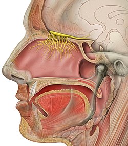
The vomeronasal organ (VNO), or Jacobson's organ, is the paired auxiliary olfactory (smell) sense organ located in the soft tissue of the nasal septum, in the nasal cavity just above the roof of the mouth in various tetrapods. The name is derived from the fact that it lies adjacent to the unpaired vomer bone in the nasal septum. It is present and functional in all snakes and lizards, and in many mammals, including cats, dogs, cattle, pigs, and some primates. Some humans may have physical remnants of a VNO, but it is vestigial and non-functional.

<span class="mw-page-title-main">Nasal cavity</span> Large, air-filled space above and behind the nose in the middle of the face

The nasal cavity is a large, air-filled space above and behind the nose in the middle of the face. The nasal septum divides the cavity into two cavities, also known as fossae. Each cavity is the continuation of one of the two nostrils. The nasal cavity is the uppermost part of the respiratory system and provides the nasal passage for inhaled air from the nostrils to the nasopharynx and rest of the respiratory tract.

<span class="mw-page-title-main">Olfactory bulb</span> Neural structure

The olfactory bulb is a neural structure of the vertebrate forebrain involved in olfaction, the sense of smell. It sends olfactory information to be further processed in the amygdala, the orbitofrontal cortex (OFC) and the hippocampus where it plays a role in emotion, memory and learning. The bulb is divided into two distinct structures: the main olfactory bulb and the accessory olfactory bulb. The main olfactory bulb connects to the amygdala via the piriform cortex of the primary olfactory cortex and directly projects from the main olfactory bulb to specific amygdala areas. The accessory olfactory bulb resides on the dorsal-posterior region of the main olfactory bulb and forms a parallel pathway. Destruction of the olfactory bulb results in ipsilateral anosmia, while irritative lesions of the uncus can result in olfactory and gustatory hallucinations.

A chemoreceptor, also known as chemosensor, is a specialized sensory receptor which transduces a chemical substance to generate a biological signal. This signal may be in the form of an action potential, if the chemoreceptor is a neuron, or in the form of a neurotransmitter that can activate a nerve fiber if the chemoreceptor is a specialized cell, such as taste receptors, or an internal peripheral chemoreceptor, such as the carotid bodies. In physiology, a chemoreceptor detects changes in the normal environment, such as an increase in blood levels of carbon dioxide (hypercapnia) or a decrease in blood levels of oxygen (hypoxia), and transmits that information to the central nervous system which engages body responses to restore homeostasis.

<span class="mw-page-title-main">Stimulus (physiology)</span> Detectable change in the internal or external surroundings

In physiology, a stimulus is a detectable change in the physical or chemical structure of an organism's internal or external environment. The ability of an organism or organ to detect external stimuli, so that an appropriate reaction can be made, is called sensitivity (excitability). Sensory receptors can receive information from outside the body, as in touch receptors found in the skin or light receptors in the eye, as well as from inside the body, as in chemoreceptors and mechanoreceptors. When a stimulus is detected by a sensory receptor, it can elicit a reflex via stimulus transduction. An internal stimulus is often the first component of a homeostatic control system. External stimuli are capable of producing systemic responses throughout the body, as in the fight-or-flight response. In order for a stimulus to be detected with high probability, its level of strength must exceed the absolute threshold; if a signal does reach threshold, the information is transmitted to the central nervous system (CNS), where it is integrated and a decision on how to react is made. Although stimuli commonly cause the body to respond, it is the CNS that finally determines whether a signal causes a reaction or not.

<span class="mw-page-title-main">Olfactory system</span> Sensory system used for smelling

The olfactory system or sense of smell is the sensory system used for smelling (olfaction). Olfaction is one of the special senses, that have directly associated specific organs. Most mammals and reptiles have a main olfactory system and an accessory olfactory system. The main olfactory system detects airborne substances, while the accessory system senses fluid-phase stimuli.

<span class="mw-page-title-main">Olfactory receptor neuron</span> Transduction nerve cell within the olfactory system

An olfactory receptor neuron (ORN), also called an olfactory sensory neuron (OSN), is a sensory neuron within the olfactory system.

<span class="mw-page-title-main">Olfactory epithelium</span> Specialised epithelial tissue in the nasal cavity that detects odours

The olfactory epithelium is a specialized epithelial tissue inside the nasal cavity that is involved in smell. In humans, it measures 5 cm2 (0.78 sq in) and lies on the roof of the nasal cavity about 7 cm (2.8 in) above and behind the nostrils. The olfactory epithelium is the part of the olfactory system directly responsible for detecting odors.

The olfactory mucosa is the neuroepithelialial mucosa lining the roof and upper parts of the septum and lateral wall of the nasal cavity which contains bipolar neurons of the primary receptor neurons of the olfactory pathway, as well as supporting cells. The neurons' dendrites project towards the nasal cavity while their axons ascend through the cribriform plate as the olfactory nerves.

<span class="mw-page-title-main">Glomerulus (olfaction)</span>

The glomerulus is a spherical structure located in the olfactory bulb of the brain where synapses form between the terminals of the olfactory nerve and the dendrites of mitral, periglomerular and tufted cells. Each glomerulus is surrounded by a heterogeneous population of juxtaglomerular neurons and glial cells.

<span class="mw-page-title-main">Sensory neuron</span> Nerve cell that converts environmental stimuli into corresponding internal stimuli

Sensory neurons, also known as afferent neurons, are neurons in the nervous system, that convert a specific type of stimulus, via their receptors, into action potentials or graded receptor potentials. This process is called sensory transduction. The cell bodies of the sensory neurons are located in the dorsal ganglia of the spinal cord.

In medicine and anatomy, the special senses are the senses that have specialized organs devoted to them:

<span class="mw-page-title-main">Mitral cell</span> Neurons that are part of the olfactory system

Mitral cells are neurons that are part of the olfactory system. They are located in the olfactory bulb in the mammalian central nervous system. They receive information from the axons of olfactory receptor neurons, forming synapses in neuropils called glomeruli. Axons of the mitral cells transfer information to a number of areas in the brain, including the piriform cortex, entorhinal cortex, and amygdala. Mitral cells receive excitatory input from olfactory sensory neurons and external tufted cells on their primary dendrites, whereas inhibitory input arises either from granule cells onto their lateral dendrites and soma or from periglomerular cells onto their dendritic tuft. Mitral cells together with tufted cells form an obligatory relay for all olfactory information entering from the olfactory nerve. Mitral cell output is not a passive reflection of their input from the olfactory nerve. In mice, each mitral cell sends a single primary dendrite into a glomerulus receiving input from a population of olfactory sensory neurons expressing identical olfactory receptor proteins, yet the odor responsiveness of the 20-40 mitral cells connected to a single glomerulus is not identical to the tuning curve of the input cells, and also differs between sister mitral cells. Odorant response properties of individual neurons in an olfactory glomerular module. The exact type of processing that mitral cells perform with their inputs is still a matter of controversy. One prominent hypothesis is that mitral cells encode the strength of an olfactory input into their firing phases relative to the sniff cycle. A second hypothesis is that the olfactory bulb network acts as a dynamical system that decorrelates to differentiate between representations of highly similar odorants over time. Support for the second hypothesis comes primarily from research in zebrafish.

A topographic map is the ordered projection of a sensory surface, like the retina or the skin, or an effector system, like the musculature, to one or more structures of the central nervous system. Topographic maps can be found in all sensory systems and in many motor systems.

<span class="mw-page-title-main">Anterior olfactory nucleus</span> Portion of the forebrain of vertebrates

The anterior olfactory nucleus is a portion of the forebrain of vertebrates.

Olfactory fatigue, also known as odor fatigue, olfactory adaptation, and noseblindness, is the temporary, normal inability to distinguish a particular odor after a prolonged exposure to that airborne compound. For example, when entering a restaurant initially the odor of food is often perceived as being very strong, but after time the awareness of the odor normally fades to the point where the smell is not perceptible or is much weaker. After leaving the area of high odor, the sensitivity is restored with time. Anosmia is the permanent loss of the sense of smell, and is different from olfactory fatigue.

Dysosmia is a disorder described as any qualitative alteration or distortion of the perception of smell. Qualitative alterations differ from quantitative alterations, which include anosmia and hyposmia. Dysosmia can be classified as either parosmia or phantosmia. Parosmia is a distortion in the perception of an odorant. Odorants smell different from what one remembers. Phantosmia is the perception of an odor when no odorant is present. The cause of dysosmia still remains a theory. It is typically considered a neurological disorder and clinical associations with the disorder have been made. Most cases are described as idiopathic and the main antecedents related to parosmia are URTIs, head trauma, and nasal and paranasal sinus disease. Dysosmia tends to go away on its own but there are options for treatment for patients that want immediate relief.

<span class="mw-page-title-main">Sense of smell</span> Sense that detects smells

The sense of smell, or olfaction, is the special sense through which smells are perceived. The sense of smell has many functions, including detecting desirable foods, hazards, and pheromones, and plays a role in taste.

<span class="mw-page-title-main">Insect olfaction</span> Function of chemical receptors

Insect olfaction refers to the function of chemical receptors that enable insects to detect and identify volatile compounds for foraging, predator avoidance, finding mating partners and locating oviposition habitats. Thus, it is the most important sensation for insects. Most important insect behaviors must be timed perfectly which is dependent on what they smell and when they smell it. For example, olfaction is essential for locating host plants and hunting prey in many species of insects, such as the moth Deilephila elpenor and the wasp Polybia sericea, respectively.

Retronasal smell, retronasal olfaction, is the ability to perceive flavor dimensions of foods and drinks. Retronasal smell is a sensory modality that produces flavor. It is best described as a combination of traditional smell and taste modalities. Retronasal smell creates flavor from smell molecules in foods or drinks shunting up through the nasal passages as one is chewing. When people use the term "smell", they are usually referring to "orthonasal smell", or the perception of smell molecules that enter directly through the nose and up the nasal passages. Retronasal smell is critical for experiencing the flavor of foods and drinks. Flavor should be contrasted with taste, which refers to five specific dimensions: (1) sweet, (2) salty, (3) bitter, (4) sour, and (5) umami. Perceiving anything beyond these five dimensions, such as distinguishing the flavor of an apple from a pear for example, requires the sense of retronasal smell.

References

  1. Mcgraw Hill's Anatomy and Physiology Revealed
  2. 1 2 3 4 Vilensky J, Robertson W, Suarez-Quian C (2015). The Clinical Anatomy of the Cranial Nerves: The Nerves of "On Old Olympus Towering Top". Ames, Iowa: Wiley-Blackwell. ISBN   978-1118492017.
  3. Saladin K (2012). "The Cranial Nerves". Anatomy and Physiology: The Unity of Form and Function (6th ed.). New York City: Mcgraw-Hill. p. 548. ISBN   978-1-61906-437-9.
  4. 1 2 3 4 5 6 7 8 9 10 11 12 13 14 15 16 17 18 19 20 21 22 23 24 25 26 Purves D, Augustine GJ, Fitzpatrick D (2018). Neuroscience (Sixth ed.). New York Oxford: Oxford University Press, Sinauer Associates is an imprint of Oxford University Press. ISBN   978-1-60535-380-7.
  5. 1 2 3 4 5 6 7 8 9 10 11 12 13 Branigan B, Tadi P (2023). "Physiology, Olfactory". StatPearls. Treasure Island (FL): StatPearls Publishing. PMID   31194396 . Retrieved 2023-12-07.
  6. 1 2 3 4 5 6 7 8 9 10 Bhatia-Dey N, Heinbockel T (June 2021). "The Olfactory System as Marker of Neurodegeneration in Aging, Neurological and Neuropsychiatric Disorders". International Journal of Environmental Research and Public Health. 18 (13): 6976. doi: 10.3390/ijerph18136976 . PMC   8297221 . PMID   34209997.
  7. 1 2 3 4 Mermelstein S, Pereira VE, Serrano PL, Pastor RA, Araujo AQ (January 2022). "Olfactory nerve: from ugly duckling to swan". Arquivos de Neuro-Psiquiatria. 80 (1): 75–83. doi:10.1590/0004-282X-ANP-2020-0529. PMC   9651502 . PMID   35239810.
  8. Gänger S, Schindowski K (August 2018). "Tailoring Formulations for Intranasal Nose-to-Brain Delivery: A Review on Architecture, Physico-Chemical Characteristics and Mucociliary Clearance of the Nasal Olfactory Mucosa". Pharmaceutics. 10 (3): 116. doi: 10.3390/pharmaceutics10030116 . PMC   6161189 . PMID   30081536.
  9. Matsui Y, Sakai N, Tsuda A, Terada Y, Takaoka M, Fujimaki H, Uchiyama I (2009). "Tracking the pathway of diesel exhaust particles from the nose to the brain by X-ray florescence analysis". Spectrochimica Acta Part B: Atomic Spectroscopy. 64 (8): 796–801. Bibcode:2009AcSpe..64..796M. doi:10.1016/j.sab.2009.06.017.
  10. Maher BA, Ahmed IA, Karloukovski V, MacLaren DA, Foulds PG, Allsop D, et al. (September 2016). "Magnetite pollution nanoparticles in the human brain". Proceedings of the National Academy of Sciences of the United States of America. 113 (39): 10797–10801. Bibcode:2016PNAS..11310797M. doi: 10.1073/pnas.1605941113 . PMC   5047173 . PMID   27601646.
  11. Stevens AS (17 December 2014). "Nano air pollutants strike a blow to the brain". Science News for Students.