Cytoskeleton

Last updated
Cell biology
Animal cell diagram
Animal Cell.svg
Components of a typical animal cell:
  1. Nucleolus
  2. Nucleus
  3. Ribosome (dots as part of 5)
  4. Vesicle
  5. Rough endoplasmic reticulum
  6. Golgi apparatus (or, Golgi body)
  7. Cytoskeleton
  8. Smooth endoplasmic reticulum
  9. Mitochondrion
  10. Vacuole
  11. Cytosol (fluid that contains organelles; with which, comprises cytoplasm)
  12. Lysosome
  13. Centrosome
  14. Cell membrane
The cytoskeleton consists of (a) microtubules, (b) microfilaments, and (c) intermediate filaments. 0317 Cytoskeletal Components.jpg
The cytoskeleton consists of (a) microtubules, (b) microfilaments, and (c) intermediate filaments.

The cytoskeleton is a complex, dynamic network of interlinking protein filaments present in the cytoplasm of all cells, including those of bacteria and archaea. [2] In eukaryotes, it extends from the cell nucleus to the cell membrane and is composed of similar proteins in the various organisms. It is composed of three main components: microfilaments, intermediate filaments, and microtubules, and these are all capable of rapid growth or disassembly depending on the cell's requirements. [3]

Contents

A multitude of functions can be performed by the cytoskeleton. Its primary function is to give the cell its shape and mechanical resistance to deformation, and through association with extracellular connective tissue and other cells it stabilizes entire tissues. [4] [5] The cytoskeleton can also contract, thereby deforming the cell and the cell's environment and allowing cells to migrate. [6] Moreover, it is involved in many cell signaling pathways and in the uptake of extracellular material (endocytosis), [7] the segregation of chromosomes during cellular division, [4] the cytokinesis stage of cell division, [8] as scaffolding to organize the contents of the cell in space [6] and in intracellular transport (for example, the movement of vesicles and organelles within the cell) [4] and can be a template for the construction of a cell wall. [4] Furthermore, it can form specialized structures, such as flagella, cilia, lamellipodia and podosomes. The structure, function and dynamic behavior of the cytoskeleton can be very different, depending on organism and cell type. [4] [9] [8] Even within one cell, the cytoskeleton can change through association with other proteins and the previous history of the network. [6]

A large-scale example of an action performed by the cytoskeleton is muscle contraction. This is carried out by groups of highly specialized cells working together. A main component in the cytoskeleton that helps show the true function of this muscle contraction is the microfilament. Microfilaments are composed of the most abundant cellular protein known as actin. [10] During contraction of a muscle, within each muscle cell, myosin molecular motors collectively exert forces on parallel actin filaments. Muscle contraction starts from nerve impulses which then causes increased amounts of calcium to be released from the sarcoplasmic reticulum. Increases in calcium in the cytosol allows muscle contraction to begin with the help of two proteins, tropomyosin and troponin. [10] Tropomyosin inhibits the interaction between actin and myosin, while troponin senses the increase in calcium and releases the inhibition. [11] This action contracts the muscle cell, and through the synchronous process in many muscle cells, the entire muscle.

History

In 1903, Nikolai K. Koltsov proposed that the shape of cells was determined by a network of tubules that he termed the cytoskeleton. The concept of a protein mosaic that dynamically coordinated cytoplasmic biochemistry was proposed by Rudolph Peters in 1929 [12] while the term (cytosquelette, in French) was first introduced by French embryologist Paul Wintrebert in 1931. [13]

When the cytoskeleton was first introduced, it was thought to be an uninteresting gel-like substance that helped organelles stay in place. [14] Much research took place to try to understand the purpose of the cytoskeleton and its components.

Initially, it was thought that the cytoskeleton was exclusive to eukaryotes but in 1992 it was discovered to be present in prokaryotes as well. This discovery came after the realization that bacteria possess proteins that are homologous to tubulin and actin; the main components of the eukaryotic cytoskeleton. [15]

Eukaryotic cytoskeleton

Eukaryotic cells contain three main kinds of cytoskeletal filaments: microfilaments, microtubules, and intermediate filaments. In neurons the intermediate filaments are known as neurofilaments. [16] Each type is formed by the polymerization of a distinct type of protein subunit and has its own characteristic shape and intracellular distribution. Microfilaments are polymers of the protein actin and are 7 nm in diameter. Microtubules are composed of tubulin and are 25 nm in diameter. Intermediate filaments are composed of various proteins, depending on the type of cell in which they are found; they are normally 8-12 nm in diameter. [2] The cytoskeleton provides the cell with structure and shape, and by excluding macromolecules from some of the cytosol, it adds to the level of macromolecular crowding in this compartment. [17] Cytoskeletal elements interact extensively and intimately with cellular membranes. [18]

Research into neurodegenerative disorders such as Parkinson's disease, Alzheimer's disease, Huntington's disease, and amyotrophic lateral sclerosis (ALS) indicate that the cytoskeleton is affected in these diseases. [19] Parkinson's disease is marked by the degradation of neurons, resulting in tremors, rigidity, and other non-motor symptoms. Research has shown that microtubule assembly and stability in the cytoskeleton is compromised causing the neurons to degrade over time. [20] In Alzheimer's disease, tau proteins which stabilize microtubules malfunction in the progression of the illness causing pathology of the cytoskeleton. [21] Excess glutamine in the Huntington protein involved with linking vesicles onto the cytoskeleton is also proposed to be a factor in the development of Huntington's Disease. [22] Amyotrophic lateral sclerosis results in a loss of movement caused by the degradation of motor neurons, and also involves defects of the cytoskeleton. [23]

Stuart Hameroff and Roger Penrose suggest a role of microtubule vibrations in neurons in the origin of consciousness. [24] [25]

Accessory proteins including motor proteins regulate and link the filaments to other cell compounds and each other and are essential for controlled assembly of cytoskeletal filaments in particular locations. [26]

A number of small-molecule cytoskeletal drugs have been discovered that interact with actin and microtubules. These compounds have proven useful in studying the cytoskeleton, and several have clinical applications.

Microfilaments

Microfilament Structure.svg
Structure of a microfilament
MEF microfilaments.jpg
Actin cytoskeleton of mouse embryo fibroblasts, stained with phalloidin

Microfilaments, also known as actin filaments, are composed of linear polymers of G-actin proteins, and generate force when the growing (plus) end of the filament pushes against a barrier, such as the cell membrane. They also act as tracks for the movement of myosin molecules that affix to the microfilament and "walk" along them. In general, the major component or protein of microfilaments are actin. The G-actin monomer combines to form a polymer which continues to form the microfilament (actin filament). These subunits then assemble into two chains that intertwine into what are called F-actin chains. [27] Myosin motoring along F-actin filaments generates contractile forces in so-called actomyosin fibers, both in muscle as well as most non-muscle cell types. [28] Actin structures are controlled by the Rho family of small GTP-binding proteins such as Rho itself for contractile acto-myosin filaments ("stress fibers"), Rac for lamellipodia and Cdc42 for filopodia.

Functions include:

Intermediate filaments

Intermediate filaments.svg
Structure of an intermediate filament
KeratinF9.png
Microscopy of keratin filaments inside cells

Intermediate filaments are a part of the cytoskeleton of many eukaryotic cells. These filaments, averaging 10 nanometers in diameter, are more stable (strongly bound) than microfilaments, and heterogeneous constituents of the cytoskeleton. Like actin filaments, they function in the maintenance of cell-shape by bearing tension (microtubules, by contrast, resist compression but can also bear tension during mitosis and during the positioning of the centrosome). Intermediate filaments organize the internal tridimensional structure of the cell, anchoring organelles and serving as structural components of the nuclear lamina. They also participate in some cell-cell and cell-matrix junctions. Nuclear lamina exist in all animals and all tissues. Some animals like the fruit fly do not have any cytoplasmic intermediate filaments. In those animals that express cytoplasmic intermediate filaments, these are tissue specific. [5] Keratin intermediate filaments in epithelial cells provide protection for different mechanical stresses the skin may endure. They also provide protection for organs against metabolic, oxidative, and chemical stresses. Strengthening of epithelial cells with these intermediate filaments may prevent onset of apoptosis, or cell death, by reducing the probability of stress. [29]

Intermediate filaments are most commonly known as the support system or "scaffolding" for the cell and nucleus while also playing a role in some cell functions. In combination with proteins and desmosomes, the intermediate filaments form cell-cell connections and anchor the cell-matrix junctions that are used in messaging between cells as well as vital functions of the cell. These connections allow the cell to communicate through the desmosome of multiple cells to adjust structures of the tissue based on signals from the cells environment. Mutations in the IF proteins have been shown to cause serious medical issues such as premature aging, desmin mutations compromising organs, Alexander Disease, and muscular dystrophy. [5]

Different intermediate filaments are:

Microtubules

Microtubule Structure.svg
Structure of a microtubule
Btub.jpg
Microtubules in a gel-fixated cell

Microtubules are hollow cylinders about 23 nm in diameter (lumen diameter of approximately 15 nm), most commonly comprising 13 protofilaments that, in turn, are polymers of alpha and beta tubulin. They have a very dynamic behavior, binding GTP for polymerization. They are commonly organized by the centrosome.

In nine triplet sets (star-shaped), they form the centrioles, and in nine doublets oriented about two additional microtubules (wheel-shaped), they form cilia and flagella. The latter formation is commonly referred to as a "9+2" arrangement, wherein each doublet is connected to another by the protein dynein. As both flagella and cilia are structural components of the cell, and are maintained by microtubules, they can be considered part of the cytoskeleton. There are two types of cilia: motile and non-motile cilia. Cilia are short and more numerous than flagella. The motile cilia have a rhythmic waving or beating motion compared to the non-motile cilia which receive sensory information for the cell; processing signals from the other cells or the fluids surrounding it. Additionally, the microtubules control the beating (movement) of the cilia and flagella. [31] Also, the dynein arms attached to the microtubules function as the molecular motors. The motion of the cilia and flagella is created by the microtubules sliding past one another, which requires ATP. [31] They play key roles in:

In addition to the roles described above, Stuart Hameroff and Roger Penrose have proposed that microtubules function in consciousness. [32]

Comparison

Cytoskeleton
type [33]
Diameter
(nm) [34]
StructureSubunit examples [33]
Microfilaments 6 Double helix Actin
Intermediate
filaments
10Two anti-parallel helices/dimers, forming tetramers
Microtubules 23 Protofilaments, in turn consisting of tubulin subunits in complex with stathmin [35] α- and β-Tubulin

Septins

Septins are a group of the highly conserved GTP binding proteins found in eukaryotes. Different septins form protein complexes with each other. These can assemble to filaments and rings. Therefore, septins can be considered part of the cytoskeleton. [36] The function of septins in cells include serving as a localized attachment site for other proteins, and preventing the diffusion of certain molecules from one cell compartment to another. [36] In yeast cells, they build scaffolding to provide structural support during cell division and compartmentalize parts of the cell. Recent research in human cells suggests that septins build cages around bacterial pathogens, immobilizing the harmful microbes and preventing them from invading other cells. [37]

Spectrin

Spectrin is a cytoskeletal protein that lines the intracellular side of the plasma membrane in eukaryotic cells. Spectrin forms pentagonal or hexagonal arrangements, forming a scaffolding and playing an important role in maintenance of plasma membrane integrity and cytoskeletal structure. [38]

Yeast cytoskeleton

In budding yeast (an important model organism), actin forms cortical patches, actin cables, and a cytokinetic ring and the cap. Cortical patches are discrete actin bodies on the membrane and are vital for endocytosis, especially the recycling of glucan synthase which is important for cell wall synthesis. Actin cables are bundles of actin filaments and are involved in the transport of vesicles towards the cap (which contains a number of different proteins to polarize cell growth) and in the positioning of mitochondria. The cytokinetic ring forms and constricts around the site of cell division. [39]

Prokaryotic cytoskeleton

Prior to the work of Jones et al., 2001, the cell wall was believed to be the deciding factor for many bacterial cell shapes, including rods and spirals. When studied, many misshapen bacteria were found to have mutations linked to development of a cell envelope. [40] The cytoskeleton was once thought to be a feature only of eukaryotic cells, but homologues to all the major proteins of the eukaryotic cytoskeleton have been found in prokaryotes. [41] Harold Erickson notes that before 1992, only eukaryotes were believed to have cytoskeleton components. However, research in the early '90s suggested that bacteria and archaea had homologues of actin and tubulin, and that these were the basis of eukaryotic microtubules and microfilaments. [42] Although the evolutionary relationships are so distant that they are not obvious from protein sequence comparisons alone, the similarity of their three-dimensional structures and similar functions in maintaining cell shape and polarity provides strong evidence that the eukaryotic and prokaryotic cytoskeletons are truly homologous. [43] Three laboratories independently discovered that FtsZ, a protein already known as a key player in bacterial cytokinesis, had the "tubulin signature sequence" present in all α-, β-, and γ-tubulins. [42] However, some structures in the bacterial cytoskeleton may not have been identified as of yet. [28] [44]

FtsZ

FtsZ was the first protein of the prokaryotic cytoskeleton to be identified. Like tubulin, FtsZ forms filaments in the presence of guanosine triphosphate (GTP), but these filaments do not group into tubules. During cell division, FtsZ is the first protein to move to the division site, and is essential for recruiting other proteins that synthesize the new cell wall between the dividing cells.

MreB and ParM

Prokaryotic actin-like proteins, such as MreB, are involved in the maintenance of cell shape. All non-spherical bacteria have genes encoding actin-like proteins, and these proteins form a helical network beneath the cell membrane that guides the proteins involved in cell wall biosynthesis. [45]

Some plasmids encode a separate system that involves an actin-like protein ParM. Filaments of ParM exhibit dynamic instability, and may partition plasmid DNA into the dividing daughter cells by a mechanism analogous to that used by microtubules during eukaryotic mitosis. [28] [46]

Crescentin

The bacterium Caulobacter crescentus contains a third protein, crescentin, that is related to the intermediate filaments of eukaryotic cells. Crescentin is also involved in maintaining cell shape, such as helical and vibrioid forms of bacteria, but the mechanism by which it does this is currently unclear. [47] Additionally, curvature could be described by the displacement of crescentic filaments, after the disruption of peptidoglycan synthesis. [48]

The cytoskeleton and cell mechanics

The cytoskeleton is a highly anisotropic and dynamic network, constantly remodeling itself in response to the changing cellular microenvironment. The network influences cell mechanics and dynamics by differentially polymerizing and depolymerizing its constituent filaments (primarily actin and myosin, but microtubules and intermediate filaments also play a role). [49] This generates forces, which play an important role in informing the cell of its microenvironment. Specifically, forces such as tension, stiffness, and shear forces have all been shown to influence cell fate, differentiation, migration, and motility. [49] Through a process called “mechanotransduction,” the cell remodels its cytoskeleton to sense and respond to these forces.

Mechanotransduction relies heavily on focal adhesions, which essentially connect the intracellular cytoskeleton with the extracellular matrix (ECM). Through focal adhesions, the cell is able to integrate extracellular forces into intracellular ones as the proteins present at focal adhesions undergo conformational changes to initiate signaling cascades. Proteins such as focal adhesion kinase (FAK) and Src have been shown to transduce force signals in response to cellular activities such as proliferation and differentiation, and are hypothesized to be key sensors in the mechanotransduction pathway. [50] As a result of mechanotransduction, the cytoskeleton changes its composition and/or orientation to accommodate the force stimulus and ensure the cell responds accordingly.

The cytoskeleton changes the mechanics of the cell in response to detected forces. For example, increasing tension within the plasma membrane makes it more likely that ion channels will open, which increases ion conductance and makes cellular change ion influx or efflux much more likely. [50] Moreover, the mechanical properties of cells determine how far and where, directionally, a force will propagate throughout the cell and how it will change cell dynamics. [51] A membrane protein that is not closely coupled to the cytoskeleton, for instance, will not produce a significant effect on the cortical actin network if it is subjected to a specifically directed force. However, membrane proteins that are more closely associated with the cytoskeleton will induce a more significant response. [50] In this way, the anisotropy of the cytoskeleton serves to more keenly direct cell responses to intra or extracellular signals.

Long-range order

The specific pathways and mechanisms by which the cytoskeleton senses and responds to forces are still under investigation. However, the long-range order generated by the cytoskeleton is known to contribute to mechanotransduction. [52] Cells, which are around 10–50 μm in diameter, are several thousand times larger than the molecules found within the cytoplasm that are essential to coordinate cellular activities. Because cells are so large in comparison to essential biomolecules, it is difficult, in the absence of an organizing network, for different parts of the cytoplasm to communicate. [53] Moreover, biomolecules must polymerize to lengths comparable to the length of the cell, but resulting polymers can be highly disorganized and unable to effectively transmit signals from one part of the cytoplasm to another. Thus, it is necessary to have the cytoskeleton to organize the polymers and ensure that they can effectively communicate across the entirety of the cell.

Common features and differences between prokaryotes and eukaryotes

By definition, the cytoskeleton is composed of proteins that can form longitudinal arrays (fibres) in all organisms. These filament forming proteins have been classified into 4 classes. Tubulin-like, actin-like, Walker A cytoskeletal ATPases (WACA-proteins), and intermediate filaments. [8] [28]

Tubulin-like proteins are tubulin in eukaryotes and FtsZ, TubZ, RepX in prokaryotes. Actin-like proteins are actin in eukaryotes and MreB, FtsA in prokaryotes. An example of a WACA-proteins, which are mostly found in prokaryotes, is MinD. Examples for intermediate filaments, which have almost exclusively been found in animals (i.e. eukaryotes) are the lamins, keratins, vimentin, neurofilaments, and desmin. [8]

Although tubulin-like proteins share some amino acid sequence similarity, their equivalence in protein-fold and the similarity in the GTP binding site is more striking. The same holds true for the actin-like proteins and their structure and ATP binding domain. [8] [28]

Cytoskeletal proteins are usually correlated with cell shape, DNA segregation and cell division in prokaryotes and eukaryotes. Which proteins fulfill which task is very different. For example, DNA segregation in all eukaryotes happens through use of tubulin, but in prokaryotes either WACA proteins, actin-like or tubulin-like proteins can be used. Cell division is mediated in eukaryotes by actin, but in prokaryotes usually by tubulin-like (often FtsZ-ring) proteins and sometimes (Thermoproteota) ESCRT-III, which in eukaryotes still has a role in the last step of division. [8]

Cytoplasmic streaming

Movement of organelles in Tradescantia stamen hair cells

Cytoplasmic streaming, also known as cyclosis, is the active movement of a cell's contents along the components of the cytoskeleton. While mainly seen in plants, all cell types use this process for transportation of waste, nutrients, and organelles to other parts of the cell.  [54] Plant and algae cells are generally larger than many other cells; so cytoplasmic streaming is important in these types of cells. This is because the cell's extra volume requires cytoplasmic streaming in order to move organelles throughout the entire cell. [55] Organelles move along microfilaments in the cytoskeleton driven by myosin motors binding and pushing along actin filament bundles. [54]  

See also

Related Research Articles

<span class="mw-page-title-main">Microtubule</span> Polymer of tubulin that forms part of the cytoskeleton

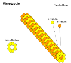
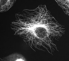
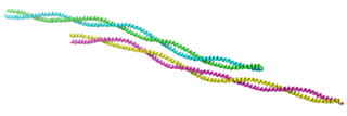
Microtubules are polymers of tubulin that form part of the cytoskeleton and provide structure and shape to eukaryotic cells. Microtubules can be as long as 50 micrometres, as wide as 23 to 27 nm and have an inner diameter between 11 and 15 nm. They are formed by the polymerization of a dimer of two globular proteins, alpha and beta tubulin into protofilaments that can then associate laterally to form a hollow tube, the microtubule. The most common form of a microtubule consists of 13 protofilaments in the tubular arrangement.

<span class="mw-page-title-main">Microfilament</span> Filament in the cytoplasm of eukaryotic cells

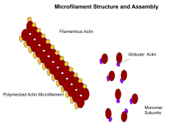
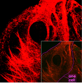
Microfilaments, also called actin filaments, are protein filaments in the cytoplasm of eukaryotic cells that form part of the cytoskeleton. They are primarily composed of polymers of actin, but are modified by and interact with numerous other proteins in the cell. Microfilaments are usually about 7 nm in diameter and made up of two strands of actin. Microfilament functions include cytokinesis, amoeboid movement, cell motility, changes in cell shape, endocytosis and exocytosis, cell contractility, and mechanical stability. Microfilaments are flexible and relatively strong, resisting buckling by multi-piconewton compressive forces and filament fracture by nanonewton tensile forces. In inducing cell motility, one end of the actin filament elongates while the other end contracts, presumably by myosin II molecular motors. Additionally, they function as part of actomyosin-driven contractile molecular motors, wherein the thin filaments serve as tensile platforms for myosin's ATP-dependent pulling action in muscle contraction and pseudopod advancement. Microfilaments have a tough, flexible framework which helps the cell in movement.

<span class="mw-page-title-main">Cleavage furrow</span> Plasma membrane invagination at the cell division site

In cell biology, the cleavage furrow is the indentation of the cell's surface that begins the progression of cleavage, by which animal and some algal cells undergo cytokinesis, the final splitting of the membrane, in the process of cell division. The same proteins responsible for muscle contraction, actin and myosin, begin the process of forming the cleavage furrow, creating an actomyosin ring. Other cytoskeletal proteins and actin binding proteins are involved in the procedure.

<span class="mw-page-title-main">Actin</span> Family of proteins

Actin is a family of globular multi-functional proteins that form microfilaments in the cytoskeleton, and the thin filaments in muscle fibrils. It is found in essentially all eukaryotic cells, where it may be present at a concentration of over 100 μM; its mass is roughly 42 kDa, with a diameter of 4 to 7 nm.

<span class="mw-page-title-main">Intermediate filament</span> Cytoskeletal structure

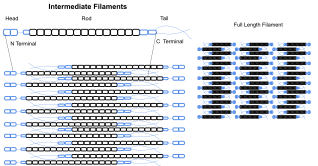
Intermediate filaments (IFs) are cytoskeletal structural components found in the cells of vertebrates, and many invertebrates. Homologues of the IF protein have been noted in an invertebrate, the cephalochordate Branchiostoma.

<span class="mw-page-title-main">Tubulin</span> Superfamily of proteins that make up microtubules

Tubulin in molecular biology can refer either to the tubulin protein superfamily of globular proteins, or one of the member proteins of that superfamily. α- and β-tubulins polymerize into microtubules, a major component of the eukaryotic cytoskeleton. Microtubules function in many essential cellular processes, including mitosis. Tubulin-binding drugs kill cancerous cells by inhibiting microtubule dynamics, which are required for DNA segregation and therefore cell division.

<span class="mw-page-title-main">Tropomyosin</span> Protein

Tropomyosin is a two-stranded alpha-helical, coiled coil protein found in many animal and fungal cells. In animals, it is an important component of the muscular system which works in conjunction with troponin to regulate muscle contraction. It is present in smooth and striated muscle tissues, which can be found in various organs and body systems, including the heart, blood vessels, respiratory system, and digestive system. In fungi, tropomyosin is found in cell walls and helps maintain the structural integrity of cells.

Cell migration is a central process in the development and maintenance of multicellular organisms. Tissue formation during embryonic development, wound healing and immune responses all require the orchestrated movement of cells in particular directions to specific locations. Cells often migrate in response to specific external signals, including chemical signals and mechanical signals. Errors during this process have serious consequences, including intellectual disability, vascular disease, tumor formation and metastasis. An understanding of the mechanism by which cells migrate may lead to the development of novel therapeutic strategies for controlling, for example, invasive tumour cells.

<span class="mw-page-title-main">Motor protein</span> Class of molecular proteins

Motor proteins are a class of molecular motors that can move along the cytoplasm of cells. They convert chemical energy into mechanical work by the hydrolysis of ATP. Flagellar rotation, however, is powered by a proton pump.

<span class="mw-page-title-main">Growth cone</span> Large actin extension of a developing neurite seeking its synaptic target

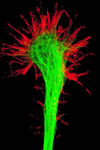
A growth cone is a large actin-supported extension of a developing or regenerating neurite seeking its synaptic target. It is the growth cone that drives axon growth. Their existence was originally proposed by Spanish histologist Santiago Ramón y Cajal based upon stationary images he observed under the microscope. He first described the growth cone based on fixed cells as "a concentration of protoplasm of conical form, endowed with amoeboid movements". Growth cones are situated on the tips of neurites, either dendrites or axons, of the nerve cell. The sensory, motor, integrative, and adaptive functions of growing axons and dendrites are all contained within this specialized structure.

<span class="mw-page-title-main">Cell cortex</span> Layer on the inner face of a cell membrane

The cell cortex, also known as the actin cortex, cortical cytoskeleton or actomyosin cortex, is a specialized layer of cytoplasmic proteins on the inner face of the cell membrane. It functions as a modulator of membrane behavior and cell surface properties. In most eukaryotic cells lacking a cell wall, the cortex is an actin-rich network consisting of F-actin filaments, myosin motors, and actin-binding proteins. The actomyosin cortex is attached to the cell membrane via membrane-anchoring proteins called ERM proteins that plays a central role in cell shape control. The protein constituents of the cortex undergo rapid turnover, making the cortex both mechanically rigid and highly plastic, two properties essential to its function. In most cases, the cortex is in the range of 100 to 1000 nanometers thick.

<span class="mw-page-title-main">Protein filament</span> Long chain of protein monomers

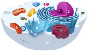
In biology, a protein filament is a long chain of protein monomers, such as those found in hair, muscle, or in flagella. Protein filaments form together to make the cytoskeleton of the cell. They are often bundled together to provide support, strength, and rigidity to the cell. When the filaments are packed up together, they are able to form three different cellular parts. The three major classes of protein filaments that make up the cytoskeleton include: actin filaments, microtubules and intermediate filaments.

<span class="mw-page-title-main">Prokaryotic cytoskeleton</span> Structural filaments in prokaryotes

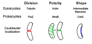
The prokaryotic cytoskeleton is the collective name for all structural filaments in prokaryotes. It was once thought that prokaryotic cells did not possess cytoskeletons, but advances in visualization technology and structure determination led to the discovery of filaments in these cells in the early 1990s. Not only have analogues for all major cytoskeletal proteins in eukaryotes been found in prokaryotes, cytoskeletal proteins with no known eukaryotic homologues have also been discovered. Cytoskeletal elements play essential roles in cell division, protection, shape determination, and polarity determination in various prokaryotes.

<span class="mw-page-title-main">ANLN</span> Mammalian protein found in Homo sapiens

Anillin is a conserved protein implicated in cytoskeletal dynamics during cellularization and cytokinesis. The ANLN gene in humans and the scraps gene in Drosophila encode Anillin. In 1989, anillin was first isolated in embryos of Drosophila melanogaster. It was identified as an F-actin binding protein. Six years later, the anillin gene was cloned from cDNA originating from a Drosophila ovary. Staining with anti-anillin antibody showed the anillin localizes to the nucleus during interphase and to the contractile ring during cytokinesis. These observations agree with further research that found anillin in high concentrations near the cleavage furrow coinciding with RhoA, a key regulator of contractile ring formation.

<span class="mw-page-title-main">Stress fiber</span> Contractile actin bundles found in non-muscle cells

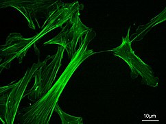
Stress fibers are contractile actin bundles found in non-muscle cells. They are composed of actin (microfilaments) and non-muscle myosin II (NMMII), and also contain various crosslinking proteins, such as α-actinin, to form a highly regulated actomyosin structure within non-muscle cells. Stress fibers have been shown to play an important role in cellular contractility, providing force for a number of functions such as cell adhesion, migration and morphogenesis.

<span class="mw-page-title-main">Amoeboid movement</span> Mode of locomotion in eukaryotic cells

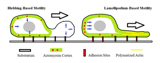
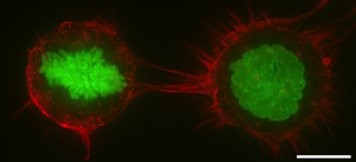
Amoeboid movement is the most typical mode of locomotion in adherent eukaryotic cells. It is a crawling-like type of movement accomplished by protrusion of cytoplasm of the cell involving the formation of pseudopodia ("false-feet") and posterior uropods. One or more pseudopodia may be produced at a time depending on the organism, but all amoeboid movement is characterized by the movement of organisms with an amorphous form that possess no set motility structures.

Actin remodeling is the biochemical process that allows for the dynamic alterations of cellular organization. The remodeling of actin filaments occurs in a cyclic pattern on cell surfaces and exists as a fundamental aspect to cellular life. During the remodeling process, actin monomers polymerize in response to signaling cascades that stem from environmental cues. The cell's signaling pathways cause actin to affect intracellular organization of the cytoskeleton and often consequently, the cell membrane. Again triggered by environmental conditions, actin filaments break back down into monomers and the cycle is completed. Actin-binding proteins (ABPs) aid in the transformation of actin filaments throughout the actin remodeling process. These proteins account for the diverse structure and changes in shape of Eukaryotic cells. Despite its complexity, actin remodeling may result in complete cytoskeletal reorganization in under a minute.

<span class="mw-page-title-main">Cytoskeletal drugs</span> Substances or medications that interact with actin or tubulin

Cytoskeletal drugs are small molecules that interact with actin or tubulin. These drugs can act on the cytoskeletal components within a cell in three main ways. Some cytoskeletal drugs stabilize a component of the cytoskeleton, such as taxol, which stabilizes microtubules, or Phalloidin, which stabilizes actin filaments. Others, such as Cytochalasin D, bind to actin monomers and prevent them from polymerizing into filaments. Drugs such as demecolcine act by enhancing the depolymerisation of already formed microtubules. Some of these drugs have multiple effects on the cytoskeleton: for example, Latrunculin both prevents actin polymerization as well as enhancing its rate of depolymerization. Typically the microtubule targeting drugs can be found in the clinic where they are used therapeutically in the treatment of some forms of cancer. As a result of the lack of specificity for specific type of actin, the use of these drugs in animals results in unacceptable off-target effects. Despite this, the actin targeting compounds are still useful tools that can be used on a cellular level to help further our understanding of how this complex part of the cells' internal machinery operates. For example, Phalloidin that has been conjugated with a fluorescent probe can be used for visualizing the filamentous actin in fixed samples.

Cell mechanics is a sub-field of biophysics that focuses on the mechanical properties and behavior of living cells and how it relates to cell function. It encompasses aspects of cell biophysics, biomechanics, soft matter physics and rheology, mechanobiology and cell biology.

Edwin W. Taylor is an adjunct professor of cell and developmental biology at Northwestern University. He was elected to the National Academy of Sciences in 2001. Taylor received a BA in physics and chemistry from the University of Toronto in 1952; an MSc in physical chemistry from McMaster University in 1955, and a PhD in biophysics from the University of Chicago in 1957. In 2001 Taylor was elected to the National Academy of Scineces in Cellular and Developmental Biology and Biochemistry.

References

  1. Creative Commons by small.svg  This article incorporates text available under the CC BY 4.0 license.Betts, J Gordon; Desaix, Peter; Johnson, Eddie; Johnson, Jody E; Korol, Oksana; Kruse, Dean; Poe, Brandon; Wise, James; Womble, Mark D; Young, Kelly A (June 8, 2023). Anatomy & Physiology. Houston: OpenStax CNX. 3.2 The cytoplasm and cellular organelles. ISBN   978-1-947172-04-3.
  2. 1 2 Hardin J, Bertoni G, Kleinsmith LJ (2015). Becker's World of the Cell (8th ed.). New York: Pearson. pp. 422–446. ISBN   978013399939-6.
  3. McKinley, Michael; Dean O'Loughlin, Valerie; Pennefather-O'Brien, Elizabeth; Harris, Ronald (2015). Human Anatomy (4th ed.). New York: McGraw Hill Education. p. 29. ISBN   978-0-07-352573-0.
  4. 1 2 3 4 5 Alberts B, et al. (2008). Molecular Biology of the Cell (5th ed.). New York: Garland Science. ISBN   978-0-8153-4105-5.
  5. 1 2 3 Herrmann H, Bär H, Kreplak L, Strelkov SV, Aebi U (July 2007). "Intermediate filaments: from cell architecture to nanomechanics". Nature Reviews. Molecular Cell Biology. 8 (7): 562–73. doi:10.1038/nrm2197. PMID   17551517. S2CID   27115011.
  6. 1 2 3 Fletcher DA, Mullins RD (January 2010). "Cell mechanics and the cytoskeleton". Nature. 463 (7280): 485–92. Bibcode:2010Natur.463..485F. doi:10.1038/nature08908. PMC   2851742 . PMID   20110992.
  7. Geli MI, Riezman H (April 1998). "Endocytic internalization in yeast and animal cells: similar and different". Journal of Cell Science. 111 ( Pt 8) (8): 1031–7. doi:10.1242/jcs.111.8.1031. PMID   9512499.
  8. 1 2 3 4 5 6 Wickstead B, Gull K (August 2011). "The evolution of the cytoskeleton". The Journal of Cell Biology. 194 (4): 513–25. doi:10.1083/jcb.201102065. PMC   3160578 . PMID   21859859.
  9. Fuchs, E.; Karakesisoglou, I. (2001). "Bridging cytoskeletal intersections". Genes & Development. 15 (1): 1–14. doi: 10.1101/gad.861501 . PMID   11156599.
  10. 1 2 Cooper, Geoffrey M. (2000). "Actin, Myosin, and Cell Movement". The Cell: A Molecular Approach. 2nd Edition. Archived from the original on 2018-04-28.
  11. Berg JM, Tymoczko JL, Stryer L (2002). "Myosins Move Along Actin Filaments". Biochemistry. 5th Edition. Archived from the original on 2018-05-02.
  12. Peters RA. "The Harben Lectures, 1929. Reprinted in: Peters, R. A. (1963) Biochemical lesions and lethal synthesis, p. 216. Pergamon Press, Oxford".{{cite journal}}: Cite journal requires |journal= (help)
  13. Frixione E (June 2000). "Recurring views on the structure and function of the cytoskeleton: a 300-year epic". Cell Motility and the Cytoskeleton. 46 (2): 73–94. doi:10.1002/1097-0169(200006)46:2<73::AID-CM1>3.0.CO;2-0. PMID   10891854. S2CID   16728876.
  14. Hardin J (2015-12-03). Becker's World of the Cell (9th ed.). Pearson. p. 351. ISBN   978-0-321-93492-5.
  15. Wickstead B, Gull K (August 2011). "The evolution of the cytoskeleton". The Journal of Cell Biology. 194 (4): 513–25. doi:10.1083/jcb.201102065. PMC   3160578 . PMID   21859859.
  16. Taran, AS; Shuvalova, LD; Lagarkova, MA; Alieva, IB (22 June 2020). "Huntington's Disease-An Outlook on the Interplay of the HTT Protein, Microtubules and Actin Cytoskeletal Components". Cells. 9 (6): 1514. doi: 10.3390/cells9061514 . PMC   7348758 . PMID   32580314.
  17. Minton AP (October 1992). "Confinement as a determinant of macromolecular structure and reactivity". Biophysical Journal. 63 (4): 1090–100. Bibcode:1992BpJ....63.1090M. doi:10.1016/S0006-3495(92)81663-6. PMC   1262248 . PMID   1420928.
  18. Doherty GJ, McMahon HT (2008). "Mediation, modulation, and consequences of membrane-cytoskeleton interactions". Annual Review of Biophysics. 37: 65–95. doi:10.1146/annurev.biophys.37.032807.125912. PMID   18573073. S2CID   17352662.
  19. Pelucchi, Silvia; Stringhi, Ramona; Marcello, Elena (2020). "Dendritic Spines in Alzheimer's Disease: How the Actin Cytoskeleton Contributes to Synaptic Failure". International Journal of Molecular Sciences. 21 (3): 908. doi: 10.3390/ijms21030908 . ISSN   1422-0067. PMC   7036943 . PMID   32019166.
  20. Pellegrini L, Wetzel A, Grannó S, Heaton G, Harvey K (February 2017). "Back to the tubule: microtubule dynamics in Parkinson's disease". Cellular and Molecular Life Sciences. 74 (3): 409–434. doi:10.1007/s00018-016-2351-6. PMC   5241350 . PMID   27600680.
  21. Bamburg JR, Bloom GS (August 2009). "Cytoskeletal pathologies of Alzheimer's Disease". Cell Motility and the Cytoskeleton. 66 (8): 635–49. doi:10.1002/cm.20388. PMC   2754410 . PMID   19479823.
  22. Caviston JP, Holzbaur EL (April 2009). "Huntingtin protein is an essential integrator of intracellular vesicular trafficking". Trends in Cell Biology. 19 (4): 147–55. doi:10.1016/j.tcb.2009.01.005. PMC   2930405 . PMID   19269181.
  23. Julien JP, Millecamps S, Kriz J (2005). Cytoskeletal Defects in Amyotrophic Lateral Sclerosis (motor neuron disease). Novartis Foundation Symposia. Vol. 264. pp. 183–92, discussion 192–6, 227–30. doi:10.1002/0470093765.ch12. ISBN   978-0-470-09373-3. PMID   15773754.{{cite book}}: |journal= ignored (help)
  24. Elsevier. "Discovery of Quantum Vibrations in "Microtubules" Inside Brain Neurons Corroborates Controversial 20-Year-Old Theory of Consciousness". www.elsevier.com. Archived from the original on 2016-11-07. Retrieved 2017-11-20.
  25. Hameroff, Stuart; Penrose, Roger (March 2014). "Consciousness in the universe". Physics of Life Reviews. 11 (1): 39–78. doi: 10.1016/j.plrev.2013.08.002 . PMID   24070914.
  26. Alberts, Bruce (2015). Molecular Biology of the Cell. Garland Science. p. 889. ISBN   978-0-8153-4464-3.
  27. 1 2 Cooper, Geoffrey M. (2000). "Structure and Organization of Actin Filaments". The Cell: A Molecular Approach. 2nd Edition. Archived from the original on 2018-05-02.
  28. 1 2 3 4 5 Gunning PW, Ghoshdastider U, Whitaker S, Popp D, Robinson RC (June 2015). "The evolution of compositionally and functionally distinct actin filaments". Journal of Cell Science. 128 (11): 2009–19. doi: 10.1242/jcs.165563 . PMID   25788699.
  29. Pan X, Hobbs RP, Coulombe PA (February 2013). "The expanding significance of keratin intermediate filaments in normal and diseased epithelia". Current Opinion in Cell Biology. 25 (1): 47–56. doi:10.1016/j.ceb.2012.10.018. PMC   3578078 . PMID   23270662.
  30. Paulin D, Li Z (November 2004). "Desmin: a major intermediate filament protein essential for the structural integrity and function of muscle". Experimental Cell Research. 301 (1): 1–7. doi:10.1016/j.yexcr.2004.08.004. PMID   15501438.
  31. 1 2 Lodish, Harvey; Berk, Arnold; Zipursky, S. Lawrence; Matsudaira, Paul; Baltimore, David; Darnell, James (2 May 2018). "Cilia and Flagella: Structure and Movement". Archived from the original on 2 May 2018. Retrieved 2 May 2018 via www.ncbi.nlm.nih.gov.{{cite journal}}: Cite journal requires |journal= (help)
  32. Hameroff, S. and Penrose, R. Physics of Life Reviews 2014, 11, 39-78
  33. 1 2 Unless else specified in boxes, then ref is:Boron WF (2003). Medical Physiology: A Cellular And Molecular Approaoch. Elsevier/Saunders. p. 1300. ISBN   978-1-4160-2328-9. Page 25
  34. Fuchs E, Cleveland DW (January 1998). "A structural scaffolding of intermediate filaments in health and disease". Science. 279 (5350): 514–9. Bibcode:1998Sci...279..514F. doi:10.1126/science.279.5350.514. PMID   9438837.
  35. Steinmetz MO (May 2007). "Structure and thermodynamics of the tubulin-stathmin interaction". Journal of Structural Biology. 158 (2): 137–47. doi:10.1016/j.jsb.2006.07.018. PMID   17029844.
  36. 1 2 Mostowy S, Cossart P (February 2012). "Septins: the fourth component of the cytoskeleton". Nature Reviews. Molecular Cell Biology. 13 (3): 183–94. doi:10.1038/nrm3284. PMID   22314400. S2CID   2418522.
  37. Mascarelli A (December 2011). "Septin proteins take bacterial prisoners: A cellular defence against microbial pathogens holds therapeutic potential". Nature. doi:10.1038/nature.2011.9540. S2CID   85080734.
  38. Huh GY, Glantz SB, Je S, Morrow JS, Kim JH (December 2001). "Calpain proteolysis of alpha II-spectrin in the normal adult human brain". Neuroscience Letters. 316 (1): 41–4. doi:10.1016/S0304-3940(01)02371-0. PMID   11720774. S2CID   53270680.
  39. Pruyne D, Bretscher A (February 2000). "Polarization of cell growth in yeast". Journal of Cell Science. 113 ( Pt 4) (4): 571–85. doi: 10.1242/jcs.113.4.571 . PMID   10652251.
  40. Jones, Laura J. F.; Carballido-López, Rut; Errington, Jeffery (2001-03-23). "Control of Cell Shape in Bacteria: Helical, Actin-like Filaments in Bacillus subtilis". Cell. 104 (6): 913–922. doi: 10.1016/S0092-8674(01)00287-2 . PMID   11290328. S2CID   14207533.
  41. Shih YL, Rothfield L (September 2006). "The bacterial cytoskeleton". Microbiology and Molecular Biology Reviews. 70 (3): 729–54. doi:10.1128/MMBR.00017-06. PMC   1594594 . PMID   16959967.
  42. 1 2 Erickson HP (February 2017). "The discovery of the prokaryotic cytoskeleton: 25th anniversary". Molecular Biology of the Cell. 28 (3): 357–358. doi:10.1091/mbc.E16-03-0183. PMC   5341718 . PMID   28137947.
  43. Michie KA, Löwe J (2006). "Dynamic filaments of the bacterial cytoskeleton" (PDF). Annual Review of Biochemistry. 75: 467–92. doi:10.1146/annurev.biochem.75.103004.142452. PMID   16756499.
  44. Briegel A, Dias DP, Li Z, Jensen RB, Frangakis AS, Jensen GJ (October 2006). "Multiple large filament bundles observed in Caulobacter crescentus by electron cryotomography". Molecular Microbiology. 62 (1): 5–14. doi: 10.1111/j.1365-2958.2006.05355.x . PMID   16987173.
  45. Popp D, Narita A, Maeda K, Fujisawa T, Ghoshdastider U, Iwasa M, Maéda Y, Robinson RC (May 2010). "Filament structure, organization, and dynamics in MreB sheets". The Journal of Biological Chemistry. 285 (21): 15858–65. doi: 10.1074/jbc.M109.095901 . PMC   2871453 . PMID   20223832.
  46. Popp D, Narita A, Lee LJ, Ghoshdastider U, Xue B, Srinivasan R, Balasubramanian MK, Tanaka T, Robinson RC (June 2012). "Novel actin-like filament structure from Clostridium tetani". The Journal of Biological Chemistry. 287 (25): 21121–9. doi: 10.1074/jbc.M112.341016 . PMC   3375535 . PMID   22514279.
  47. Ausmees N, Kuhn JR, Jacobs-Wagner C (December 2003). "The bacterial cytoskeleton: an intermediate filament-like function in cell shape". Cell. 115 (6): 705–13. doi: 10.1016/S0092-8674(03)00935-8 . PMID   14675535. S2CID   14459851.
  48. Esue, Osigwe (January 2010). "Dynamics of the Bacterial Intermediate Filament Crescentin In Vitro and In Vivo". PLOS ONE. 5 (1): e8855. Bibcode:2010PLoSO...5.8855E. doi: 10.1371/journal.pone.0008855 . PMC   2816638 . PMID   20140233.
  49. 1 2 Chen, Christopher S. (2008-10-15). "Mechanotransduction – a field pulling together?". Journal of Cell Science. 121 (20): 3285–3292. doi:10.1242/jcs.023507. ISSN   1477-9137. PMID   18843115. S2CID   1287523.
  50. 1 2 3 Orr, A. Wayne; Helmke, Brian P.; Blackman, Brett R.; Schwartz, Martin A. (January 2006). "Mechanisms of Mechanotransduction". Developmental Cell. 10 (1): 11–20. doi: 10.1016/j.devcel.2005.12.006 . PMID   16399074.
  51. Janmey, Paul A.; McCulloch, Christopher A. (2007-08-15). "Cell Mechanics: Integrating Cell Responses to Mechanical Stimuli". Annual Review of Biomedical Engineering. 9 (1): 1–34. doi:10.1146/annurev.bioeng.9.060906.151927. ISSN   1523-9829. PMID   17461730.
  52. Fletcher, Daniel A.; Mullins, R. Dyche (January 2010). "Cell mechanics and the cytoskeleton". Nature. 463 (7280): 485–492. Bibcode:2010Natur.463..485F. doi:10.1038/nature08908. ISSN   0028-0836. PMC   2851742 . PMID   20110992.
  53. Mullins, R. D. (2010-01-01). "Cytoskeletal Mechanisms for Breaking Cellular Symmetry". Cold Spring Harbor Perspectives in Biology. 2 (1): a003392. doi:10.1101/cshperspect.a003392. ISSN   1943-0264. PMC   2827899 . PMID   20182610.
  54. 1 2 Woodhouse FG, Goldstein RE (August 2013). "Cytoplasmic streaming in plant cells emerges naturally by microfilament self-organization". Proceedings of the National Academy of Sciences of the United States of America. 110 (35): 14132–7. arXiv: 1308.6422 . Bibcode:2013PNAS..11014132W. doi: 10.1073/pnas.1302736110 . PMC   3761564 . PMID   23940314.
  55. Goldstein RE, van de Meent JW (August 2015). "A physical perspective on cytoplasmic streaming". Interface Focus. 5 (4): 20150030. doi:10.1098/rsfs.2015.0030. PMC   4590424 . PMID   26464789.