Lamin

Last updated
Confocal microscopic analysis of a dermal fibroblast in primary culture from a control (a and b) and the subject with HGPS (c and d). Labelling was performed with anti-lamin A/C antibodies. Note the presence of irregularly shaped nuclear envelopes in many of the subject's fibroblasts Laminopathic nuclei.jpg
Confocal microscopic analysis of a dermal fibroblast in primary culture from a control (a and b) and the subject with HGPS (c and d). Labelling was performed with anti-lamin A/C antibodies. Note the presence of irregularly shaped nuclear envelopes in many of the subject's fibroblasts

Lamins, also known as nuclear lamins are fibrous proteins in type V intermediate filaments, providing structural function and transcriptional regulation in the cell nucleus. Nuclear lamins interact with inner nuclear membrane proteins to form the nuclear lamina on the interior of the nuclear envelope. Lamins have elastic and mechanosensitive properties, and can alter gene regulation in a feedback response to mechanical cues. [1] Lamins are present in all animals but are not found in microorganisms, plants or fungi. [2] [3] Lamin proteins are involved in the disassembling and reforming of the nuclear envelope during mitosis, the positioning of nuclear pores, and programmed cell death. Mutations in lamin genes can result in several genetic laminopathies, which may be life-threatening.

Contents

History

Lamins were first identified in the cell nucleus, using electron-microscopy. However, they were not recognized as vital components of nuclear structural support until 1975. [4] During this time period, investigations of rat liver nuclei revealed that lamins have an architectural relationship with chromatin and nuclear pores. [5] Later in 1978, immunolabeling techniques revealed that lamins are localized at the nuclear envelope under the inner nuclear membrane. It wasn't until 1986 that an analysis of lamin cDNA clones across a variety of species supported that lamins belong to the intermediate filament (IF) protein family. [4] Further investigations found evidence that supports that all IF proteins arose from a common lamin-like ancestor. This theory is based on the observation that organisms that contain IF proteins necessarily contain lamins as well; however, the presence of lamins is not a requirement for simultaneously containing IF proteins. Furthermore, sequence comparisons between lamins and IF proteins support that an amino-acid sequence that is characteristic of lamins is found in early forms of IF proteins. This sequence is lost in later forms of IF proteins, suggesting that the structure of later intermediate filaments diverged. [6] [7] After this research, investigations of lamins slowed. Studies of lamins became more popular in the 1990s when it was discovered that mutations in the genes that code for lamins can be related to muscular dystrophies, cardiomyopathies, and neuropathies. [8] [9] Current research is being performed to develop treatment methods for the aforementioned laminopathies and to investigate the role lamins play in the aging process.

Structure

The structure of lamins is composed of three units that are common among intermediate filaments: a central α-helical rod domain containing heptad repeats surrounded by globular N and C-terminal domains. The N-terminal is shorter and located at the top (head) while the C-terminal is longer and located at the end (tail). [2] [10] Lamins have a unique structure of the heptad repeats that is continuous in nature and contains an additional six heptads. [11] While the head domain of lamins is fairly consistent, the composition of the tail domain varies based on the type of lamin. However, all C-terminal domains contain a nuclear localization sequence (NLS). Similar to other IF proteins, lamins self-assemble into more complex structures. The basic unit of these structures is a coiled-coil dimer. The dimers arrange themselves in a head-to-tail manner, allowing for the formation of a protofilament. As these protofilaments aggregate, they form lamin filaments. Lamins of higher level organisms, such as vertebrates, continue to assemble into paracrystalline arrays. [2] These complex structures allow nuclear lamins to perform their specialized functions in maintaining the shape of the nucleus as well as roles during mitosis and apoptosis.

A- and B-types

Lamins are divided into two major categories: A- and B-types. These subdivisions are based on similarities in cDNA sequences, structural features, isoelectric points, and expression trends. [2] [5]

A-type lamins

A-type lamins are characterized by a neutral isoelectric point, and they are typically displayed during later stages of embryonic development. Expressed in differentiated cells, A-type lamins originate from the LMNA gene. [12] Two isoforms, lamins A and C, can be created from this gene via alternative splicing. This creates a high amount of homology between the isoforms. [4] Unlike lamin C, Lamin A is generated in a precursor form called prelamin A. Prelamin A and lamin C differ in structure only at the carboxyl-terminus. Here, prelamin A contains two extra exons that lamin C lacks. Furthermore, lamin C contains six unique amino-acid residues while prelamin A contains ninety-eight residues not found in the other isoform. [9] A CaaX motif is found within the unique residues in prelamin A. Due to the presence of the CaaX motif, prelamin A undergoes a series of posttranslational modifications to become mature lamin A. These steps include farnesylation of the carboxyl-terminal cysteine, endoproteolytic release of the terminal amino acids, carboxymethalation of the accessible farnesylcysteine, and removal of the final fifteen residues by a zinc metalloprotease. The very first modification involving farnesylation of prelamin A is crucial to the development of mature lamin A. Isoform lamin C does not undergo posttranslational modifications. [9] [13] Some studies have demonstrated that lamins A and C are not required for the formation of the nuclear lamina, yet disruptions in the LMNA gene can contribute to physical and mental limitations. [14]

B-type lamins

B-type lamins are characterized by an acidic isoelectric point, and they are typically expressed in every cell. [12] [15] As with A-type lamins, there are multiple isoforms of B-type lamins, the most common being lamin B1 and lamin B2. They are produced from two separate genes, LMNB1 and LMNB2. [9] Similar to prelamin A, B-type lamins also contain a CaaX motif at the carboxyl-terminus. This marker triggers the same sequence of posttranslational modifications previously described for prelamin A except for the final cleavage step involving a zinc metalloprotease. [9] [13] Further investigations of B-type lamins across multiple species have found evidence that supports that B-type lamins existed before A-type lamins. This stems from the similarity in structure of B-type lamins between invertebrates and vertebrates. Furthermore, organisms that only contain a single lamin contain a B-type lamin. [6] Other studies that have investigated the structural similarities and differences between A- and B-type lamins have found that the positions of introns/exons in B-type lamins have been conserved in A-type lamins, with more variations in the A-type lamins. This suggests that the common ancestor of these lamin types was a B-type lamin.

Function

Maintenance of nuclear shape

Due to their properties as a type of IF protein, lamins provide support for maintaining the shape of the nucleus. They also play an indirect role in anchoring the nucleus to the endoplasmic reticulum, forming a continuous unit within the cell. This is accomplished by lamin and lamin-interacting proteins (SUN1/SUN2) connecting with proteins on the outer nuclear membrane. These proteins in turn interact with cytoskeletal elements of the endoplasmic reticulum, forming a strong complex that can withstand mechanical stress. [6] Nuclei that lack lamins or have mutated versions have a deformed shape and do not function properly. [2]

Mitosis

During mitosis, lamins are phosphorylated by Mitosis-Promoting Factor (MPF), which drives the disassembly of the lamina and the nuclear envelope. This allows chromatin to condense and the DNA to be replicated. After chromosome segregation, dephosphorylation of nuclear lamins by a phosphatase promotes reassembly of the nuclear envelope.

Apoptosis

Apoptosis is a highly organized process of programmed cell death. Lamins are crucial targets for this process due to their close associations with chromatin and the nuclear envelope. Apoptotic enzymes called caspases target lamins and cleave both A- and B-types. [15] This allows chromatin to separate from the nuclear lamina in order to be condensed. As apoptosis continues, cell structures slowly shrink into compartmentalized "blebs." Finally, these apoptotic bodies are digested by phagocytes. [3] Studies of apoptosis involving mutant A- and B-type lamins that are resistant to cleavage by caspases show decreased DNA condensation and apoptotic “blebbing” formation, thereby underscoring the important role of lamins in apoptosis. [10]

Clinical significance

Mutations in the LMNA gene, encoding Lamins A and C, can produce a series of disorders ranging from muscular dystrophies, neuropathies, cardiomyopathies, and premature ageing syndromes. Collectively, these conditions are known as laminopathies.

Hutchinson-Gilford progeria syndrome

One specific laminopathy is Hutchinson-Gilford progeria syndrome (HGPS), characterized by premature ageing. Those affected by the condition appear normal at birth, but show signs of premature ageing including hair-loss, thinness, joint abnormalities, and weak motor skills as they develop. Furthermore, health problems usually seen in older persons such as atherosclerosis and high blood pressure occur at a much younger age. Those with HGPS typically die in their early teens, usually following a heart attack or stroke. [3] [16]

HGPS is caused by a point mutation in the LMNA gene that codes for lamin A. The genetic alteration results in an alternative splice, creating a mutated form of prelamin A that is much shorter and lacks the cleavage site for a zinc metalloprotease. Because prelamin A cannot be properly processed during posttranslational modifications, it retains its lipid modification (farnesylation) and remains in the inner nuclear membrane. This disrupts the mechanical stability of the nucleus, resulting in a higher rate of cell death and therefore a higher rate of aging. [3] Current studies are investigating the effects of farnesyl-transferase inhibitors (FTIs) to see if farnesyl attachment can be inhibited during posttranslational modification of prelamin A in order to treat patients with HGPS. [8]

Lamin A/C heart disease

Some laminopathies affect heart muscle. These mutations cause a spectrum of heart disease ranging from no apparent effect to severe dilated cardiomyopathy leading to heart failure. Laminopathies frequently cause heart rhythm problems at an early stage in the disease process including abnormally slow heart rhythms such as sinus node dysfunction and atrioventricular block, and abnormally rapid heart rhythms such as ventricular tachycardia. As a result, those with Lamin A/C heart disease are often treated with pacemakers or implantable defibrillators in addition to medication. [17]

Related Research Articles

<span class="mw-page-title-main">Cell nucleus</span> Eukaryotic membrane-bounded organelle containing DNA

The cell nucleus is a membrane-bound organelle found in eukaryotic cells. Eukaryotic cells usually have a single nucleus, but a few cell types, such as mammalian red blood cells, have no nuclei, and a few others including osteoclasts have many. The main structures making up the nucleus are the nuclear envelope, a double membrane that encloses the entire organelle and isolates its contents from the cellular cytoplasm; and the nuclear matrix, a network within the nucleus that adds mechanical support.

<span class="mw-page-title-main">Progeria</span> Genetic disorder that causes early aging

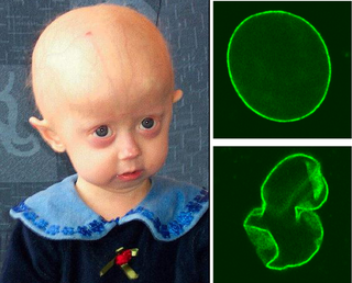
Progeria is a specific type of progeroid syndrome, also known as Hutchinson–Gilford syndrome or Hutchinson–Gilford progeroid syndrome (HGPS). A single gene mutation is responsible for causing progeria. The gene, known as lamin A (LMNA), makes a protein necessary for holding the nucleus of the cell together. When this gene gets mutated, an abnormal form of lamin A protein called progerin is produced. Progeroid syndromes are a group of diseases that causes individuals to age faster than usual, leading to them appearing older than they actually are. Patients born with progeria typically live to an age of mid-teens to early twenties.

<span class="mw-page-title-main">Nucleoplasm</span> Protoplasm that permeates a cells nucleus

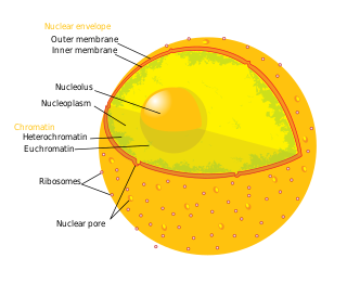
The nucleoplasm, also known as karyoplasm, is the type of protoplasm that makes up the cell nucleus, the most prominent organelle of the eukaryotic cell. It is enclosed by the nuclear envelope, also known as the nuclear membrane. The nucleoplasm resembles the cytoplasm of a eukaryotic cell in that it is a gel-like substance found within a membrane, although the nucleoplasm only fills out the space in the nucleus and has its own unique functions. The nucleoplasm suspends structures within the nucleus that are not membrane-bound and is responsible for maintaining the shape of the nucleus. The structures suspended in the nucleoplasm include chromosomes, various proteins, nuclear bodies, the nucleolus, nucleoporins, nucleotides, and nuclear speckles.

<span class="mw-page-title-main">Intermediate filament</span> Cytoskeletal structure

Intermediate filaments (IFs) are cytoskeletal structural components found in the cells of vertebrates, and many invertebrates. Homologues of the IF protein have been noted in an invertebrate, the cephalochordate Branchiostoma.

<span class="mw-page-title-main">Nuclear lamina</span>

The nuclear lamina is a dense fibrillar network inside the nucleus of eukaryote cells. It is composed of intermediate filaments and membrane associated proteins. Besides providing mechanical support, the nuclear lamina regulates important cellular events such as DNA replication and cell division. Additionally, it participates in chromatin organization and it anchors the nuclear pore complexes embedded in the nuclear envelope.

<span class="mw-page-title-main">Emery–Dreifuss muscular dystrophy</span> Medical condition

Emery–Dreifuss muscular dystrophy (EDMD) is a type of muscular dystrophy, a group of heritable diseases that cause progressive impairment of muscles. EDMD affects muscles used for movement, causing atrophy, weakness and contractures. It almost always affects the heart, causing abnormal rhythms, heart failure, or sudden cardiac death. It is rare, affecting 0.39 per 100,000 people. It is named after Alan Eglin H. Emery and Fritz E. Dreifuss.

<span class="mw-page-title-main">Thymopoietin</span>

Lamina-associated polypeptide 2 (LAP2), isoforms beta/gamma is a protein that in humans is encoded by the TMPO gene. LAP2 is an inner nuclear membrane (INM) protein.

<span class="mw-page-title-main">Laminopathy</span> Medical condition

Laminopathies are a group of rare genetic disorders caused by mutations in genes encoding proteins of the nuclear lamina. They are included in the more generic term nuclear envelopathies that was coined in 2000 for diseases associated with defects of the nuclear envelope. Since the first reports of laminopathies in the late 1990s, increased research efforts have started to uncover the vital role of nuclear envelope proteins in cell and tissue integrity in animals.

<span class="mw-page-title-main">Prelamin-A/C</span> Filament protein

Prelamin-A/C, or lamin A/C is a protein that in humans is encoded by the LMNA gene. Lamin A/C belongs to the lamin family of proteins.

<span class="mw-page-title-main">Lamin B receptor</span> Protein-coding gene in the species Homo sapiens

Lamin-B receptor is a protein, and in humans, it is encoded by the LBR gene.

<span class="mw-page-title-main">Nuclear envelope</span> Nuclear membrane surrounding the nucleus in eukaryotic cells

The nuclear envelope, also known as the nuclear membrane, is made up of two lipid bilayer membranes that in eukaryotic cells surround the nucleus, which encloses the genetic material.

<span class="mw-page-title-main">Promyelocytic leukemia protein</span> Protein-coding gene in the species Homo sapiens

Promyelocytic leukemia protein (PML) is the protein product of the PML gene. PML protein is a tumor suppressor protein required for the assembly of a number of nuclear structures, called PML-nuclear bodies, which form amongst the chromatin of the cell nucleus. These nuclear bodies are present in mammalian nuclei, at about 1 to 30 per cell nucleus. PML-NBs are known to have a number of regulatory cellular functions, including involvement in programmed cell death, genome stability, antiviral effects and controlling cell division. PML mutation or loss, and the subsequent dysregulation of these processes, has been implicated in a variety of cancers.

<span class="mw-page-title-main">Restrictive dermopathy</span> Medical condition

Restrictive dermopathy (RD) is a rare, lethal autosomal recessive skin condition characterized by syndromic facies, tight skin, sparse or absent eyelashes, and secondary joint changes.

<span class="mw-page-title-main">Nuclear prelamin A recognition factor</span> Protein-coding gene in the species Homo sapiens

Nuclear prelamin A recognition factor, also known as NARF, is a protein which in humans is encoded by the NARF gene.

<span class="mw-page-title-main">Lamin B1</span> Protein-coding gene in the species Homo sapiens

Lamin-B1 is a protein that in humans is encoded by the LMNB1 gene.

<span class="mw-page-title-main">Progerin</span>

Progerin is a truncated version of the lamin A protein involved in the pathology of Hutchinson–Gilford progeria syndrome. Progerin is most often generated by a sporadic single point nucleotide polymorphism c.1824 C>T in the gene that codes for matured Lamin A. This mutation activates a cryptic splice site that induces a mutation in premature Lamin A with the deletion of a 50 amino acids group near the C-terminus. The endopeptidase ZMPSTE24 cannot cleave between the missing RSY - LLG amino acid sequence during the maturation of Lamin A, due to the deletion of the 50 amino acids which included that sequence. This leaves the intact premature Lamin A bonded to the methylated carboxyl farnesyl group creating the defective protein Progerin, rather than the desired protein matured Lamin A. Approximately 90% of all Hutchinson–Gilford progeria syndrome cases are heterozygous for this deleterious single nucleotide polymorphism within exon 11 of the LMNA gene causing the post-translational modifications to produce Progerin.

<span class="mw-page-title-main">Inner nuclear membrane protein</span> Protein embedded in inner membrane of nuclear envelope

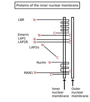
Inner nuclear membrane proteins are membrane proteins that are embedded in or associated with the inner membrane of the nuclear envelope. There are about 60 INM proteins, most of which are poorly characterized with respect to structure and function. Among the few well-characterized INM proteins are lamin B receptor (LBR), lamina-associated polypeptide 1 (LAP1), lamina-associated polypeptide-2 (LAP2), emerin and MAN1.

Progeroid syndromes (PS) are a group of rare genetic disorders that mimic physiological aging, making affected individuals appear to be older than they are. The term progeroid syndrome does not necessarily imply progeria, which is a specific type of progeroid syndrome.

Veena Krishnaji Parnaik is an Indian cell biologist and the current Chief Scientist at the Centre for Cellular and Molecular Biology. She obtained her Masters in Science in medicinal biochemistry from the University of Mumbai and received her PhD from Ohio State University before moving back to India to work at the CCMB. Her research is focused on understanding the functional role of the nuclear lamina and how defects in it may lead to disorders such as progeria and muscular dystrophy.

<span class="mw-page-title-main">LMNA-related congenital muscular dystrophy</span> Medical condition

Lamin A/C congenital muscular dystrophy (CMD) is a disease that it is included in laminopathies. Laminopathies are caused, among other mutations, to mutations in LMNA, a gene that synthesizes lamins A and C.

References

  1. Dutta, S; Bhattacharyya, M; Sengupta, K (14 October 2016). "Implications and Assessment of the Elastic Behavior of Lamins in Laminopathies". Cells. 5 (4): 37. doi: 10.3390/cells5040037 . PMC   5187521 . PMID   27754432.
  2. 1 2 3 4 5 Dechat, Thomas; Adam, Stephen A.; Taimen, Pekka; Shimi, Takeshi; Goldman, Robert D. (2016-11-24). "Nuclear Lamins". Cold Spring Harbor Perspectives in Biology. 2 (11): a000547. doi:10.1101/cshperspect.a000547. ISSN   1943-0264. PMC   2964183 . PMID   20826548.
  3. 1 2 3 4 Hardin Jeff, and Bertoni Gregory (2016). Becker's World of the Cell, 9th Edition. Boston, MA: Pearson. ISBN   9780321934925.
  4. 1 2 3 Moir, Robert D.; Spann, Timothy P.; Lopez-Soler, Reynold I.; Yoon, Miri; Goldman, Anne E.; Khuon, Satya; Goldman, Robert D. (2000-04-01). "Review: The Dynamics of the Nuclear Lamins during the Cell Cycle— Relationship between Structure and Function". Journal of Structural Biology. 129 (2): 324–334. doi:10.1006/jsbi.2000.4251. PMID   10806083.
  5. 1 2 Eriksson, John E.; Dechat, Thomas; Grin, Boris; Helfand, Brian; Mendez, Melissa; Pallari, Hanna-Mari; Goldman, Robert D. (2009-07-01). "Introducing intermediate filaments: from discovery to disease". The Journal of Clinical Investigation. 119 (7): 1763–71. doi:10.1172/JCI38339. ISSN   0021-9738. PMC   2701876 . PMID   19587451.
  6. 1 2 3 Dittmer, Travis A; Misteli, Tom (2011-01-01). "The lamin protein family". Genome Biology. 12 (5): 222. doi: 10.1186/gb-2011-12-5-222 . ISSN   1465-6906. PMC   3219962 . PMID   21639948.
  7. Cooper, Geoffrey M. (2000-01-01). "Intermediate Filaments". The Cell: A Molecular Approach. 2nd Edition.
  8. 1 2 Simpkins, Beth. "A Comeback for the Ages: Lamin's connection with aging has reinvigorated research" . Retrieved 2016-11-24.
  9. 1 2 3 4 5 Young, Stephen G.; Jung, Hea-Jin; Lee, John M.; Fong, Loren G. (2016-11-24). "Nuclear Lamins and Neurobiology". Molecular and Cellular Biology. 34 (15): 2776–2785. doi:10.1128/MCB.00486-14. ISSN   0270-7306. PMC   4135577 . PMID   24842906.
  10. 1 2 Dechat, Thomas; Pfleghaar, Katrin; Sengupta, Kaushik; Shimi, Takeshi; Shumaker, Dale K.; Solimando, Liliana; Goldman, Robert D. (2008-04-01). "Nuclear lamins: major factors in the structural organization and function of the nucleus and chromatin". Genes & Development. 22 (7): 832–853. doi:10.1101/gad.1652708. ISSN   0890-9369. PMC   2732390 . PMID   18381888.
  11. Goldman, Robert D.; Gruenbaum, Yosef; Moir, Robert D.; Shumaker, Dale K.; Spann, Timothy P. (2002-03-01). "Nuclear lamins: building blocks of nuclear architecture". Genes & Development. 16 (5): 533–547. doi: 10.1101/gad.960502 . ISSN   0890-9369. PMID   11877373.
  12. 1 2 Stuurman, Nico; Heins, Susanne; Aebi, Ueli (1998-01-01). "Nuclear Lamins: Their Structure, Assembly, and Interactions". Journal of Structural Biology. 122 (1): 42–66. doi:10.1006/jsbi.1998.3987. PMID   9724605.
  13. 1 2 Dechat, Thomas; Adam, Stephen A.; Goldman, Robert D. (2009-01-01). "Nuclear lamins and chromatin: When structure meets function". Advances in Enzyme Regulation. 49 (1): 157–166. doi:10.1016/j.advenzreg.2008.12.003. PMC   3253622 . PMID   19154754.
  14. Burke, Brian (2001-04-30). "Lamins and Apoptosis". The Journal of Cell Biology. 153 (3): f5–f7. doi:10.1083/jcb.153.3.f5. ISSN   0021-9525. PMC   2190563 . PMID   11331313.
  15. 1 2 Gruenbaum, Yosef; Wilson, Katherine L.; Harel, Amnon; Goldberg, Michal; Cohen, Merav (2000-04-01). "Review: Nuclear Lamins—Structural Proteins with Fundamental Functions". Journal of Structural Biology. 129 (2): 313–323. doi:10.1006/jsbi.2000.4216. PMID   10806082.
  16. Reference, Genetics Home. "Hutchinson-Gilford progeria syndrome". Genetics Home Reference. Retrieved 2016-11-24.
  17. Captur, Gabriella; Arbustini, Eloisa; Bonne, Gisèle; Syrris, Petros; Mills, Kevin; Wahbi, Karim; Mohiddin, Saidi A.; McKenna, William J.; Pettit, Stephen (2017-11-25). "Lamin and the heart" (PDF). Heart. 104 (6): 468–479. doi:10.1136/heartjnl-2017-312338. ISSN   1468-201X. PMID   29175975. S2CID   3563474.