Laminopathy

Last updated
Laminopathy
Laminopathic nuclei.jpg
Normal nuclear lamina (a and b) and mutant nuclear lamina (c and d) from a patient with HGPS, visualized by immunofluorescence - note the irregular and bumpy shape of the laminopathic nuclei [1]
Specialty Clinical Genetics
Symptoms Muscle weakness, reduced sensation, shortness of breath, syncope
Complications Diabetes, heart failure, arrhythmias
Usual onsetVariable
DurationLifelong
CausesGenetic
Diagnostic method Clinical, genetic testing
Treatment Physiotherapy, orthopaedic surgery, pacemaker, implantable defibrillator
Medication ACE inhibitor, beta blocker, aldosterone antagonist
Prognosis Variable

Laminopathies ( lamino- + -pathy ) are a group of rare genetic disorders caused by mutations in genes encoding proteins of the nuclear lamina. Since the first reports of laminopathies in the late 1990s, increased research efforts have started to uncover the vital role of nuclear envelope proteins in cell and tissue integrity in animals. Laminopathies are a group of degenerative diseases, other disorders associated with inner nuclear membrane proteins are known as nuclear envelopathies. [2]

Contents

Symptoms and signs

Laminopathies and other nuclear envelopathies have a large variety of clinical symptoms including skeletal and/or cardiac muscular dystrophy, lipodystrophy and diabetes, dysplasia, dermo- or neuropathy, leukodystrophy, and progeria (premature aging). Most of these symptoms develop after birth, typically during childhood or adolescence. Some laminopathies however may lead to an early death, and mutations of lamin B1 (LMNB1 gene) may be lethal before or at birth. [3]

Genetics

Patients with classical laminopathy have mutations in the gene coding for lamin A/C (LMNA gene).[ citation needed ]

Mutations in the gene coding for lamin B2 (LMNB2 gene) have been linked to Barraquer-Simons syndrome [4] and duplication in the gene coding for lamin B1 (LMNB1 gene) cause autosomal dominant leukodystrophy. [5]

Mutations implicated in other nuclear envelopathies were found in genes coding for lamin-binding proteins such as lamin B receptor (LBR gene), [6] [7] [8] [9] [10] emerin (EMD gene) and LEM domain-containing protein 3 (LEMD3 gene) and prelamin A-processing enzymes such as the zinc metalloproteinase STE24 (ZMPSTE24 gene).

Mutations causing laminopathies include recessive as well as dominant alleles with rare de novo mutations creating dominant alleles that do not allow their carriers to reproduce before death.[ citation needed ]

The nuclear envelopathy with the highest frequency in human populations is Emery–Dreifuss muscular dystrophy caused by an X-linked mutation in the EMD gene coding for emerin and affecting an estimated 1 in 100,000 people.[ citation needed ]

Molecular mechanism

Lamins are intermediate filament proteins that form the nuclear lamina scaffold underneath the nuclear envelope in animal cells. They are attached to the nuclear envelope membrane via farnesyl anchors and interaction with inner nuclear membrane proteins such as lamin B receptor and emerin. The nuclear lamina appears to be an adaptation to mobility in animals as sessile organisms such as plants or fungi do not have lamins [11] and the symptoms of many laminopathies include muscle defects. Mutations in these genes might lead to defects in filament assembly and/or attachment to the nuclear envelope and thus jeopardize nuclear envelope stability in physically stressed tissues such as muscle fibers, bone, skin and connective tissue. [12]

Messenger RNA produced from the LMNA gene undergoes alternative splicing and is translated into lamins A and C. Lamin A undergoes farnesylation to attach a membrane anchor to the protein. This version of the protein is also referred to as prelamin A. Farnesylated prelamin A is further processed into mature lamin A by a metalloproteinase removing the last 15 amino acids and its farnesylated cysteine. This allows lamin A to dissociate from the nuclear envelope membrane and fulfill nuclear functions. Mutations causing laminopathies interfere with these processes on different levels.[ citation needed ]

Nonsense and missense mutations

Missense mutations in the lamin A/C rod and tail domains are the cause for a wide array of genetic disorders, suggesting that lamin A/C protein contains distinct functional domains that are essential for the maintenance and integrity of different cell lineages. Interaction between lamin A and the nuclear envelope protein emerin appears to be crucial in muscle cells, with certain mutations in lamin mimicking mutations in emerin and causing Emery–Dreifuss muscular dystrophy. Different mutations lead to dominant-negative and recessive alleles. Mutations in the lamin rod domain leading to mislocalization of both lamin A and emerin occur in patients with autosomal dominant forms of muscular dystrophy and cardiomyopathy.[ citation needed ]

Most lamin B mutations appear to be lethal with mutations in lamin B1 causing death at birth in mice. [3] In 2006, lamin B2 missense mutations were identified in patients with acquired partial lipodystrophy. [13]

Point mutations

The most common mutation in the lamin A/C is the homozygous Arg527His (arginine replaced by histidine at position 527) substitution in exon 9 of the LMNA gene [14] Other known mutations are Ala529Val and Arg527His/Val440Met. [15] Additionally, some mutations such as Arg527Cys, Lys542Asn, Arg471Cys, Thr528Met/Met540Thr, and Arg471Cys/Arg527Cys, Arg527Leu result in mandibuloacral dysplasia with progeria-like features. [16]

Splicing defects

Mutations causing progeria are defective in splicing LMNA mRNA, therefore producing abnormal lamin A protein, also known as progerin. The mutations activate a cryptic splice site within exon 11 of the gene, thereby causing the deletion of the processing site on prelamin A. [17] This results in an accumulation of progerin that is unable to mature into lamin A, leading to misshapen nuclei. Missplicing also leads to the complete or partial loss of exon 11 and results in a truncated prelamin A protein in the neonatal lethal tight skin contracture syndrome. [18]

Processing defects

Since the metalloproteinase STE24 is required to process prelamin A into mature lamin A, mutations in this gene abolishing protease activity cause defects similar to laminopathies caused by prelamin A with truncated processing sites. Symptoms in patients with ZMPSTE24 mutation range from mandibuloacral dysplasia, progeroid appearance, and generalized lipodystrophy to infant-lethal restrictive dermopathy.[ citation needed ]

Gene dosage effects

In the case of autosomal dominant leukodystrophy, the disease is associated with a duplication of the lamin B gene LMNB1. The exact dosage of lamin B in cells appears to be crucial for nuclear integrity as increased expression of lamin B causes a degenerative phenotype in fruit flies and leads to abnormal nuclear morphology. [19]

Autoimmune antibodies

Antibodies against lamins are detected in the sera of some individuals with autoimmune diseases. [20]

DNA repair

A-type lamins promote genetic stability by maintaining the levels of proteins that have key roles in DNA double-strand break repair during the processes of non-homologous end joining and homologous recombination. [21] Mutations in lamin A (LMNA) cause Hutchinson–Gilford progeria syndrome, a dramatic form of premature aging. [17] Mouse cells deficient for maturation of prelamin A show increased DNA damage and chromosome aberrations and are more sensitive to DNA damaging agents. [22] The inability to adequately repair DNA damages when A-type lamins are defective is likely responsible for some of the aspects of premature aging.[ citation needed ]

Diagnosis

Types of known laminopathies and other nuclear envelopathies

SyndromeOMIM IDSymptomsMutation inIdentified in
Atypical Werner syndrome 277700 Progeria with increased severity compared to normal Werner syndrome Lamin A/C2003 [23]
Barraquer–Simons syndrome 608709 Lipodystrophy Lamin B22006 [13]
Buschke–Ollendorff syndrome 166700 Skeletal dysplasia, skin lesions LEM domain containing protein 3 (lamin-binding protein)2004 [24]
Cardiomyopathy, dilated, with quadriceps myopathy 607920 Cardiomyopathy Lamin A/C2003 [26]
Charcot–Marie–Tooth disease, axonal, type 2B1 605588 Neuropathy Lamin A/C2002 [27]
Emery–Dreifuss muscular dystrophy, X-linked (EDMD) 310300 Skeletal and cardiac muscular dystrophy Emerin (lamin-binding protein)1996, [28] 2000 [29]
Emery–Dreifuss muscular dystrophy, autosomal dominant (EDMD2) 181350 Skeletal and cardiac muscular dystrophy Lamin A/C1999 [30]
Emery–Dreifuss muscular dystrophy, autosomal recessive (EDMD3) 604929 Skeletal and cardiac muscular dystrophy Lamin A/C2000 [31]
Familial partial lipodystrophy of the Dunnigan type (FPLD) 151660 Lipoatrophic diabetes Lamin A/C2002 [32]
Greenberg dysplasia 215140 Skeletal dysplasia Lamin B receptor2003 [33]
Hutchinson–Gilford progeria syndrome (HGPS) 176670 Progeria Lamin A/C2003 [17]
Leukodystrophy, demyelinating, adult-onset, autosomal dominant (ADLD) 169500 Progressive demyelinating disorder affecting the central nervous system Lamin B1 (tandem gene duplication)2006 [19]
Limb-girdle muscular dystrophy type 1B (LGMD1B) 159001 Muscular dystrophy of hips and shoulders, cardiomyopathy Lamin A/C2000 [34]
Lipoatrophy with diabetes, hepatic steatosis, hypertrophic cardiomyopathy, and leukomelanodermic papules (LDHCP) 608056 Lipoatrophic diabetes, fatty liver, hypertrophic cardiomyopathy, skin lesions Lamin A/C2003 [35]
Mandibuloacral dysplasia with type A lipodystrophy (MADA) 248370 Dysplasia and lipodystrophy Lamin A/C2002 [14]
Mandibuloacral dysplasia with type B lipodystrophy (MADB) 608612 Dysplasia and lipodystrophy Zinc metalloprotease STE24 (prelamin-processing enzyme)2003 [36]
Pelger–Huet anomaly (PHA) 169400 Myelodysplasia Lamin B receptor2002 [37]
Restrictive dermopathy, lethal 275210 Dermopathy Lamin A/C or Zinc metalloprotease STE24 (prelamin-processing enzyme)2004 [18]

Treatment

Currently, there is no cure for laminopathies and treatment is largely symptomatic and supportive. Physical therapy and/or corrective orthopedic surgery may be helpful for patients with muscular dystrophies. Laminopathies affecting heart muscle may cause heart failure requiring treatment with medications including ACE inhibitors, beta blockers and aldosterone antagonists, while the abnormal heart rhythms that frequently occur in these patients may require a pacemaker or implantable defibrillator. [38] Treatment for neuropathies may include medication for seizures and spasticity.[ citation needed ]

Research

The recent progress in uncovering the molecular mechanisms of toxic progerin formation in laminopathies leading to premature aging has opened up the potential for the development of targeted treatment. The farnesylation of prelamin A and its pathological form progerin is carried out by the enzyme farnesyl transferase. Farnesyl transferase inhibitors (FTIs) can be used effectively to reduce symptoms in two mouse model systems for progeria and to revert the abnormal nuclear morphology in progeroid cell cultures. Two oral FTIs, lonafarnib and tipifarnib, are already in use as anti-tumor medication in humans and may become avenues of treatment for children with laminopathic progeria. Nitrogen-containing bisphosphate drugs used in the treatment of osteoporosis reduce farnesyldiphosphate production and thus prelamin A farnesylation. Testing of these drugs may prove them to be useful in treating progeria as well. The use of antisense oligonucleotides to inhibit progerin synthesis in affected cells is another avenue of current research into the development of anti-progerin drugs. [39] [40]

Related Research Articles

<span class="mw-page-title-main">Progeria</span> Genetic disorder that causes early aging

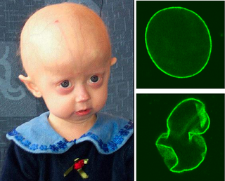
Progeria is a specific type of progeroid syndrome, also known as Hutchinson–Gilford syndrome or Hutchinson–Gilford progeroid syndrome (HGPS). A single gene mutation is responsible for causing progeria. The gene, known as lamin A (LMNA), makes a protein necessary for holding the nucleus of the cell together. When this gene gets mutated, an abnormal form of lamin A protein called progerin is produced. Progeroid syndromes are a group of diseases that cause individuals to age faster than usual, leading to them appearing older than they actually are. People born with progeria typically live until their mid to late teens or early twenties.

<span class="mw-page-title-main">Lamin</span>

Lamins, also known as nuclear lamins are fibrous proteins in type V intermediate filaments, providing structural function and transcriptional regulation in the cell nucleus. Nuclear lamins interact with inner nuclear membrane proteins to form the nuclear lamina on the interior of the nuclear envelope. Lamins have elastic and mechanosensitive properties, and can alter gene regulation in a feedback response to mechanical cues. Lamins are present in all animals but are not found in microorganisms, plants or fungi. Lamin proteins are involved in the disassembling and reforming of the nuclear envelope during mitosis, the positioning of nuclear pores, and programmed cell death. Mutations in lamin genes can result in several genetic laminopathies, which may be life-threatening.

In genetics, a missense mutation is a point mutation in which a single nucleotide change results in a codon that codes for a different amino acid. It is a type of nonsynonymous substitution.

Malouf syndrome is a congenital disorder that causes one or more of the following symptoms: mental retardation, ovarian dysgenesis, congestive cardiomyopathy, broad nasal base, blepharoptosis, and bone abnormalities, and occasionally marfanoid habitus.

<span class="mw-page-title-main">Emery–Dreifuss muscular dystrophy</span> Medical condition

Emery–Dreifuss muscular dystrophy (EDMD) is a type of muscular dystrophy, a group of heritable diseases that cause progressive impairment of muscles. EDMD affects muscles used for movement, causing atrophy, weakness and contractures. It almost always affects the heart, causing abnormal rhythms, heart failure, or sudden cardiac death. It is rare, affecting 0.39 per 100,000 people. It is named after Alan Eglin H. Emery and Fritz E. Dreifuss.

<span class="mw-page-title-main">Emerin</span> Protein-coding gene in humans

Emerin is a protein that in humans is encoded by the EMD gene, also known as the STA gene. Emerin, together with LEMD3, is a LEM domain-containing integral protein of the inner nuclear membrane in vertebrates. Emerin is highly expressed in cardiac and skeletal muscle. In cardiac muscle, emerin localizes to adherens junctions within intercalated discs where it appears to function in mechanotransduction of cellular strain and in beta-catenin signaling. Mutations in emerin cause X-linked recessive Emery–Dreifuss muscular dystrophy, cardiac conduction abnormalities and dilated cardiomyopathy.

<span class="mw-page-title-main">Prelamin-A/C</span> Filament protein

Prelamin-A/C, or lamin A/C is a protein that in humans is encoded by the LMNA gene. Lamin A/C belongs to the lamin family of proteins.

<span class="mw-page-title-main">Restrictive dermopathy</span> Medical condition

Restrictive dermopathy (RD) is a rare, lethal autosomal recessive skin condition characterized by syndromic facies, tight skin, sparse or absent eyelashes, and secondary joint changes.

<span class="mw-page-title-main">PABPN1</span> Protein-coding gene in the species Homo sapiens

Polyadenylate-binding protein 2 (PABP-2) also known as polyadenylate-binding nuclear protein 1 (PABPN1) is a protein that in humans is encoded by the PABPN1 gene. PABN1 is a member of a larger family of poly(A)-binding proteins in the human genome.

<span class="mw-page-title-main">Delta-sarcoglycan</span> Mammalian protein found in Homo sapiens

Delta-sarcoglycan is a protein that in humans is encoded by the SGCD gene.

<span class="mw-page-title-main">SGCG</span> Protein-coding gene in the species Homo sapiens

Gamma-sarcoglycan is a protein that in humans is encoded by the SGCG gene. The α to δ-sarcoglycans are expressed predominantly (β) or exclusively in striated muscle. A mutation in any of the sarcoglycan genes may lead to a secondary deficiency of the other sarcoglycan proteins, presumably due to destabilisation of the sarcoglycan complex. The disease-causing mutations in the α to δ genes cause disruptions within the dystrophin-associated protein (DAP) complex in the muscle cell membrane. The transmembrane components of the DAP complex link the cytoskeleton to the extracellular matrix in adult muscle fibres, and are essential for the preservation of the integrity of the muscle cell membrane.

<span class="mw-page-title-main">TOR1AIP1</span> Protein-coding gene in the species Homo sapiens

Torsin-1A-interacting protein 1 is a protein that in humans is encoded by the TOR1AIP1 gene. More commonly known as lamina associated polypeptide 1 (LAP1), it is a type II integral membrane protein that resides in the inner nuclear membrane. The luminal domain of LAP1 interacts with Torsin A and is necessary for the ATPase activity of Torsin A. LAP1 plays a critical role in skeletal and heart muscle. Mutations in TOR1AIP1 have been linked to muscular dystrophy and cardiomyopathy. It's deletion from mouse hepatocytes leads to defected very-low density lipoprotein secretion and causes non-alcoholic fatty liver disease and non-alcoholic steatohepatitis

ZMPSTE24 is a human gene. The protein encoded by this gene is a metallopeptidase. It is involved in the processing of lamin A. Defects in the ZMPSTE24 gene lead to similar laminopathies as defects in lamin A, because the latter is a substrate for the former. In humans, a mutation abolishing the ZMPSTE24 cleavage site in prelamin A causes a progeroid disorder. Failure to correctly process prelamin A leads to deficient ability to repair DNA double-strand breaks.

<span class="mw-page-title-main">Progerin</span>

Progerin is a truncated version of the lamin A protein involved in the pathology of Hutchinson–Gilford progeria syndrome. Progerin is most often generated by a sporadic single point nucleotide polymorphism c.1824 C>T in the gene that codes for matured Lamin A. This mutation activates a cryptic splice site that induces a mutation in premature Lamin A with the deletion of a 50 amino acids group near the C-terminus. The endopeptidase ZMPSTE24 cannot cleave between the missing RSY - LLG amino acid sequence during the maturation of Lamin A, due to the deletion of the 50 amino acids which included that sequence. This leaves the intact premature Lamin A bonded to the methylated carboxyl farnesyl group creating the defective protein Progerin, rather than the desired protein matured Lamin A. Approximately 90% of all Hutchinson–Gilford progeria syndrome cases are heterozygous for this deleterious single nucleotide polymorphism within exon 11 of the LMNA gene causing the post-translational modifications to produce Progerin.

<span class="mw-page-title-main">Inner nuclear membrane protein</span> Protein embedded in inner membrane of nuclear envelope

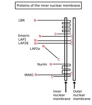
Inner nuclear membrane proteins are membrane proteins that are embedded in or associated with the inner membrane of the nuclear envelope. There are about 60 INM proteins, most of which are poorly characterized with respect to structure and function. Among the few well-characterized INM proteins are lamin B receptor (LBR), lamina-associated polypeptide 1 (LAP1), lamina-associated polypeptide-2 (LAP2), emerin and MAN1.

Mandibuloacral dysplasia (MAD) is a rare autosomal recessive syndrome characterized by mandibular hypoplasia, delayed cranial suture closure, dysplastic clavicles, abbreviated and club-shaped terminal phalanges, acroosteolysis, atrophy of the skin of the hands and feet, and typical facial changes.

Familial partial lipodystrophy, also known as Köbberling–Dunnigan syndrome, is a rare genetic metabolic condition characterized by the loss of subcutaneous fat.

Progeroid syndromes (PS) are a group of rare genetic disorders that mimic physiological aging, making affected individuals appear to be older than they are. The term progeroid syndrome does not necessarily imply progeria, which is a specific type of progeroid syndrome.

Veena Krishnaji Parnaik is an Indian cell biologist and the current Chief Scientist at the Centre for Cellular and Molecular Biology. She obtained her Masters in Science in medicinal biochemistry from the University of Mumbai and received her PhD from Ohio State University before moving back to India to work at the CCMB. Her research is focused on understanding the functional role of the nuclear lamina and how defects in it may lead to disorders such as progeria and muscular dystrophy.

<span class="mw-page-title-main">LMNA-related congenital muscular dystrophy</span> Medical condition

Lamin A/C congenital muscular dystrophy (CMD) is a disease that it is included in laminopathies. Laminopathies are caused, among other mutations, to mutations in LMNA, a gene that synthesizes lamins A and C.

References

  1. Paradisi M, McClintock D, Boguslavsky RL, Pedicelli C, Worman HJ, Djabali K (2005). "Dermal fibroblasts in Hutchinson–Gilford progeria syndrome with the lamin A G608G mutation have dysmorphic nuclei and are hypersensitive to heat stress". BMC Cell Biol. 6: 27. doi: 10.1186/1471-2121-6-27 . PMC   1183198 . PMID   15982412.
  2. Chi, YH; Chen, ZJ; Jeang, KT (22 October 2009). "The nuclear envelopathies and human diseases". Journal of Biomedical Science. 16 (1): 96. doi: 10.1186/1423-0127-16-96 . PMC   2770040 . PMID   19849840.
  3. 1 2 Vergnes L, Peterfy M, Bergo MO, Young SG, Reue K (2004). "Lamin B1 is required for mouse development and nuclear integrity". Proc. Natl. Acad. Sci. U.S.A. 101 (28): 10428–33. Bibcode:2004PNAS..10110428V. doi: 10.1073/pnas.0401424101 . PMC   478588 . PMID   15232008.
  4. Hegele RA, Cao H, Liu DM, Costain GA, Charlton-Menys V, Rodger NW, Durrington PN (2006). "Sequencing of the reannotated LMNB2 gene reveals novel mutations in patients with acquired partial lipodystrophy". Am J Hum Genet. 79 (2): 383–389. doi:10.1086/505885. PMC   1559499 . PMID   16826530.{{cite journal}}: CS1 maint: multiple names: authors list (link)
  5. Padiath QS, Saigoh K, Schiffmann R, Asahara H, Yamada T, Koeppen A, Hogan K, Ptácek LJ, Fu YH (2006). "Lamin B1 duplications cause autosomal dominant leukodystrophy". Nat Genet. 38 (10): 1114–1123. doi:10.1038/ng1872. PMID   16951681. S2CID   25336497.{{cite journal}}: CS1 maint: multiple names: authors list (link)
  6. Hoffmann, Katrin; Dreger, Christine K.; Olins, Ada L.; Olins, Donald E.; Shultz, Leonard D.; Lucke, Barbara; Karl, Hartmut; Kaps, Reinhard; Müller, Dietmar; Vayá, Amparo; Aznar, Justo (2002-07-15). "Mutations in the gene encoding the lamin B receptor produce an altered nuclear morphology in granulocytes (Pelger–Huët anomaly)". Nature Genetics. 31 (4): 410–414. doi:10.1038/ng925. ISSN   1061-4036. PMID   12118250. S2CID   6020153.
  7. Waterham, Hans R.; Koster, Janet; Mooyer, Petra; Noort, Gerard van; Kelley, Richard I.; Wilcox, William R.; Ronald Wanders, J.A.; Raoul Hennekam, C.M.; Jan Oosterwijk, C. (April 2003). "Autosomal Recessive HEM/Greenberg Skeletal Dysplasia Is Caused by 3β-Hydroxysterol Δ14-Reductase Deficiency Due to Mutations in the Lamin B Receptor Gene". The American Journal of Human Genetics. 72 (4): 1013–1017. doi:10.1086/373938. ISSN   0002-9297. PMC   1180330 . PMID   12618959.
  8. Greenberg, Cheryl R.; Rimoin, David L.; Gruber, Helen E.; DeSa, D. J. B.; Reed, M.; Lachman, Ralph S.; Optiz, John M.; Reynolds, James F. (March 1988). "A new autosomal recessive lethal chondrodystrophy with congenital hydrops". American Journal of Medical Genetics. 29 (3): 623–632. doi:10.1002/ajmg.1320290321. ISSN   0148-7299. PMID   3377005.
  9. Shultz, L. D. (2003-01-01). "Mutations at the mouse ichthyosis locus are within the lamin B receptor gene: a single gene model for human Pelger-Huet anomaly". Human Molecular Genetics. 12 (1): 61–69. doi: 10.1093/hmg/ddg003 . ISSN   1460-2083. PMID   12490533.
  10. Young, Alexander Neil; Perlas, Emerald; Ruiz-Blanes, Nerea; Hierholzer, Andreas; Pomella, Nicola; Martin-Martin, Belen; Liverziani, Alessandra; Jachowicz, Joanna W.; Giannakouros, Thomas; Cerase, Andrea (2021-04-12). "Deletion of LBR N-terminal domains recapitulates Pelger-Huet anomaly phenotypes in mouse without disrupting X chromosome inactivation". Communications Biology. 4 (1): 478. doi:10.1038/s42003-021-01944-2. ISSN   2399-3642. PMC   8041748 . PMID   33846535.
  11. Mans BJ, Anantharaman V, Aravind L, Koonin EV (2004). "Comparative genomics, evolution and origins of the nuclear envelope and nuclear pore complex". Cell Cycle. 3 (12): 1612–37. doi: 10.4161/cc.3.12.1316 . PMID   15611647.
  12. Houben F, Ramaekers FC, Snoeckx LH, Broers JL (May 2007). "Role of nuclear lamina-cytoskeleton interactions in the maintenance of cellular strength". Biochim. Biophys. Acta. 1773 (5): 675–86. doi: 10.1016/j.bbamcr.2006.09.018 . PMID   17050008.
  13. 1 2 Hegele RA, Cao H, Liu DM, Costain GA, Charlton-Menys V, Rodger NW, Durrington PN (2006). "Sequencing of the reannotated LMNB2 gene reveals novel mutations in patients with acquired partial lipodystrophy". Am. J. Hum. Genet. 79 (2): 383–9. doi:10.1086/505885. PMC   1559499 . PMID   16826530.
  14. 1 2 Novelli G, Muchir A, Sangiuolo F, Helbling-Leclerc A, D'Apice MR, Massart C, Capon F, Sbraccia P, Federici M, Lauro R, Tudisco C, Pallotta R, Scarano G, Dallapiccola B, Merlini L, Bonne G (2002). "Mandibuloacral dysplasia is caused by a mutation in LMNA-encoding lamin A/C". Am. J. Hum. Genet. 71 (2): 426–31. doi:10.1086/341908. PMC   379176 . PMID   12075506.
  15. Zirn B, Kress W, Grimm T, Berthold LD, et al. (2008). "Association of homozygous LMNA mutation R471C with new phenotype: mandibuloacral dysplasia, progeria, and rigid spine muscular dystrophy". Am J Med Genet A. 146A (8): 1049–1054. doi:10.1002/ajmg.a.32259. PMID   18348272. S2CID   205309256.
  16. Al-Haggar M, Madej-Pilarczyk A, Kozlowski L, Bujnicki JM, Yahia S, Abdel-Hadi D, Shams A, Ahmad N, Hamed S, Puzianowska-Kuznicka M (2012). "A novel homozygous p.Arg527Leu LMNA mutation in two unrelated Egyptian families causes overlapping mandibuloacral dysplasia and progeria syndrome". Eur J Hum Genet. 20 (11): 1134–40. doi:10.1038/ejhg.2012.77. PMC   3476705 . PMID   22549407.
  17. 1 2 3 Eriksson M, Brown WT, Gordon LB, Glynn MW, Singer J, Scott L, Erdos MR, Robbins CM, Moses TY, Berglund P, Dutra A, Pak E, Durkin S, Csoka AB, Boehnke M, Glover TW, Collins FS (2003). "Recurrent de novo point mutations in lamin A cause Hutchinson–Gilford progeria syndrome". Nature. 423 (6937): 293–8. Bibcode:2003Natur.423..293E. doi:10.1038/nature01629. hdl: 2027.42/62684 . PMC   10540076 . PMID   12714972. S2CID   4420150.
  18. 1 2 Navarro CL, De Sandre-Giovannoli A, Bernard R, Boccaccio I, Boyer A, Genevieve D, Hadj-Rabia S, Gaudy-Marqueste C, Smitt HS, Vabres P, Faivre L, Verloes A, Van Essen T, Flori E, Hennekam R, Beemer FA, Laurent N, Le Merrer M, Cau P, Levy N (2004). "Lamin A and ZMPSTE24 (FACE-1) defects cause nuclear disorganization and identity restrictive dermopathy as a lethal neonatal laminopathy". Hum. Mol. Genet. 13 (20): 2493–2503. doi: 10.1093/hmg/ddh265 . PMID   15317753.
  19. 1 2 Padiath QS, Saigoh K, Schiffmann R, Asahara H, Yamada T, Koeppen A, Hogan K, Ptacek LJ, Fu YH (2006). "Lamin B1 duplications cause autosomal dominant leukodystrophy". Nature Genetics. 38 (10): 1114–1123. doi:10.1038/ng1872. PMID   16951681. S2CID   25336497.
  20. Lassoued K, Guilly MN, Danon F, Andre C, Dhumeaux D, Clauvel JP, Brouet JC, Seligmann M, Courvalin JC (1988). "Antinuclear autoantibodies specific for lamins. Characterization and clinical significance". Ann Intern Med. 108 (6): 829–3. doi:10.7326/0003-4819-108-6-829. PMID   3285745.{{cite journal}}: CS1 maint: multiple names: authors list (link)
  21. Redwood AB, Perkins SM, Vanderwaal RP, Feng Z, Biehl KJ, Gonzalez-Suarez I, Morgado-Palacin L, Shi W, Sage J, Roti-Roti JL, Stewart CL, Zhang J, Gonzalo S (2011). "A dual role for A-type lamins in DNA double-strand break repair". Cell Cycle. 10 (15): 2549–60. doi:10.4161/cc.10.15.16531. PMC   3180193 . PMID   21701264.
  22. Liu B, Wang J, Chan KM, Tjia WM, Deng W, Guan X, Huang JD, Li KM, Chau PY, Chen DJ, Pei D, Pendas AM, Cadiñanos J, López-Otín C, Tse HF, Hutchison C, Chen J, Cao Y, Cheah KS, Tryggvason K, Zhou Z (2005). "Genomic instability in laminopathy-based premature aging". Nat. Med. 11 (7): 780–5. doi:10.1038/nm1266. PMID   15980864. S2CID   11798376.
  23. Chen L, Lee L, Kudlow BA, Dos Santos HG, Sletvold O, Shafeghati Y, Botha EG, Garg E, Hanson NB, Martin GM, Mian IS, Kennedy BK, Oshima J (2003). "LMNA mutations in atypical Werner's syndrome". Lancet. 362 (9382): 440–5. doi:10.1016/S0140-6736(03)14069-X. PMID   12927431. S2CID   21980784.
  24. Hellemans J, Preobrazhenska O, Willaert A, Debeer P, Verdonk PCM, Costa T, Janssens K, Menten B, Van Roy N, Vermeulen SJT, Savarirayan R, Van Hul W, et al. (2004). "Loss-of-function mutations in LEMD3 result in osteopoikilosis, Buschke–Ollendorff syndrome and melorheostosis". Nature Genetics. 36 (11): 1213–8. doi: 10.1038/ng1453 . PMID   15489854.
  25. Fatkin D, MacRae C, Sasaki T, Wolff MR, Porcu M, Frenneaux M, Atherton J, Vidaillet HJ, Spudich S, De Girolami U, Seidman JG, Seidman CE (1999). "Missense mutations in the rod domain of the lamin A/C gene as causes of dilated cardiomyopathy and conduction-system disease" (PDF). N. Engl. J. Med. 341 (23): 1715–24. doi:10.1056/NEJM199912023412302. PMID   10580070. S2CID   22942654.
  26. Charniot JC, Pascal C, Bouchier C, Sebillon P, Salama J, Duboscq-Bidot L, Peuchmaurd M, Desnos M, Artigou JY, Komajda M (2003). "Functional consequences of an LMNA mutation associated with a new cardiac and non-cardiac phenotype". Hum. Mutat. 21 (5): 473–81. doi:10.1002/humu.10170. PMID   12673789. S2CID   32614095.
  27. De Sandre-Giovannoli A, Chaouch M, Kozlov S, Vallat JM, Tazir M, Kassouri N, Szepetowski P, Hammadouche T, Vandenberghe A, Stewart CL, Grid D, Levy N (2002). "Homozygous defects in LMNA, encoding lamin A/C nuclear-envelope proteins, cause autosomal recessive axonal neuropathy in human (Charcot–Marie–Tooth disorder type 2) and mouse". Am. J. Hum. Genet. 70 (3): 726–36. doi:10.1086/339274. PMC   384949 . PMID   11799477.
  28. Manilal S, Nguyen TM, Sewry CA, Morris GE (1996). "The Emery–Dreifuss muscular dystrophy protein, emerin, is a nuclear membrane protein". Hum. Mol. Genet. 5 (6): 801–8. doi:10.1093/hmg/5.6.801. PMID   8776595.
  29. Clements L, Manilal S, Love DR, Morris GE (2000). "Direct interaction between emerin and lamin A". Biochem. Biophys. Res. Commun. 267 (3): 709–14. doi:10.1006/bbrc.1999.2023. PMID   10673356.
  30. Bonne G, Di Barletta MR, Varnous S, Becane HM, Hammouda EH, Merlini L, Muntoni F, Greenberg CR, Gary F, Urtizberea JA, Duboc D, Fardeau M, Toniolo D, Schwartz K (1999). "Mutations in the gene encoding lamin A/C cause autosomal dominant Emery–Dreifuss muscular dystrophy". Nature Genetics. 21 (3): 285–8. doi:10.1038/6799. PMID   10080180. S2CID   7327176.
  31. Raffaele di Barletta M, Ricci E, Galluzzi G, Tonali P, Mora M, Morandi L, Romorini A, Voit T, Orstavik KH, Merlini L, Trevisan C, Biancalana V, Hausmanowa-Petrusewicz I, Bione S, Ricotti R, Schwartz K, Bonne G, Toniolo D (2000). "Different mutations in the LMNA gene cause autosomal dominant and autosomal recessive Emery–Dreifuss muscular dystrophy". Am. J. Hum. Genet. 66 (4): 1407–12. doi:10.1086/302869. PMC   1288205 . PMID   10739764.
  32. Cao H, Hegele RA (2002). "Nuclear lamin A/C R482Q mutation in Canadian kindreds with Dunnigan-type familial partial lipodystrophy". Hum. Mol. Genet. 9 (1): 109–12. doi: 10.1093/hmg/9.1.109 . PMID   10587585.
  33. Waterham HR, Koster J, Mooyer P, van Noort G, Kelley RI, Wilcox WR, Wanders RJ, Hennekam RC, Oosterwijk JC (2003). "Autosomal recessive HEM/Greenberg skeletal dysplasia is caused by 3-beta-hydroxysterol delta(14)-reductase deficiency due to mutations in the lamin B receptor gene". Am. J. Hum. Genet. 72 (4): 1013–17. doi:10.1086/373938. PMC   1180330 . PMID   12618959.
  34. Muchir A, Bonne G, van der Kooi AJ, van Meegen M, Baas F, Bolhuis PA, de Visser M, Schwartz K (2000). "Identification of mutations in the gene encoding lamins A/C in autosomal dominant limb girdle muscular dystrophy with atrioventricular conduction disturbances (LGMD1B)". Hum. Mol. Genet. 9 (9): 1453–9. doi: 10.1093/hmg/9.9.1453 . PMID   10814726.
  35. Caux F, Dubosclard E, Lascols O, Buendia B, Chazouilleres O, Cohen A, Courvalin JC, Laroche L, Capeau J, Vigouroux C, Christin-Maitre S (2003). "A new clinical condition linked to a novel mutation in lamins A and C with generalized lipoatrophy, insulin-resistant diabetes, disseminated leukomelanodermic papules, liver steatosis, and cardiomyopathy". J. Clin. Endocrinol. Metab. 88 (3): 1006–13. doi:10.1210/jc.2002-021506. PMID   12629077.
  36. Agarwal AK, Fryns JP, Auchus RJ, Garg A (2003). "Zinc metalloproteinase, ZMPSTE24, is mutated in mandibuloacral dysplasia". Hum. Mol. Genet. 12 (16): 1995–2001. doi: 10.1093/hmg/ddg213 . PMID   12913070.
  37. Hoffmann K, Dreger CK, Olins AL, Olins DE, Shultz LD, Lucke B, Karl H, Kaps R, Muller D, Vaya A, Aznar J, Ware RE, Cruz NS, Lindner TH, Herrmann H, Reis A, Sperling K (2002). "Mutations in the gene encoding the lamin B receptor produce an altered nuclear morphology in granulocytes (Pelger-Huet anomaly)". Nature Genetics. 31 (4): 410–4. doi:10.1038/ng925. PMID   12118250. S2CID   6020153.
  38. Captur, Gabriella; Arbustini, Eloisa; Bonne, Gisèle; Syrris, Petros; Mills, Kevin; Wahbi, Karim; Mohiddin, Saidi A.; McKenna, William J.; Pettit, Stephen (2017-11-25). "Lamin and the heart" (PDF). Heart. 104 (6): 468–479. doi:10.1136/heartjnl-2017-312338. ISSN   1468-201X. PMID   29175975. S2CID   3563474.
  39. Rusinal AE, Sinensky MS (2006). "Farnesylated lamins, progeroid syndromes and farnesyl transferase inhibitors". J. Cell Sci. 119 (Pt 16): 3265–72. doi: 10.1242/jcs.03156 . PMID   16899817.
  40. Meta M, Yang SH, Bergo MO, Fong LG, Young SG (2006). "Protein farnesyltransferase inhibitors and progeria". Trends Mol. Med. 12 (10): 480–7. doi:10.1016/j.molmed.2006.08.006. PMID   16942914.