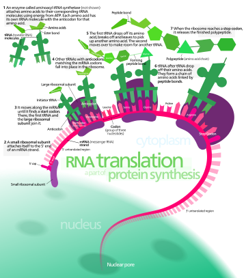
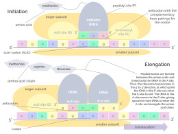
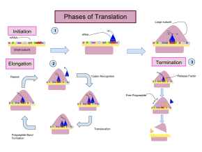
Overview of eukaryotic messenger RNA (mRNA) translationTranslation of mRNA and ribosomal protein synthesisInitiation and elongation stages of translation involving RNA nucleobases, the ribosome, transfer RNA, and amino acidsThe three phases of translation: (1) in initiation, the small ribosomal subunit binds to the RNA strand and the initiator tRNA–amino acid complex binds to the start codon, culminating in attachment of the large subunit; (2) elongation occurs as a cycle, in which codons are sequentially recognized by charged tRNAs, followed by peptide bond formation with transfer of the polypeptide between tRNAs within the ribosome and finally translocation of the ribosome to the next codon; (3) termination, when a stop codon is reached, a release factor binds and the polypeptide is released (note that labels for translocation and peptide bond formation are reversed in this image)
In biology, translation is the process in living cells in which proteins are produced using RNA molecules as templates. The generated protein is a sequence of amino acids. This sequence is determined by the sequence of nucleotides in the RNA. The nucleotides are considered three at a time. Each such triple results in addition of one specific amino acid to the protein being generated. The matching from nucleotide triple to amino acid is called the genetic code. The translation is performed by a large complex of functional RNA and proteins called ribosomes. The entire process is called gene expression.
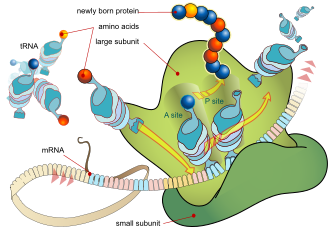
In translation, messenger RNA (mRNA) is decoded in a ribosome, outside the nucleus, to produce a specific amino acid chain, or polypeptide. The polypeptide later folds into an activeprotein and performs its functions in the cell. The ribosome facilitates decoding by inducing the binding of complementarytransfer RNA (tRNA) anticodon sequences to mRNA codons. The tRNAs carry specific amino acids that are chained together into a polypeptide as the mRNA passes through and is "read" by the ribosome.
Translation proceeds in three phases:
Initiation: The ribosome assembles around the target mRNA. The first tRNA is attached at the start codon.
Elongation: The last tRNA validated by the small ribosomal subunit (accommodation) transfers the amino acid. It carries to the large ribosomal subunit which binds it to the one of the preceding admitted tRNA (transpeptidation). The ribosome then moves to the next mRNA codon to continue the process (translocation), creating an amino acid chain.
Termination: When a stop codon is reached, the ribosome releases the polypeptide. The ribosomal complex remains intact and moves on to the next mRNA to be translated.
In prokaryotes (bacteria and archaea), translation occurs in the cytosol, where the large and small subunits of the ribosome bind to the mRNA. In eukaryotes, translation occurs in the cytoplasm or across the membrane of the endoplasmic reticulum in a process called co-translational translocation. In co-translational translocation, the entire ribosome/mRNA complex binds to the outer membrane of the rough endoplasmic reticulum (ER), and the new protein is synthesized and released into the ER; the newly created polypeptide can be stored inside the ER for future vesicle transport and secretion outside the cell, or immediately secreted.
Many types of transcribed RNA, such as tRNA, ribosomal RNA, and small nuclear RNA, do not undergo a translation into proteins.
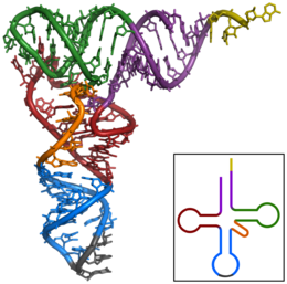
A ribosome translating a protein that is secreted into the endoplasmic reticulum (tRNAs colored dark blue)Tertiary structure of tRNA (CCA tail in yellow, Acceptor stem in purple, Variable loop in orange, Darm in red, Anticodon arm in blue with Anticodon in black, Tarm in green)
The basic process of protein production is the addition of one amino acid at a time to the end of a protein. This operation is performed by a ribosome.[1] A ribosome is made up of two subunits, a small subunit, and a large subunit. These subunits come together before the translation of mRNA into a protein to provide a location for translation to be carried out and a polypeptide to be produced.[2] The choice of amino acid type to add is determined by a messenger RNA (mRNA) molecule. Each amino acid added is matched to a three-nucleotide subsequence of the mRNA. For each such triplet possible, the corresponding amino acid is accepted. The successive amino acids added to the chain are matched to successive nucleotide triplets in the mRNA. In this way, the sequence of nucleotides in the template mRNA chain determines the sequence of amino acids in the generated amino acid chain.[3] The addition of an amino acid occurs at the C-terminus of the peptide; thus, translation is said to be amine-to-carboxyl directed.[4]
The mRNA carries genetic information encoded as a ribonucleotide sequence from the chromosomes to the ribosomes. The ribonucleotides are "read" by translational machinery in a sequence of nucleotide triplets called codons. Each of those triplets codes for a specific amino acid.[citation needed]
The ribosome molecules translate this code to a specific sequence of amino acids. The ribosome is a multisubunit structure containing ribosomal RNA (rRNA) and proteins. It is the "factory" where amino acids are assembled into proteins.
Transfer RNAs (tRNAs) are small noncoding RNA chains (74–93 nucleotides) that transport amino acids to the ribosome. The repertoire of tRNA genes varies widely between species, with some bacteria having between 20 and 30 genes while complex eukaryotes could have thousands.[5] tRNAs have a site for amino acid attachment, and a site called an anticodon. The anticodon is an RNA triplet complementary to the mRNA triplet that codes for their cargo amino acid.
Aminoacyl tRNA synthetases (enzymes) catalyze the bonding between specific tRNAs and the amino acids that their anticodon sequences call for. The product of this reaction is an aminoacyl-tRNA. The amino acid is joined by its carboxyl group to the 3' OH of the tRNA by an ester bond. When the tRNA has an amino acid linked to it, the tRNA is termed "charged". In bacteria, this aminoacyl-tRNA is carried to the ribosome by EF-Tu, where mRNA codons are matched through complementary base pairing to specific tRNA anticodons. Aminoacyl-tRNA synthetases that mispair tRNAs with the wrong amino acids can produce mischarged aminoacyl-tRNAs, which can result in inappropriate amino acids at the respective position in the protein. This "mistranslation"[6] of the genetic code naturally occurs at low levels in most organisms, but certain cellular environments cause an increase in permissive mRNA decoding, sometimes to the benefit of the cell.
The ribosome has two binding sites for tRNA. They are the aminoacyl site (abbreviated A), and the peptidyl site/ exit site (abbreviated P/E). Concerning the mRNA, the three sites are oriented 5' to 3' E-P-A, because ribosomes move toward the 3' end of mRNA. The A-site binds the incoming tRNA with the complementary codon on the mRNA. The P/E-site holds the tRNA with the growing polypeptide chain. When an aminoacyl-tRNA initially binds to its corresponding codon on the mRNA, it is in the A site. Then, a peptide bond forms between the amino acid of the tRNA in the A site and the amino acid of the charged tRNA in the P/E site. The growing polypeptide chain is transferred to the tRNA in the A site. Translocation occurs, moving the tRNA to the P/E site, now without an amino acid; the tRNA that was in the A site, now charged with the polypeptide chain, is moved to the P/E site and the uncharged tRNA leaves, and another aminoacyl-tRNA enters the A site to repeat the process.[7]
After the new amino acid is added to the chain, and after the tRNA is released out of the ribosome and into the cytosol, the energy provided by the hydrolysis of a GTP bound to the translocaseEF-G (in bacteria) and a/eEF-2 (in eukaryotes and archaea) moves the ribosome down one codon towards the 3' end. The energy required for translation of proteins is significant. For a protein containing n amino acids, the number of high-energy phosphate bonds required to translate it is 4n-1.[8] The rate of translation varies; it is significantly higher in prokaryotic cells (up to 17–21 amino acid residues per second) than in eukaryotic cells (up to 6–9 amino acid residues per second).[9]
Initiation and termination of translation
Initiation involves the small subunit of the ribosome binding to the 5' end of mRNA with the help of initiation factors (IF). In bacteria and a minority of archaea, initiation of protein synthesis involves the recognition of a purine-rich initiation sequence on the mRNA called the Shine–Dalgarno sequence. The Shine–Dalgarno sequence binds to a complementary pyrimidine-rich sequence on the 3' end of the 16S rRNA part of the 30S ribosomal subunit. The binding of these complementary sequences ensures that the 30S ribosomal subunit is bound to the mRNA and is aligned such that the initiation codon is placed in the 30S portion of the P-site. Once the mRNA and 30S subunit are properly bound, an initiation factor brings the initiator tRNA–amino acid complex, f-Met-tRNA, to the 30S P site. The initiation phase is completed once a 50S subunit joins the 30S subunit, forming an active 70S ribosome.[10] Termination of the polypeptide occurs when the A site of the ribosome is occupied by a stop codon (UAA, UAG, or UGA) on the mRNA, creating the primary structure of a protein. tRNA usually cannot recognize or bind to stop codons. Instead, the stop codon induces the binding of a release factor protein[11] (RF1 & RF2) that prompts the disassembly of the entire ribosome/mRNA complex by the hydrolysis of the polypeptide chain from the peptidyl transferase center [1] of the ribosome.[12] Drugs or special sequence motifs on the mRNA can change the ribosomal structure so that near-cognate tRNAs are bound to the stop codon instead of the release factors. In such cases of 'translational readthrough', translation continues until the ribosome encounters the next stop codon.[13]
Errors in translation
Even though the ribosomes are usually considered accurate and processive machines, the translation process is subject to errors that can lead either to the synthesis of erroneous proteins or to the premature abandonment of translation, either because a tRNA couples to a wrong codon or because a tRNA is coupled to the wrong amino acid. [14] The rate of error in synthesizing proteins has been estimated to be between 1 in 105 and 1 in 103 misincorporated amino acids, depending on the experimental conditions.[15] The rate of premature translation abandonment, instead, has been estimated to be of the order of magnitude of 10−4 events per translated codon.[16]
Regulation
The process of translation is highly regulated in both eukaryotic and prokaryotic organisms. Regulation of translation can impact the global rate of protein synthesis which is closely coupled to the metabolic and proliferative state of a cell.
To delve deeper into this intricate process, scientists typically use a technique known as ribosome profiling.[17] This method enables researchers to take a snapshot of the translatome, showing which parts of the mRNA are being translated into proteins by ribosomes at a given time. Ribosome profiling provides valuable insights into translation dynamics, revealing the complex interplay between gene sequence, mRNA structure, and translation regulation. For example, research utilizing this method has revealed that genetic differences and their subsequent expression as mRNAs can also impact translation rate in an RNA-specific manner.[18]
Expanding on this concept, a more recent development is single-cell ribosome profiling, a technique that allows us to study the translation process at the resolution of individual cells.[19] This is particularly significant as cells, even those of the same type, can exhibit considerable variability in their protein synthesis. Single-cell ribosome profiling has the potential to shed light on the heterogeneous nature of cells, leading to a more nuanced understanding of how translation regulation can impact cell behavior, metabolic state, and responsiveness to various stimuli or conditions.
Clinical significance
Translational control is critical for the development and survival of cancer. Cancer cells must frequently regulate the translation phase of gene expression, though it is not fully understood why translation is targeted over steps like transcription. While cancer cells often have genetically altered translation factors, it is much more common for cancer cells to modify the levels of existing translation factors.[20] Several major oncogenic signaling pathways, including the RAS–MAPK, PI3K/AKT/mTOR, MYC, and WNT–β-catenin pathways, ultimately reprogram the genome via translation.[21] Cancer cells also control translation to adapt to cellular stress. During stress, the cell translates mRNAs that can mitigate the stress and promote survival. An example of this is the expression of AMPK in various cancers; its activation triggers a cascade that can ultimately allow the cancer to escape apoptosis (programmed cell death) triggered by nutrition deprivation. Future cancer therapies may involve disrupting the translation machinery of the cell to counter the downstream effects of cancer.[20]
Mathematical modeling of translation
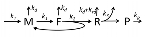
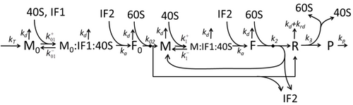
Figure M0. Basic and the simplest model M0 of protein synthesis. Here, * M – amount of mRNA with translation initiation site not occupied by assembling ribosome, * F – amount of mRNA with translation initiation site occupied by assembling ribosome, * R – amount of ribosomes sitting on mRNA synthesizing proteins, * P – amount of synthesized proteins.Figure M1'. The extended model of protein synthesis M1 with explicit presentation of 40S, 60S and initiation factors (IF) binding.
The transcription-translation process description, mentioning only the most basic "elementary" processes, consists of:
production of mRNA molecules (including splicing),
initiation of these molecules with help of initiation factors (e.g., the initiation can include the circularization step though it is not universally required),
initiation of translation, recruiting the small ribosomal subunit,
assembly of full ribosomes,
elongation, (i.e. movement of ribosomes along mRNA with production of protein),
termination of translation,
degradation of mRNA molecules,
degradation of proteins.
The process of amino acid building to create protein in translation is a subject of various physic models for a long time starting from the first detailed kinetic models such as[23] or others taking into account stochastic aspects of translation and using computer simulations. Many chemical kinetics-based models of protein synthesis have been developed and analyzed in the last four decades.[24][25] Beyond chemical kinetics, various modeling formalisms such as Totally Asymmetric Simple Exclusion Process,[25]Probabilistic Boolean Networks, Petri Nets and max-plus algebra have been applied to model the detailed kinetics of protein synthesis or some of its stages. A basic model of protein synthesis that takes into account all eight 'elementary' processes has been developed,[22] following the paradigm that "useful models are simple and extendable".[26] The simplest model M0 is represented by the reaction kinetic mechanism (Figure M0). It was generalised to include 40S, 60S and initiation factors (IF) binding (Figure M1'). It was extended further to include effect of microRNA on protein synthesis.[27] Most of models in this hierarchy can be solved analytically. These solutions were used to extract 'kinetic signatures' of different specific mechanisms of synthesis regulation.
It is also possible to translate either by hand (for short sequences) or by computer (after first programming one appropriately, see section below); this allows biologists and chemists to draw out the chemical structure of the encoded protein on paper.
First, convert each template DNA base to its RNA complement (note that the complement of A is now U), as shown below. Note that the template strand of the DNA is the one the RNA is polymerized against; the other DNA strand would be the same as the RNA, but with thymine instead of uracil.
DNA -> RNA A -> U T -> A C -> G G -> C A=T-> A=U
Then split the RNA into triplets (groups of three bases). Note that there are 3 translation "windows", or reading frames, depending on where you start reading the code. Finally, use the table at Genetic code to translate the above into a structural formula as used in chemistry.
Whereas other aspects such as the 3D structure, called tertiary structure, of protein can only be predicted using sophisticated algorithms, the amino acid sequence, called primary structure, can be determined solely from the nucleic acid sequence with the aid of a translation table.
This approach may not give the correct amino acid composition of the protein, in particular if unconventional amino acids such as selenocysteine are incorporated into the protein, which is coded for by a conventional stop codon in combination with a downstream hairpin (SElenoCysteine Insertion Sequence, or SECIS).
There are many computer programs capable of translating a DNA/RNA sequence into a protein sequence. Normally this is performed using the Standard Genetic Code, however, few programs can handle all the "special" cases, such as the use of the alternative initiation codons which are biologically significant. For instance, the rare alternative start codon CTG codes for Methionine when used as a start codon, and for Leucine in all other positions.
Example: Condensed translation table for the Standard Genetic Code (from the NCBI Taxonomy webpage).[28]
The "Starts" row indicate three start codons, UUG, CUG, and the very common AUG. It also indicates the first amino acid residue when interpreted as a start: in this case it is all methionine.
Even when working with ordinary eukaryotic sequences such as the Yeast genome, it is often desired to be able to use alternative translation tables—namely for translation of the mitochondrial genes. Currently the following translation tables are defined by the NCBI Taxonomy Group for the translation of the sequences in GenBank:[28]
The genetic code is the set of rules used by living cells to translate information encoded within genetic material into proteins. Translation is accomplished by the ribosome, which links proteinogenic amino acids in an order specified by messenger RNA (mRNA), using transfer RNA (tRNA) molecules to carry amino acids and to read the mRNA three nucleotides at a time. The genetic code is highly similar among all organisms and can be expressed in a simple table with 64 entries.
Protein biosynthesis is a core biological process, occurring inside cells, balancing the loss of cellular proteins through the production of new proteins. Proteins perform a number of critical functions as enzymes, structural proteins or hormones. Protein synthesis is a very similar process for both prokaryotes and eukaryotes but there are some distinct differences.
Ribosomes are macromolecular machines, found within all cells, that perform biological protein synthesis. Ribosomes link amino acids together in the order specified by the codons of messenger RNA molecules to form polypeptide chains. Ribosomes consist of two major components: the small and large ribosomal subunits. Each subunit consists of one or more ribosomal RNA molecules and many ribosomal proteins. The ribosomes and associated molecules are also known as the translational apparatus.
Transfer RNA is an adaptor molecule composed of RNA, typically 76 to 90 nucleotides in length, that serves as the physical link between the mRNA and the amino acid sequence of proteins. Transfer RNA (tRNA) does this by carrying an amino acid to the protein-synthesizing machinery of a cell called the ribosome. Complementation of a 3-nucleotide codon in a messenger RNA (mRNA) by a 3-nucleotide anticodon of the tRNA results in protein synthesis based on the mRNA code. As such, tRNAs are a necessary component of translation, the biological synthesis of new proteins in accordance with the genetic code.
The Shine–Dalgarno (SD) sequence is a ribosomal binding site in bacterial and archaeal messenger RNA, generally located around 8 bases upstream of the start codon AUG. The RNA sequence helps recruit the ribosome to the messenger RNA (mRNA) to initiate protein synthesis by aligning the ribosome with the start codon. Once recruited, tRNA may add amino acids in sequence as dictated by the codons, moving downstream from the translational start site.
Ribosomal ribonucleic acid (rRNA) is a type of non-coding RNA which is the primary component of ribosomes, essential to all cells. rRNA is a ribozyme which carries out protein synthesis in ribosomes. Ribosomal RNA is transcribed from ribosomal DNA (rDNA) and then bound to ribosomal proteins to form small and large ribosome subunits. rRNA is the physical and mechanical factor of the ribosome that forces transfer RNA (tRNA) and messenger RNA (mRNA) to process and translate the latter into proteins. Ribosomal RNA is the predominant form of RNA found in most cells; it makes up about 80% of cellular RNA despite never being translated into proteins itself. Ribosomes are composed of approximately 60% rRNA and 40% ribosomal proteins, though this ratio differs between prokaryotes and eukaryotes.
Bacterial translation is the process by which messenger RNA is translated into proteins in bacteria.
Eukaryotic translation is the biological process by which messenger RNA is translated into proteins in eukaryotes. It consists of four phases: initiation, elongation, termination, and recapping.
The Kozak consensus sequence is a nucleic acid motif that functions as the protein translation initiation site in most eukaryotic mRNA transcripts. Regarded as the optimum sequence for initiating translation in eukaryotes, the sequence is an integral aspect of protein regulation and overall cellular health as well as having implications in human disease. It ensures that a protein is correctly translated from the genetic message, mediating ribosome assembly and translation initiation. A wrong start site can result in non-functional proteins. As it has become more studied, expansions of the nucleotide sequence, bases of importance, and notable exceptions have arisen. The sequence was named after the scientist who discovered it, Marilyn Kozak. Kozak discovered the sequence through a detailed analysis of DNA genomic sequences.
Aminoacyl-tRNA is tRNA to which its cognate amino acid is chemically bonded (charged). The aa-tRNA, along with particular elongation factors, deliver the amino acid to the ribosome for incorporation into the polypeptide chain that is being produced during translation.
EF-Tu is a prokaryotic elongation factor responsible for catalyzing the binding of an aminoacyl-tRNA (aa-tRNA) to the ribosome. It is a G-protein, and facilitates the selection and binding of an aa-tRNA to the A-site of the ribosome. As a reflection of its crucial role in translation, EF-Tu is one of the most abundant and highly conserved proteins in prokaryotes. It is found in eukaryotic mitochondria as TUFM.
Protein metabolism denotes the various biochemical processes responsible for the synthesis of proteins and amino acids (anabolism), and the breakdown of proteins by catabolism.
50S is the larger subunit of the 70S ribosome of prokaryotes, i.e. bacteria and archaea. It is the site of inhibition for antibiotics such as macrolides, chloramphenicol, clindamycin, and the pleuromutilins. It includes the 5S ribosomal RNA and 23S ribosomal RNA.
The prokaryotic small ribosomal subunit, or 30S subunit, is the smaller subunit of the 70S ribosome found in prokaryotes. It is a complex of the 16S ribosomal RNA (rRNA) and 19 proteins. This complex is implicated in the binding of transfer RNA to messenger RNA (mRNA). The small subunit is responsible for the binding and the reading of the mRNA during translation. The small subunit, both the rRNA and its proteins, complexes with the large 50S subunit to form the 70S prokaryotic ribosome in prokaryotic cells. This 70S ribosome is then used to translate mRNA into proteins.
Ribosomal frameshifting, also known as translational frameshifting or translational recoding, is a biological phenomenon that occurs during translation that results in the production of multiple, unique proteins from a single mRNA. The process can be programmed by the nucleotide sequence of the mRNA and is sometimes affected by the secondary, 3-dimensional mRNA structure. It has been described mainly in viruses, retrotransposons and bacterial insertion elements, and also in some cellular genes.
EF-G is a prokaryotic elongation factor involved in protein translation. As a GTPase, EF-G catalyzes the movement (translocation) of transfer RNA (tRNA) and messenger RNA (mRNA) through the ribosome.
An expanded genetic code is an artificially modified genetic code in which one or more specific codons have been re-allocated to encode an amino acid that is not among the 22 common naturally-encoded proteinogenic amino acids.
The P-site is the second binding site for tRNA in the ribosome. The other two sites are the A-site (aminoacyl), which is the first binding site in the ribosome, and the E-site (exit), the third. During protein translation, the P-site holds the tRNA which is linked to the growing polypeptide chain. When a stop codon is reached, the peptidyl-tRNA bond of the tRNA located in the P-site is cleaved releasing the newly synthesized protein. During the translocation step of the elongation phase, the mRNA is advanced by one codon, coupled to movement of the tRNAs from the ribosomal A to P and P to E sites, catalyzed by elongation factor EF-G.
In molecular biology, VAR1 protein domain, otherwise known as variant protein 1, is a ribosomal protein that forms part of the small ribosomal subunit in yeast mitochondria. Mitochondria possess their own ribosomes responsible for the synthesis of a small number of proteins encoded by the mitochondrial genome. VAR1 is the only protein in the yeast mitochondrial ribosome to be encoded in the mitochondria - the remaining approximately 80 ribosomal proteins are encoded in the nucleus. VAR1 along with 15S rRNA are necessary for the formation of mature 37S subunits.
Ribosomal L28e protein family is a family of evolutionarily related proteins. Members include 60S ribosomal protein L28.
↑ Brooker RJ, Widmaier EP, Graham LE, Stiling PD (2014). Biology (Third international studented.). New York, NY: McGraw Hill Education. p.249. ISBN978-981-4581-85-1.
↑ Neill C (1996). Biology (Fourthed.). The Benjamin/Cummings Publishing Company. pp.309–310. ISBN0-8053-1940-9.
↑ Nakamoto T (February 2011). "Mechanisms of the initiation of protein synthesis: in reading frame binding of ribosomes to mRNA". Molecular Biology Reports. 38 (2): 847–55. doi:10.1007/s11033-010-0176-1. PMID20467902. S2CID22038744.
↑ Heinrich R, Rapoport TA (September 1980). "Mathematical modelling of translation of mRNA in eucaryotes; steady state, time-dependent processes and application to reticulocytes". Journal of Theoretical Biology. 86 (2): 279–313. Bibcode:1980JThBi..86..279H. doi:10.1016/0022-5193(80)90008-9. PMID7442295.
1 2 Skjøndal-Bar N, Morris DR (January 2007). "Dynamic model of the process of protein synthesis in eukaryotic cells". Bulletin of Mathematical Biology. 69 (1): 361–93. doi:10.1007/s11538-006-9128-2. PMID17031456. S2CID83701439.
Champe PC, Harvey RA, Ferrier DR (2004). Lippincott's Illustrated Reviews: Biochemistry (3rded.). Hagerstwon, MD: Lippincott Williams & Wilkins. ISBN0-7817-2265-9.
Cox M, Nelson DR, Lehninger AL (2005). Lehninger principles of biochemistry (4thed.). San Francisco...: W.H. Freeman. ISBN0-7167-4339-6.
Malys N, McCarthy JE (March 2011). "Translation initiation: variations in the mechanism can be anticipated". Cellular and Molecular Life Sciences. 68 (6): 991–1003. doi:10.1007/s00018-010-0588-z. PMID21076851. S2CID31720000.
This page is based on this Wikipedia article Text is available under the CC BY-SA 4.0 license; additional terms may apply. Images, videos and audio are available under their respective licenses.