Vesicle (biology and chemistry)

Last updated
Scheme of a liposome formed by phospholipids in an aqueous solution Liposome scheme-en.svg
Scheme of a liposome formed by phospholipids in an aqueous solution

In cell biology, a vesicle is a structure within or outside a cell, consisting of liquid or cytoplasm enclosed by a lipid bilayer. Vesicles form naturally during the processes of secretion (exocytosis), uptake (endocytosis), and the transport of materials within the plasma membrane. Alternatively, they may be prepared artificially, in which case they are called liposomes (not to be confused with lysosomes). If there is only one phospholipid bilayer, the vesicles are called unilamellar liposomes ; otherwise they are called multilamellar liposomes. [1] The membrane enclosing the vesicle is also a lamellar phase, similar to that of the plasma membrane, and intracellular vesicles can fuse with the plasma membrane to release their contents outside the cell. Vesicles can also fuse with other organelles within the cell. A vesicle released from the cell is known as an extracellular vesicle.

Contents

Vesicles perform a variety of functions. Because it is separated from the cytosol, the inside of the vesicle can be made to be different from the cytosolic environment. For this reason, vesicles are a basic tool used by the cell for organizing cellular substances. Vesicles are involved in metabolism, transport, buoyancy control, [2] and temporary storage of food and enzymes. They can also act as chemical reaction chambers.

Sarfus image of lipid vesicles Sarfus.LipidVesicles.jpg
Sarfus image of lipid vesicles
IUPAC definition

Closed structure formed by amphiphilic molecules that contains solvent (usually water). [3]

The 2013 Nobel Prize in Physiology or Medicine was shared by James Rothman, Randy Schekman and Thomas Südhof for their roles in elucidating (building upon earlier research, some of it by their mentors) the makeup and function of cell vesicles, especially in yeasts and in humans, including information on each vesicle's parts and how they are assembled. Vesicle dysfunction is thought to contribute to Alzheimer's disease, diabetes, some hard-to-treat cases of epilepsy, some cancers and immunological disorders and certain neurovascular conditions. [4] [5]

Types of vesicular structures

Electron micrograph of a cell containing a food vacuole (fv) and transport vacuole (tv) in a malaria parasite Hemozoin in food vacuole.jpg
Electron micrograph of a cell containing a food vacuole (fv) and transport vacuole (tv) in a malaria parasite

Vacuoles

Vacuoles are cellular organelles that contain mostly water.[ citation needed ]

Lysosomes

Transport vesicles

Secretory vesicles

Secretory vesicles contain materials that are to be excreted from the cell. Cells have many reasons to excrete materials. One reason is to dispose of wastes. Another reason is tied to the function of the cell. Within a larger organism, some cells are specialized to produce certain chemicals. These chemicals are stored in secretory vesicles and released when needed.

Types

Extracellular vesicles

Extracellular vesicles (EVs) are lipid bilayer-delimited particles produced by all domains of life including complex eukaryotes, both Gram-negative and Gram-positive bacteria, mycobacteria, and fungi. [7] [8]

Types

  • Ectosomes/microvesicles are shed directly from the plasma membrane and can range in size from around 30 nm to larger than a micron in diameter [9] :Table 1). These may include large particles such as apoptotic blebs released by dying cells, [10] [9] :Table 1 large oncosomes released by some cancer cells, or "exophers," released by nematode neurons [11] and mouse cardiomyocytes.
  • Exosomes: membranous vesicles of endocytic origin (30-100 nm diameter). [9] :Table 1

Different types of EVs may be separated based on density [9] :Table 1 (by gradient differential centrifugation), size, or surface markers. [12] However, EV subtypes have an overlapping size and density ranges, and subtype-unique markers must be established on a cell-by-cell basis. Therefore, it is difficult to pinpoint the biogenesis pathway that gave rise to a particular EV after it has left the cell. [8]

In humans, endogenous extracellular vesicles likely play a role in coagulation, intercellular signaling and waste management. [9] They are also implicated in the pathophysiological processes involved in multiple diseases, including cancer. [13] Extracellular vesicles have raised interest as a potential source of biomarker discovery because of their role in intercellular communication, release into easily accessible body fluids and the resemblance of their molecular content to that of the releasing cells. [14] The extracellular vesicles of (mesenchymal) stem cells, also known as the secretome of stem cells, are being researched and applied for therapeutic purposes, predominantly degenerative, auto-immune and/or inflammatory diseases. [15]

In Gram-negative bacteria, EVs are produced by the pinching off of the outer membrane; however, how EVs escape the thick cell walls of Gram-positive bacteria, mycobacteria and fungi is still unknown. These EVs contain varied cargo, including nucleic acids, toxins, lipoproteins and enzymes and have important roles in microbial physiology and pathogenesis. In host–pathogen interactions, gram negative bacteria produce vesicles which play roles in establishing a colonization niche, carrying and transmitting virulence factors into host cells and modulating host defense and response. [16]

Ocean cyanobacteria have been found to continuously release vesicles containing proteins, DNA and RNA into the open ocean. Vesicles carrying DNA from diverse bacteria are abundant in coastal and open-ocean seawater samples. [17]

Protocells

The RNA world hypothesis assumes that the first self-replicating genomes were strands of RNA. This hypothesis contains the idea that RNA strands formed ribozymes (folded RNA molecules) capable of catalyzing RNA replication. These primordial biological catalysis were considered to be contained within vesicles (protocells) with membranes composed of fatty acids and related amphiphiles. [18] Template-directed RNA synthesis by the copying of RNA templates inside fatty acid vesicles has been demonstrated by Adamata and Szostak. [18]

Other types

Gas vesicles are used by archaea, bacteria and planktonic microorganisms, possibly to control vertical migration by regulating the gas content and thereby buoyancy, or possibly to position the cell for maximum solar light harvesting. These vesicles are typically lemon-shaped or cylindrical tubes made out of protein; [19] their diameter determines the strength of the vesicle with larger ones being weaker. The diameter of the vesicle also affects its volume and how efficiently it can provide buoyancy. In cyanobacteria, natural selection has worked to create vesicles that are at the maximum diameter possible while still being structurally stable. The protein skin is permeable to gases but not water, keeping the vesicles from flooding. [2]

Matrix vesicles are located within the extracellular space, or matrix. Using electron microscopy, they were discovered independently in 1967 by H. Clarke Anderson [20] and Ermanno Bonucci. [21] These cell-derived vesicles are specialized to initiate biomineralisation of the matrix in a variety of tissues, including bone, cartilage and dentin. During normal calcification, a major influx of calcium and phosphate ions into the cells accompanies cellular apoptosis (genetically determined self-destruction) and matrix vesicle formation. Calcium-loading also leads to formation of phosphatidylserine:calcium:phosphate complexes in the plasma membrane mediated in part by a protein called annexins. Matrix vesicles bud from the plasma membrane at sites of interaction with the extracellular matrix. Thus, matrix vesicles convey to the extracellular matrix calcium, phosphate, lipids and the annexins which act to nucleate mineral formation. These processes are precisely coordinated to bring about, at the proper place and time, mineralization of the tissue's matrix unless the Golgi are non-existent.[ citation needed ]

Multivesicular body, or MVB, is a membrane-bound vesicle containing a number of smaller vesicles.[ citation needed ]

Formation and transport

Cell biology
Animal cell diagram
Animal Cell.svg
Components of a typical animal cell:
  1. Nucleolus
  2. Nucleus
  3. Ribosome (dots as part of 5)
  4. Vesicle
  5. Rough endoplasmic reticulum
  6. Golgi apparatus (or, Golgi body)
  7. Cytoskeleton
  8. Smooth endoplasmic reticulum
  9. Mitochondrion
  10. Vacuole
  11. Cytosol (fluid that contains organelles; with which, comprises cytoplasm)
  12. Lysosome
  13. Centrosome
  14. Cell membrane

Some vesicles are made when part of the membrane pinches off the endoplasmic reticulum or the Golgi complex. Others are made when an object outside of the cell is surrounded by the cell membrane.[ citation needed ]

Vesicle coat and cargo molecules

The vesicle "coat" is a collection of proteins that serve to shape the curvature of a donor membrane, forming the rounded vesicle shape. Coat proteins can also function to bind to various transmembrane receptor proteins, called cargo receptors. These receptors help select what material is endocytosed in receptor-mediated endocytosis or intracellular transport.

There are three types of vesicle coats: clathrin, COPI and COPII. The various types of coat proteins help with sorting of vesicles to their final destination. Clathrin coats are found on vesicles trafficking between the Golgi and plasma membrane, the Golgi and endosomes and the plasma membrane and endosomes. COPI coated vesicles are responsible for retrograde transport from the Golgi to the ER, while COPII coated vesicles are responsible for anterograde transport from the ER to the Golgi.

The clathrin coat is thought to assemble in response to regulatory G protein. A protein coat assembles and disassembles due to an ADP ribosylation factor (ARF) protein.

Vesicle docking

Surface proteins called SNAREs identify the vesicle's cargo and complementary SNAREs on the target membrane act to cause fusion of the vesicle and target membrane. Such v-SNARES are hypothesised to exist on the vesicle membrane, while the complementary ones on the target membrane are known as t-SNAREs.[ citation needed ]

Often SNAREs associated with vesicles or target membranes are instead classified as Qa, Qb, Qc, or R SNAREs owing to further variation than simply v- or t-SNAREs. An array of different SNARE complexes can be seen in different tissues and subcellular compartments, with 36 isoforms currently identified in humans.[ citation needed ]

Regulatory Rab proteins are thought to inspect the joining of the SNAREs. Rab protein is a regulatory GTP-binding protein and controls the binding of these complementary SNAREs for a long enough time for the Rab protein to hydrolyse its bound GTP and lock the vesicle onto the membrane.

SNAREs proteins in plants are understudied compared to fungi and animals. The cell botanist Natasha Raikhel has done some of the basic research in this area, including Zheng et al 1999 in which she and her team found AtVTI1a to be essential to Golgivacuole transport. [22]

Vesicle fusion

Vesicle fusion can occur in one of two ways: full fusion or kiss-and-run fusion. Fusion requires the two membranes to be brought within 1.5 nm of each other. For this to occur water must be displaced from the surface of the vesicle membrane. This is energetically unfavorable and evidence suggests that the process requires ATP, GTP and acetyl-coA. Fusion is also linked to budding, which is why the term budding and fusing arises.

In receptor downregulation

Membrane proteins serving as receptors are sometimes tagged for downregulation by the attachment of ubiquitin. After arriving an endosome via the pathway described above, vesicles begin to form inside the endosome, taking with them the membrane proteins meant for degradation; When the endosome either matures to become a lysosome or is united with one, the vesicles are completely degraded. Without this mechanism, only the extracellular part of the membrane proteins would reach the lumen of the lysosome and only this part would be degraded. [23]

It is because of these vesicles that the endosome is sometimes known as a multivesicular body. The pathway to their formation is not completely understood; unlike the other vesicles described above, the outer surface of the vesicles is not in contact with the cytosol.

Preparation

Isolated vesicles

Producing membrane vesicles is one of the methods to investigate various membranes of the cell. After the living tissue is crushed into suspension, various membranes form tiny closed bubbles. Big fragments of the crushed cells can be discarded by low-speed centrifugation and later the fraction of the known origin (plasmalemma, tonoplast, etc.) can be isolated by precise high-speed centrifugation in the density gradient. Using osmotic shock, it is possible temporarily open vesicles (filling them with the required solution) and then centrifugate down again and resuspend in a different solution. Applying ionophores like valinomycin can create electrochemical gradients comparable to the gradients inside living cells.

Vesicles are mainly used in two types of research:

  • To find and later isolate membrane receptors that specifically bind hormones and various other important substances. [24]
  • To investigate transport of various ions or other substances across the membrane of the given type. [25] While transport can be more easily investigated with patch clamp techniques, vesicles can also be isolated from objects for which a patch clamp is not applicable.

Artificial vesicles

Artificial vesicles are classified into three groups based on their size: small unilamellar liposomes/vesicles (SUVs) with a size range of 20–100 nm, large unilamellar liposomes/vesicles (LUVs) with a size range of 100–1000 nm and giant unilamellar liposomes/vesicles (GUVs) with a size range of 1–200 µm. [26] Smaller vesicles in the same size range as trafficking vesicles found in living cells are frequently used in biochemistry and related fields. For such studies, a homogeneous phospholipid vesicle suspension can be prepared by extrusion or sonication, [27] or by rapid injection of a phospholipid solution into an aqueous buffer solution. [28] In this way, aqueous vesicle solutions can be prepared of different phospholipid composition, as well as different sizes of vesicles. Larger synthetically made vesicles such as GUVs are used for in vitro studies in cell biology in order to mimic cell membranes. These vesicles are large enough to be studied using traditional fluorescence light microscopy. A variety of methods exist to encapsulate biological reactants like protein solutions within such vesicles, making GUVs an ideal system for the in vitro recreation (and investigation) of cell functions in cell-like model membrane environments. [29] These methods include microfluidic methods, which allow for a high-yield production of vesicles with consistent sizes. [30]

See also

Related Research Articles

<span class="mw-page-title-main">Endomembrane system</span> Membranes in the cytoplasm of a eukaryotic cell

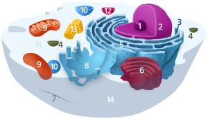
The endomembrane system is composed of the different membranes (endomembranes) that are suspended in the cytoplasm within a eukaryotic cell. These membranes divide the cell into functional and structural compartments, or organelles. In eukaryotes the organelles of the endomembrane system include: the nuclear membrane, the endoplasmic reticulum, the Golgi apparatus, lysosomes, vesicles, endosomes, and plasma (cell) membrane among others. The system is defined more accurately as the set of membranes that forms a single functional and developmental unit, either being connected directly, or exchanging material through vesicle transport. Importantly, the endomembrane system does not include the membranes of plastids or mitochondria, but might have evolved partially from the actions of the latter.

<span class="mw-page-title-main">Endocytosis</span> Cellular process

Endocytosis is a cellular process in which substances are brought into the cell. The material to be internalized is surrounded by an area of cell membrane, which then buds off inside the cell to form a vesicle containing the ingested material. Endocytosis includes pinocytosis and phagocytosis. It is a form of active transport.

<span class="mw-page-title-main">Lysosome</span> Cell membrane organelle

A lysosome is a membrane-bound organelle found in many animal cells. They are spherical vesicles that contain hydrolytic enzymes that digest many kinds of biomolecules. A lysosome has a specific composition, of both its membrane proteins and its lumenal proteins. The lumen's pH (~4.5–5.0) is optimal for the enzymes involved in hydrolysis, analogous to the activity of the stomach. Besides degradation of polymers, the lysosome is involved in cell processes of secretion, plasma membrane repair, apoptosis, cell signaling, and energy metabolism.

<span class="mw-page-title-main">Exocytosis</span> Active transport and bulk transport in which a cell transports molecules out of the cell

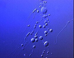
Exocytosis is a form of active transport and bulk transport in which a cell transports molecules out of the cell. As an active transport mechanism, exocytosis requires the use of energy to transport material. Exocytosis and its counterpart, endocytosis, are used by all cells because most chemical substances important to them are large polar molecules that cannot pass through the hydrophobic portion of the cell membrane by passive means. Exocytosis is the process by which a large amount of molecules are released; thus it is a form of bulk transport. Exocytosis occurs via secretory portals at the cell plasma membrane called porosomes. Porosomes are permanent cup-shaped lipoprotein structure at the cell plasma membrane, where secretory vesicles transiently dock and fuse to release intra-vesicular contents from the cell.

<span class="mw-page-title-main">Endosome</span> Vacuole to which materials ingested by endocytosis are delivered

Endosomes are a collection of intracellular sorting organelles in eukaryotic cells. They are parts of endocytic membrane transport pathway originating from the trans Golgi network. Molecules or ligands internalized from the plasma membrane can follow this pathway all the way to lysosomes for degradation or can be recycled back to the cell membrane in the endocytic cycle. Molecules are also transported to endosomes from the trans Golgi network and either continue to lysosomes or recycle back to the Golgi apparatus.

<span class="mw-page-title-main">Pinocytosis</span> Mode of endocytosis to bring small particles into a cell

In cellular biology, pinocytosis, otherwise known as fluid endocytosis and bulk-phase pinocytosis, is a mode of endocytosis in which small molecules dissolved in extracellular fluid are brought into the cell through an invagination of the cell membrane, resulting in their containment within a small vesicle inside the cell. These pinocytotic vesicles then typically fuse with early endosomes to hydrolyze the particles.

<span class="mw-page-title-main">Receptor-mediated endocytosis</span> Process by which cells absorb materials

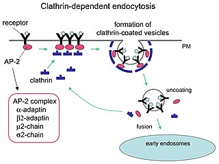
Receptor-mediated endocytosis (RME), also called clathrin-mediated endocytosis, is a process by which cells absorb metabolites, hormones, proteins – and in some cases viruses – by the inward budding of the plasma membrane (invagination). This process forms vesicles containing the absorbed substances and is strictly mediated by receptors on the surface of the cell. Only the receptor-specific substances can enter the cell through this process.

<span class="mw-page-title-main">Phagosome</span>

In cell biology, a phagosome is a vesicle formed around a particle engulfed by a phagocyte via phagocytosis. Professional phagocytes include macrophages, neutrophils, and dendritic cells (DCs).

<span class="mw-page-title-main">SNARE protein</span> Protein family

SNARE proteins – "SNAPREceptors" – are a large protein family consisting of at least 24 members in yeasts, more than 60 members in mammalian cells, and some numbers in plants. The primary role of SNARE proteins is to mediate the fusion of vesicles with the target membrane; this notably mediates exocytosis, but can also mediate the fusion of vesicles with membrane-bound compartments. The best studied SNAREs are those that mediate the release of synaptic vesicles containing neurotransmitters in neurons. These neuronal SNAREs are the targets of the neurotoxins responsible for botulism and tetanus produced by certain bacteria.

Cell physiology is the biological study of the activities that take place in a cell to keep it alive. The term physiology refers to normal functions in a living organism. Animal cells, plant cells and microorganism cells show similarities in their functions even though they vary in structure.

<span class="mw-page-title-main">Vesicular transport protein</span>

A vesicular transport protein, or vesicular transporter, is a membrane protein that regulates or facilitates the movement of specific molecules across a vesicle's membrane. As a result, vesicular transporters govern the concentration of molecules within a vesicle.

<span class="mw-page-title-main">Microvesicle</span> Type of extracellular vesicle

Microvesicles are a type of extracellular vesicle (EV) that are released from the cell membrane. In multicellular organisms, microvesicles and other EVs are found both in tissues and in many types of body fluids. Delimited by a phospholipid bilayer, microvesicles can be as small as the smallest EVs or as large as 1000 nm. They are considered to be larger, on average, than intracellularly-generated EVs known as exosomes. Microvesicles play a role in intercellular communication and can transport molecules such as mRNA, miRNA, and proteins between cells.

<span class="mw-page-title-main">STX6</span> Protein-coding gene in the species Homo sapiens

Syntaxin-6 is a protein that in humans is encoded by the STX6 gene.

<span class="mw-page-title-main">Cytosis</span> Movement of molecules into or out of cells

-Cytosis is a suffix that either refers to certain aspects of cells ie cellular process or phenomenon or sometimes refers to predominance of certain type of cells. It essentially means "of the cell". Sometimes it may be shortened to -osis and may be related to some of the processes ending with -esis or similar suffixes.

<span class="mw-page-title-main">ATG8</span>

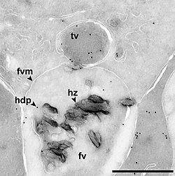
Autophagy-related protein 8 (Atg8) is a ubiquitin-like protein required for the formation of autophagosomal membranes. The transient conjugation of Atg8 to the autophagosomal membrane through a ubiquitin-like conjugation system is essential for autophagy in eukaryotes. Even though there are homologues in animals, this article mainly focuses on its role in lower eukaryotes such as Saccharomyces cerevisiae.

<span class="mw-page-title-main">Cell membrane</span> Biological membrane that separates the interior of a cell from its outside environment

The cell membrane is a biological membrane that separates and protects the interior of a cell from the outside environment. The cell membrane consists of a lipid bilayer, made up of two layers of phospholipids with cholesterols interspersed between them, maintaining appropriate membrane fluidity at various temperatures. The membrane also contains membrane proteins, including integral proteins that span the membrane and serve as membrane transporters, and peripheral proteins that loosely attach to the outer (peripheral) side of the cell membrane, acting as enzymes to facilitate interaction with the cell's environment. Glycolipids embedded in the outer lipid layer serve a similar purpose. The cell membrane controls the movement of substances in and out of a cell, being selectively permeable to ions and organic molecules. In addition, cell membranes are involved in a variety of cellular processes such as cell adhesion, ion conductivity, and cell signalling and serve as the attachment surface for several extracellular structures, including the cell wall and the carbohydrate layer called the glycocalyx, as well as the intracellular network of protein fibers called the cytoskeleton. In the field of synthetic biology, cell membranes can be artificially reassembled.

Clathrin adaptor proteins, also known as adaptins, are vesicular transport adaptor proteins associated with clathrin. These proteins are synthesized in the ribosomes, processed in the endoplasmic reticulum and transported from the Golgi apparatus to the trans-Golgi network, and from there via small carrier vesicles to their final destination compartment. The association between adaptins and clathrin are important for vesicular cargo selection and transporting. Clathrin coats contain both clathrin and adaptor complexes that link clathrin to receptors in coated vesicles. Clathrin-associated protein complexes are believed to interact with the cytoplasmic tails of membrane proteins, leading to their selection and concentration. Therefore, adaptor proteins are responsible for the recruitment of cargo molecules into a growing clathrin-coated pits. The two major types of clathrin adaptor complexes are the heterotetrameric vesicular transport adaptor proteins (AP1-5), and the monomeric GGA adaptors. Adaptins are distantly related to the other main type of vesicular transport proteins, the coatomer subunits, sharing between 16% and 26% of their amino acid sequence.

Unconventional protein secretion represents a manner in which the proteins are delivered to the surface of plasma membrane or extracellular matrix independent of the endoplasmic reticulum or Golgi apparatus. This includes cytokines and mitogens with crucial function in complex processes such as inflammatory response or tumor-induced angiogenesis. Most of these proteins are involved in processes in higher eukaryotes, however an unconventional export mechanism was found in lower eukaryotes too. Even proteins folded in their correct conformation can pass plasma membrane this way, unlike proteins transported via ER/Golgi pathway. Two types of unconventional protein secretion are these: signal-peptid-containing proteins and cytoplasmatic and nuclear proteins that are missing an ER-signal peptide (1).

Membrane vesicle trafficking in eukaryotic animal cells involves movement of biochemical signal molecules from synthesis-and-packaging locations in the Golgi body to specific release locations on the inside of the plasma membrane of the secretory cell. It takes place in the form of Golgi membrane-bound micro-sized vesicles, termed membrane vesicles (MVs).

<span class="mw-page-title-main">Intracellular transport</span> Directed movement of vesicles and substances within a cell

Intracellular transport is the movement of vesicles and substances within a cell. Intracellular transport is required for maintaining homeostasis within the cell by responding to physiological signals. Proteins synthesized in the cytosol are distributed to their respective organelles, according to their specific amino acid’s sorting sequence. Eukaryotic cells transport packets of components to particular intracellular locations by attaching them to molecular motors that haul them along microtubules and actin filaments. Since intracellular transport heavily relies on microtubules for movement, the components of the cytoskeleton play a vital role in trafficking vesicles between organelles and the plasma membrane by providing mechanical support. Through this pathway, it is possible to facilitate the movement of essential molecules such as membrane‐bounded vesicles and organelles, mRNA, and chromosomes.

References

  1. Akbarzadeh A, Rezaei-Sadabady R, Davaran S, Joo SW, Zarghami N, Hanifehpour Y, Samiei M, Kouhi M, Nejati-Koshki K (February 2013). "Liposome: classification, preparation, and applications". Nanoscale Res Lett. 8 (1): 102. Bibcode:2013NRL.....8..102A. doi: 10.1186/1556-276X-8-102 . PMC   3599573 . PMID   23432972.
  2. 1 2 Walsby AE (March 1994). "Gas vesicles". Microbiological Reviews. 58 (1): 94–144. doi:10.1128/mmbr.58.1.94-144.1994. PMC   372955 . PMID   8177173.
  3. Slomkowski S, Alemán JV, Gilbert RG, Hess M, Horie K, Jones RG, et al. (2011). "Terminology of polymers and polymerization processes in dispersed systems (IUPAC Recommendations 2011)" (PDF). Pure and Applied Chemistry . 83 (12): 2229–2259. doi:10.1351/PAC-REC-10-06-03. S2CID   96812603.
  4. "Nobel medical prize goes to 2 Americans, 1 German". CNN. 2005-10-19. Retrieved 2013-10-09.
  5. 2013 Nobel Prize in Physiology or Medicine, press release 2013-10-07
  6. Deatherage BL, Cookson BT (June 2012). "Membrane vesicle release in bacteria, eukaryotes, and archaea: a conserved yet underappreciated aspect of microbial life". Infection and Immunity. 80 (6): 1948–57. doi:10.1128/IAI.06014-11. PMC   3370574 . PMID   22409932.
  7. Yáñez-Mó M, Siljander PR, Andreu Z, Zavec AB, Borràs FE, Buzas EI, et al. (2015). "Biological properties of extracellular vesicles and their physiological functions". Journal of Extracellular Vesicles. 4: 27066. doi:10.3402/jev.v4.27066. PMC   4433489 . PMID   25979354.
  8. 1 2 Théry C, Witwer KW, Aikawa E, Alcaraz MJ, Anderson JD, Andriantsitohaina R, et al. (2018). "Minimal information for studies of extracellular vesicles 2018 (MISEV2018): a position statement of the International Society for Extracellular Vesicles and update of the MISEV2014 guidelines". Journal of Extracellular Vesicles. 7 (1): 1535750. doi:10.1080/20013078.2018.1535750. PMC   6322352 . PMID   30637094.
  9. 1 2 3 4 5 van der Pol E, Böing AN, Harrison P, Sturk A, Nieuwland R (July 2012). "Classification, functions, and clinical relevance of extracellular vesicles". Pharmacological Reviews. 64 (3): 676–705. doi:10.1124/pr.112.005983. PMID   22722893. S2CID   7764903. Free full text
  10. van der Pol E, Böing AN, Gool EL, Nieuwland R (January 2016). "Recent developments in the nomenclature, presence, isolation, detection and clinical impact of extracellular vesicles". Journal of Thrombosis and Haemostasis. 14 (1): 48–56. doi: 10.1111/jth.13190 . PMID   26564379.
  11. Melentijevic I, Toth ML, Arnold ML, Guasp RJ, Harinath G, Nguyen KC, et al. (February 2017). "C. elegans neurons jettison protein aggregates and mitochondria under neurotoxic stress". Nature. 542 (7641): 367–371. Bibcode:2017Natur.542..367M. doi:10.1038/nature21362. PMC   5336134 . PMID   28178240.
  12. Mateescu B, Kowal EJ, van Balkom BW, Bartel S, Bhattacharyya SN, Buzás EI, et al. (2017). "Obstacles and opportunities in the functional analysis of extracellular vesicle RNA - an ISEV position paper". Journal of Extracellular Vesicles. 6 (1): 1286095. doi:10.1080/20013078.2017.1286095. PMC   5345583 . PMID   28326170.
  13. Dhondt B, Rousseau Q, De Wever O, Hendrix A (September 2016). "Function of extracellular vesicle-associated miRNAs in metastasis". Cell and Tissue Research. 365 (3): 621–41. doi:10.1007/s00441-016-2430-x. hdl:1854/LU-7250365. PMID   27289232. S2CID   2746182.
  14. Dhondt B, Van Deun J, Vermaerke S, de Marco A, Lumen N, De Wever O, Hendrix A (June 2018). "Urinary extracellular vesicle biomarkers in urological cancers: From discovery towards clinical implementation". The International Journal of Biochemistry & Cell Biology. 99: 236–256. doi:10.1016/j.biocel.2018.04.009. hdl:1854/LU-8559155. PMID   29654900. S2CID   4876604.
  15. Teixeira FG, Carvalho MM, Sousa N, Salgado AJ (October 2013). "Mesenchymal stem cells secretome: a new paradigm for central nervous system regeneration?". Cellular and Molecular Life Sciences. 70 (20): 3871–82. doi:10.1007/s00018-013-1290-8. hdl: 1822/25128 . PMID   23456256. S2CID   18640402.
  16. Kuehn MJ, Kesty NC (November 2005). "Bacterial outer membrane vesicles and the host–pathogen interaction". Genes & Development. 19 (22): 2645–55. doi: 10.1101/gad.1299905 . PMID   16291643.
  17. Biller SJ, Schubotz F, Roggensack SE, Thompson AW, Summons RE, Chisholm SW (January 2014). "Bacterial vesicles in marine ecosystems". Science. 343 (6167): 183–6. Bibcode:2014Sci...343..183B. doi:10.1126/science.1243457. hdl: 1721.1/84545 . PMID   24408433. S2CID   206551356.
  18. 1 2 Adamala K, Szostak JW. Nonenzymatic template-directed RNA synthesis inside model protocells. Science. 2013 Nov 29;342(6162):1098-100. doi: 10.1126/science.1241888. PMID 24288333; PMCID: PMC4104020
  19. Pfeifer F (October 2012). "Distribution, formation and regulation of gas vesicles". Nature Reviews. Microbiology. 10 (10): 705–15. doi:10.1038/nrmicro2834. PMID   22941504. S2CID   9926129.
  20. Anderson HC (October 1967). "Electron microscopic studies of induced cartilage development and calcification". The Journal of Cell Biology. 35 (1): 81–101. doi:10.1083/jcb.35.1.81. PMC   2107116 . PMID   6061727.
  21. Bonucci E (September 1967). "Fine structure of early cartilage calcification". Journal of Ultrastructure Research. 20 (1): 33–50. doi:10.1016/S0022-5320(67)80034-0. PMID   4195919.
  22. Raikhel, Natasha V. (2017-04-28). "Firmly Planted, Always Moving". Annual Review of Plant Biology . Annual Reviews. 68 (1): 1–27. doi: 10.1146/annurev-arplant-042916-040829 . ISSN   1543-5008. PMID   27860488.
  23. Katzmann DJ, Odorizzi G, Emr SD (December 2002). "Receptor downregulation and multivesicular-body sorting". Nature Reviews. Molecular Cell Biology. 3 (12): 893–905. doi:10.1038/nrm973. PMID   12461556. S2CID   1344520.
  24. Sidhu VK, Vorhölter FJ, Niehaus K, Watt SA (June 2008). "Analysis of outer membrane vesicle associated proteins isolated from the plant pathogenic bacterium Xanthomonas campestris pv. campestris". BMC Microbiology. 8: 87. doi: 10.1186/1471-2180-8-87 . PMC   2438364 . PMID   18518965.
  25. Scherer GG, Martiny-Baron G (1985). "K+/H+ exchange transport in plantmembranevesicles is evidence for K+ transport". Plant Science. 41 (3): 161–8. doi:10.1016/0168-9452(85)90083-4.
  26. Walde P, Cosentino K, Engel H, Stano P (May 2010). "Giant vesicles: preparations and applications". ChemBioChem. 11 (7): 848–65. doi:10.1002/cbic.201000010. PMID   20336703. S2CID   30723166.
  27. Barenholz Y, Gibbes D, Litman BJ, Goll J, Thompson TE, Carlson RD (June 1977). "A simple method for the preparation of homogeneous phospholipid vesicles". Biochemistry. 16 (12): 2806–10. doi:10.1021/bi00631a035. PMID   889789.
  28. Batzri S, Korn ED (April 1973). "Single bilayer liposomes prepared without sonication". Biochimica et Biophysica Acta (BBA) - Biomembranes. 298 (4): 1015–9. doi:10.1016/0005-2736(73)90408-2. PMID   4738145.
  29. Litschel T, Schwille P (March 2021). "Protein Reconstitution Inside Giant Unilamellar Vesicles". Annual Review of Biophysics. 50: 525–548. doi:10.1146/annurev-biophys-100620-114132. PMID   33667121. S2CID   232131463.
  30. Sato Y, Takinoue M (March 2019). "Creation of Artificial Cell-Like Structures Promoted by Microfluidics Technologies". Micromachines. 10 (4): 216. doi: 10.3390/mi10040216 . PMC   6523379 . PMID   30934758.

Further reading