Tissue (biology)

Last updated
Microscopic view of a histologic specimen of human lung, consisting of various tissues: blood, connective tissue, vascular endothelium and respiratory epithelium, stained with hematoxylin and eosin. Emphysema H and E.jpg
Microscopic view of a histologic specimen of human lung, consisting of various tissues: blood, connective tissue, vascular endothelium and respiratory epithelium, stained with hematoxylin and eosin.

In biology, tissue is an assembly of similar cells and their extracellular matrix from the same embryonic origin that together carry out a specific function. [1] [2] Tissues occupy a biological organizational level between cells and a complete organ. Accordingly, organs are formed by the functional grouping together of multiple tissues. [3]

Contents

Biological organisms follow this hierarchy:

Cells < Tissue < Organ < Organ System < Organism

The English word "tissue" derives from the French word "tissu", the past participle of the verb tisser, "to weave".

The study of tissues is known as histology or, in connection with disease, as histopathology. Xavier Bichat is considered as the "Father of Histology". Plant histology is studied in both plant anatomy and physiology. The classical tools for studying tissues are the paraffin block in which tissue is embedded and then sectioned, the histological stain, and the optical microscope. Developments in electron microscopy, immunofluorescence, and the use of frozen tissue-sections have enhanced the detail that can be observed in tissues. With these tools, the classical appearances of tissues can be examined in health and disease, enabling considerable refinement of medical diagnosis and prognosis.

Plant tissue

Cross-section of a flax plant stem with several layers of different tissue types:
Pith
Protoxylem
Xylem
Phloem
Sclerenchyma (bast fiber)
Cortex
Epidermis Stem-histology-cross-section-tag.svg
Cross-section of a flax plant stem with several layers of different tissue types:

In plant anatomy, tissues are categorized broadly into three tissue systems: the epidermis, the ground tissue, and the vascular tissue.

Plant tissues can also be divided differently into two types:

  1. Meristematic tissues
  2. Permanent tissues.

Meristematic tissue

Meristematic tissue consists of actively dividing cells and leads to increase in length and thickness of the plant. The primary growth of a plant occurs only in certain specific regions, such as in the tips of stems or roots. It is in these regions that meristematic tissue is present. Cells of this type of tissue are roughly spherical or polyhedral to rectangular in shape, with thin cell walls. New cells produced by meristem are initially those of meristem itself, but as the new cells grow and mature, their characteristics slowly change and they become differentiated as components of meristematic tissue, being classified as:

There are two types of meristematic Tissue

1.Primary meristem.

2.Secondary meristem.

The cells of meristematic tissue are similar in structure and have a thin and elastic primary cell wall made of cellulose. They are compactly arranged without inter-cellular spaces between them. Each cell contains a dense cytoplasm and a prominent cell nucleus. The dense protoplasm of meristematic cells contains very few vacuoles. Normally the meristematic cells are oval, polygonal, or rectangular in shape.

Meristematic tissue cells have a large nucleus with small or no vacuoles because they have no need to store anything, as opposed to their function of multiplying and increasing the girth and length of the plant, with no intercellular spaces.

Permanent tissues

Permanent tissues may be defined as a group of living or dead cells formed by meristematic tissue and have lost their ability to divide and have permanently placed at fixed positions in the plant body. Meristematic tissues that take up a specific role lose the ability to divide. This process of taking up a permanent shape, size and a function is called cellular differentiation. Cells of meristematic tissue differentiate to form different types of permanent tissues. There are 2 types of permanent tissues:

  1. simple permanent tissues
  2. complex permanent tissues

Simple permanent tissue

Simple permanent tissue is a group of cells which are similar in origin, structure, and function. They are of three types:

  1. Parenchyma
  2. Collenchyma
  3. Sclerenchyma
Parenchyma

Parenchyma (Greek, para – 'beside'; enchyma– infusion – 'tissue') is the bulk of a substance. In plants, it consists of relatively unspecialized living cells with thin cell walls that are usually loosely packed so that intercellular spaces are found between cells of this tissue. These are generally isodiametric, in shape. They contain small number of vacuoles or sometimes they even may not contain any vacuole. Even if they do so the vacuole is of much smaller size than of normal animal cells. This tissue provides support to plants and also stores food. Chlorenchyma is a special type of parenchyma that contains chlorophyll and performs photosynthesis. In aquatic plants, aerenchyma tissues, or large air cavities, give support to float on water by making them buoyant. Parenchyma cells called idioblasts have metabolic waste. Spindle shape fiber also contained into this cell to support them and known as prosenchyma, succulent parenchyma also noted. In xerophytes, parenchyma tissues store water.

Collenchyma
Cross section of collenchyma cells Plant cell type collenchyma.png
Cross section of collenchyma cells

Collenchyma (Greek, ‘Colla’ means gum and ‘enchyma’ means infusion) is a living tissue of primary body like Parenchyma. Cells are thin-walled but possess thickening of cellulose, water and pectin substances (pectocellulose) at the corners where a number of cells join. This tissue gives tensile strength to the plant and the cells are compactly arranged and have very little inter-cellular spaces. It occurs chiefly in hypodermis of stems and leaves. It is absent in monocots and in roots.

Collenchymatous tissue acts as a supporting tissue in stems of young plants. It provides mechanical support, elasticity, and tensile strength to the plant body. It helps in manufacturing sugar and storing it as starch. It is present in the margin of leaves and resists tearing effect of the wind.

Sclerenchyma

Sclerenchyma (Greek, Sclerous means hard and enchyma means infusion) consists of thick-walled, dead cells and protoplasm is negligible. These cells have hard and extremely thick secondary walls due to uniform distribution and high secretion of lignin and have a function of providing mechanical support. They do not have inter-molecular space between them. Lignin deposition is so thick that the cell walls become strong, rigid and impermeable to water which is also known as a stone cell or sclereids. These tissues are mainly of two types: sclerenchyma fiber and sclereids. Sclerenchyma fiber cells have a narrow lumen and are long, narrow and unicellular. Fibers are elongated cells that are strong and flexible, often used in ropes. Sclereids have extremely thick cell walls and are brittle, and are found in nutshells and legumes.

Epidermis

The entire surface of the plant consists of a single layer of cells called epidermis or surface tissue. The entire surface of the plant has this outer layer of the epidermis. Hence it is also called surface tissue. Most of the epidermal cells are relatively flat. The outer and lateral walls of the cell are often thicker than the inner walls. The cells form a continuous sheet without intercellular spaces. It protects all parts of the plant. The outer epidermis is coated with a waxy thick layer called cutin which prevents loss of water. The epidermis also consists of stomata (singular:stoma) which helps in transpiration.

Complex permanent tissue

The complex permanent tissue consists of more than one type of cells having a common origin which work together as a unit. Complex tissues are mainly concerned with the transportation of mineral nutrients, organic solutes (food materials), and water. That's why it is also known as conducting and vascular tissue. The common types of complex permanent tissue are:

Xylem and phloem together form vascular bundles.

Xylem

Xylem (Greek, xylos = wood) serves as a chief conducting tissue of vascular plants. It is responsible for the conduction of water and inorganic solutes. Xylem consists of four kinds of cells:

  • Tracheids
  • Vessels (or tracheae)
  • Xylem fibers or Xylem sclerenchyma
  • Xylem parenchyma
Cross section of 2-year-old Tilia americana, highlighting xylem ray shape and orientation Xylem rays (Tilia Americana).jpg
Cross section of 2-year-old Tilia americana , highlighting xylem ray shape and orientation

Xylem tissue is organised in a tube-like fashion along the main axes of stems and roots. It consists of a combination of parenchyma cells, fibers, vessels, tracheids, and ray cells. Longer tubes made up of individual cellssels tracheids, while vessel members are open at each end. Internally, there may be bars of wall material extending across the open space. These cells are joined end to end to form long tubes. Vessel members and tracheids are dead at maturity. Tracheids have thick secondary cell walls and are tapered at the ends. They do not have end openings such as the vessels. The end overlap with each other, with pairs of pits present. The pit pairs allow water to pass from cell to cell.

Though most conduction in xylem tissue is vertical, lateral conduction along the diameter of a stem is facilitated via rays.[ citation needed ] Rays are horizontal rows of long-living parenchyma cells that arise out of the vascular cambium.

Phloem

Phloem consists of:

Phloem is an equally important plant tissue as it also is part of the 'plumbing system' of a plant. Primarily, phloem carries dissolved food substances throughout the plant. This conduction system is composed of sieve-tube member and companion cells, that are without secondary walls. The parent cells of the vascular cambium produce both xylem and phloem. This usually also includes fibers, parenchyma and ray cells. Sieve tubes are formed from sieve-tube members laid end to end. The end walls, unlike vessel members in xylem, do not have openings. The end walls, however, are full of small pores where cytoplasm extends from cell to cell. These porous connections are called sieve plates. In spite of the fact that their cytoplasm is actively involved in the conduction of food materials, sieve-tube members do not have nuclei at maturity. It is the companion cells that are nestled between sieve-tube members that function in some manner bringing about the conduction of food. Sieve-tube members that are alive contain a polymer called callose, a carbohydrate polymer, forming the callus pad/callus, the colourless substance that covers the sieve plate. Callose stays in solution as long as the cell contents are under pressure. Phloem transports food and materials in plants upwards and downwards as required.

Animal tissue

Animal tissues are grouped into four basic types: connective, muscle, nervous, and epithelial. [4] Collections of tissues joined in units to serve a common function compose organs. While most animals can generally be considered to contain the four tissue types, the manifestation of these tissues can differ depending on the type of organism. For example, the origin of the cells comprising a particular tissue type may differ developmentally for different classifications of animals. Tissue appeared for the first time in the diploblasts, but modern forms only appeared in triploblasts.

The epithelium in all animals is derived from the ectoderm and endoderm (or their precursor in sponges), with a small contribution from the mesoderm, forming the endothelium, a specialized type of epithelium that composes the vasculature. By contrast, a true epithelial tissue is present only in a single layer of cells held together via occluding junctions called tight junctions, to create a selectively permeable barrier. This tissue covers all organismal surfaces that come in contact with the external environment such as the skin, the airways, and the digestive tract. It serves functions of protection, secretion, and absorption, and is separated from other tissues below by a basal lamina.

The connective tissue and the muscular are derived from the mesoderm. The nervous tissue is derived from the ectoderm.

Epithelial tissues

The epithelial tissues are formed by cells that cover the organ surfaces, such as the surface of skin, the airways, surfaces of soft organs, the reproductive tract, and the inner lining of the digestive tract. The cells comprising an epithelial layer are linked via semi-permeable, tight junctions; hence, this tissue provides a barrier between the external environment and the organ it covers. In addition to this protective function, epithelial tissue may also be specialized to function in secretion, excretion and absorption. Epithelial tissue helps to protect organs from microorganisms, injury, and fluid loss.

Functions of epithelial tissue:

There are many kinds of epithelium, and nomenclature is somewhat variable. Most classification schemes combine a description of the cell-shape in the upper layer of the epithelium with a word denoting the number of layers: either simple (one layer of cells) or stratified (multiple layers of cells). However, other cellular features such as cilia may also be described in the classification system. Some common kinds of epithelium are listed below:

Connective tissue

Connective tissues are made up of cells separated by non-living material, which is called an extracellular matrix. This matrix can be liquid or rigid. For example, blood contains plasma as its matrix and bone's matrix is rigid. Connective tissue gives shape to organs and holds them in place. Blood, bone, tendon, ligament, adipose, and areolar tissues are examples of connective tissues. One method of classifying connective tissues is to divide them into three types: fibrous connective tissue, skeletal connective tissue, and fluid connective tissue.

Muscle tissue

Cross section through skeletal muscle and a small nerve at high magnification (H&E stain) Skeletal muscle - cross section, nerve bundle.jpg
Cross section through skeletal muscle and a small nerve at high magnification (H&E stain)

Muscle cells (myocytes) form the active contractile tissue of the body. Muscle tissue functions to produce force and cause motion, either locomotion or movement within internal organs. Muscle is formed of contractile filaments and is separated into three main types; smooth muscle, skeletal muscle and cardiac muscle. Smooth muscle has no striations when examined microscopically. It contracts slowly but maintains contractibility over a wide range of stretch lengths. It is found in such organs as sea anemone tentacles and the body wall of sea cucumbers. Skeletal muscle contracts rapidly but has a limited range of extension. It is found in the movement of appendages and jaws. Obliquely striated muscle is intermediate between the other two. The filaments are staggered and this is the type of muscle found in earthworms that can extend slowly or make rapid contractions. [5] In higher animals striated muscles occur in bundles attached to bone to provide movement and are often arranged in antagonistic sets. Smooth muscle is found in the walls of the uterus, bladder, intestines, stomach, oesophagus, respiratory airways, and blood vessels. Cardiac muscle is found only in the heart, allowing it to contract and pump blood round the body.

Nervous tissue

Cells comprising the central nervous system and peripheral nervous system are classified as nervous (or neural) tissue. In the central nervous system, neural tissues form the brain and spinal cord. In the peripheral nervous system, neural tissues form the cranial nerves and spinal nerves, inclusive of the motor neurons.

Mineralized tissues

Mineralized tissues are biological tissues that incorporate minerals into soft matrices. Such tissues may be found in both plants and animals.

History

Xavier Bichat (1771-1802) Pierre-Maximilien Delafontaine - Portrait of Marie Francois Xavier Bichat.jpg
Xavier Bichat (1771–1802)

Xavier Bichat introduced the word tissue into the study of anatomy by 1801. [6] He was "the first to propose that tissue is a central element in human anatomy, and he considered organs as collections of often disparate tissues, rather than as entities in themselves". [7] Although he worked without a microscope, Bichat distinguished 21 types of elementary tissues from which the organs of the human body are composed, [8] a number later reduced by other authors.

See also

Related Research Articles

<span class="mw-page-title-main">Plant cell</span> Type of eukaryotic cell present in green plants

Plant cells are the cells present in green plants, photosynthetic eukaryotes of the kingdom Plantae. Their distinctive features include primary cell walls containing cellulose, hemicelluloses and pectin, the presence of plastids with the capability to perform photosynthesis and store starch, a large vacuole that regulates turgor pressure, the absence of flagella or centrioles, except in the gametes, and a unique method of cell division involving the formation of a cell plate or phragmoplast that separates the new daughter cells.

<span class="mw-page-title-main">Xylem</span> Water transport tissue in vascular plants

Xylem is one of the two types of transport tissue in vascular plants, the other being phloem. The basic function of the xylem is to transport water from roots to stems and leaves, but it also transports nutrients. The word xylem is derived from the Ancient Greek word ξύλον (xylon), meaning "wood"; the best-known xylem tissue is wood, though it is found throughout a plant. The term was introduced by Carl Nägeli in 1858.

<span class="mw-page-title-main">Phloem</span> Sugar transport tissue in vascular plants

Phloem is the living tissue in vascular plants that transports the soluble organic compounds made during photosynthesis and known as photosynthates, in particular the sugar sucrose, to the rest of the plant. This transport process is called translocation. In trees, the phloem is the innermost layer of the bark, hence the name, derived from the Ancient Greek word φλοιός (phloiós), meaning "bark". The term was introduced by Carl Nägeli in 1858. Different types of phloem can be distinguished. The early phloem formed in the growth apices is called protophloem. Protophloem eventually becomes obliterated once it connects to the durable phloem in mature organs, the metaphloem. Further, secondary phloem is formed during the thickening of stem structures.

<span class="mw-page-title-main">Vascular plant</span> Phylum of plants with xylem and phloem

Vascular plants, or collectively the phylum Tracheophyta, form a large group of land plants that have lignified tissues for conducting water and minerals throughout the plant. They also have a specialized non-lignified tissue to conduct products of photosynthesis. Vascular plants include the clubmosses, horsetails, ferns, gymnosperms, and angiosperms. Scientific names for the group include Tracheophyta, Tracheobionta and Equisetopsida sensu lato. Some early land plants had less developed vascular tissue; the term eutracheophyte has been used for all other vascular plants, including all living ones.

<span class="mw-page-title-main">Bark (botany)</span> Outermost layers of stems and roots of woody plants

Bark is the outermost layer of stems and roots of woody plants. Plants with bark include trees, woody vines, and shrubs. Bark refers to all the tissues outside the vascular cambium and is a nontechnical term. It overlays the wood and consists of the inner bark and the outer bark. The inner bark, which in older stems is living tissue, includes the innermost layer of the periderm. The outer bark on older stems includes the dead tissue on the surface of the stems, along with parts of the outermost periderm and all the tissues on the outer side of the periderm. The outer bark on trees which lies external to the living periderm is also called the rhytidome.

<span class="mw-page-title-main">Vascular cambium</span> Main growth tissue in the stems, roots of plants

The vascular cambium is the main growth tissue in the stems and roots of many plants, specifically in dicots such as buttercups and oak trees, gymnosperms such as pine trees, as well as in certain other vascular plants. It produces secondary xylem inwards, towards the pith, and secondary phloem outwards, towards the bark.

<span class="mw-page-title-main">Cork cambium</span> Part of a plant

Cork cambium is a tissue found in many vascular plants as a part of the epidermis. It is one of the many layers of bark, between the cork and primary phloem. The cork cambium is a lateral meristem and is responsible for secondary growth that replaces the epidermis in roots and stems. It is found in woody and many herbaceous dicots, gymnosperms and some monocots. It is one of the plant's meristems – the series of tissues consisting of embryonic disk cells from which the plant grows. The function of cork cambium is to produce the cork, a tough protective material.

<span class="mw-page-title-main">Meristem</span> Type of plant tissue involved in cell proliferation

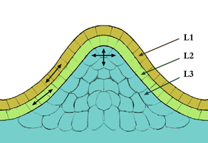
The meristem is a type of tissue found in plants. It consists of undifferentiated cells capable of cell division. Cells in the meristem can develop into all the other tissues and organs that occur in plants. These cells continue to divide until a time when they get differentiated and then lose the ability to divide.

<span class="mw-page-title-main">Epithelium</span> Tissue lining the surfaces of organs in animals

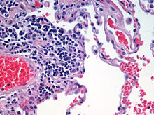
Epithelium or epithelial tissue is a thin, continuous, protective layer of compactly packed cells with a little intercellular matrix. Epithelial tissues line the outer surfaces of organs and blood vessels throughout the body, as well as the inner surfaces of cavities in many internal organs. An example is the epidermis, the outermost layer of the skin. Epithelial tissue is one of the four basic types of animal tissue, along with connective tissue, muscle tissue and nervous tissue. These tissues also lack blood or lymph supply. The tissue is supplied by nerves.

<span class="mw-page-title-main">Sclereid</span> Plant tissue type

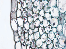
Sclereids are a reduced form of sclerenchyma cells with highly thickened, lignified cellular walls that form small bundles of durable layers of tissue in most plants. The presence of numerous sclereids form the cores of apples and produce the gritty texture of guavas.

<span class="mw-page-title-main">Loose connective tissue</span> Type of connective tissue in animals

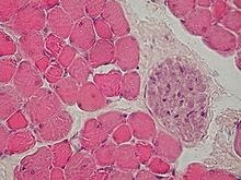
Loose connective tissue, also known as areolar tissue, is a cellular connective tissue with thin and relatively sparse collagen fibers. They have a semi-fluid matrix with lesser proportions of fibers. Its ground substance occupies more volume than the fibers do. It has a viscous to gel-like consistency and plays an important role in the diffusion of oxygen and nutrients from the capillaries that course through this connective tissue as well as in the diffusion of carbon dioxide and metabolic wastes back to the vessels. Moreover, loose connective tissue is primarily located beneath the epithelia that cover the body surfaces and line the internal surfaces of the body. It is also associated with the epithelium of glands and surrounds the smallest blood vessels. This tissue is thus the initial site where pathogenic agents, such as bacteria that have breached an epithelial surface, are challenged and destroyed by cells of the immune system.

The pericycle is a cylinder of parenchyma or sclerenchyma cells that lies just inside the endodermis and is the outer most part of the stele of plants.

Sieve elements are specialized cells that are important for the function of phloem, which is a highly organized tissue that transports organic compounds made during photosynthesis. Sieve elements are the major conducting cells in phloem. Conducting cells aid in transport of molecules especially for long-distance signaling. In plant anatomy, there are two main types of sieve elements. Companion cells and sieve cells originate from meristems, which are tissues that actively divide throughout a plant's lifetime. They are similar to the development of xylem, a water conducting tissue in plants whose main function is also transportation in the plant vascular system. Sieve elements' major function includes transporting sugars over long distance through plants by acting as a channel. Sieve elements elongate cells containing sieve areas on their walls. Pores on sieve areas allow for cytoplasmic connections to neighboring cells, which allows for the movement of photosynthetic material and other organic molecules necessary for tissue function. Structurally, they are elongated and parallel to the organ or tissue that they are located in. Sieve elements typically lack a nucleus and contain none to a very small number of ribosomes. The two types of sieve elements, sieve tube members and sieve cells, have different structures. Sieve tube members are shorter and wider with greater area for nutrient transport while sieve cells tend to be longer and narrower with smaller area for nutrient transport. Although the function of both of these kinds of sieve elements is the same, sieve cells are found in gymnosperms, non-flowering vascular plants, while sieve tube members are found in angiosperms, flowering vascular plants.

<span class="mw-page-title-main">Ground tissue</span> Category of tissue in plants

The ground tissue of plants includes all tissues that are neither dermal nor vascular. It can be divided into three types based on the nature of the cell walls. This tissue system is present between the dermal tissue and forms the main bulk of the plant body.

  1. Parenchyma cells have thin primary walls and usually remain alive after they become mature. Parenchyma forms the "filler" tissue in the soft parts of plants, and is usually present in cortex, pericycle, pith, and medullary rays in primary stem and root.
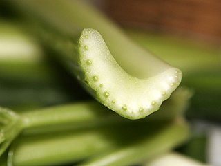
  2. Collenchyma cells have thin primary walls with some areas of secondary thickening. Collenchyma provides extra mechanical and structural support, particularly in regions of new growth.
  3. Sclerenchyma cells have thick lignified secondary walls and often die when mature. Sclerenchyma provides the main structural support to a plant.
<span class="mw-page-title-main">Vascular tissue</span> Conducting tissue in vascular plants

Vascular tissue is a complex conducting tissue, formed of more than one cell type, found in vascular plants. The primary components of vascular tissue are the xylem and phloem. These two tissues transport fluid and nutrients internally. There are also two meristems associated with vascular tissue: the vascular cambium and the cork cambium. All the vascular tissues within a particular plant together constitute the vascular tissue system of that plant.

<span class="mw-page-title-main">Secondary growth</span> Type of growth in plants

In botany, secondary growth is the growth that results from cell division in the cambia or lateral meristems and that causes the stems and roots to thicken, while primary growth is growth that occurs as a result of cell division at the tips of stems and roots, causing them to elongate, and gives rise to primary tissue. Secondary growth occurs in most seed plants, but monocots usually lack secondary growth. If they do have secondary growth, it differs from the typical pattern of other seed plants.

Important structures in plant development are buds, shoots, roots, leaves, and flowers; plants produce these tissues and structures throughout their life from meristems located at the tips of organs, or between mature tissues. Thus, a living plant always has embryonic tissues. By contrast, an animal embryo will very early produce all of the body parts that it will ever have in its life. When the animal is born, it has all its body parts and from that point will only grow larger and more mature. However, both plants and animals pass through a phylotypic stage that evolved independently and that causes a developmental constraint limiting morphological diversification.

<span class="mw-page-title-main">Lepidodendrales</span> Extinct order of vascular tree-like plants

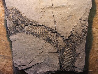
Lepidodendrales or arborescent lycophytes are an extinct order of primitive, vascular, heterosporous, arborescent (tree-like) plants belonging to Lycopodiopsida. Members of Lepidodendrales are the best understood of the fossil lycopsids due to the vast diversity of Lepidodendrales specimens and the diversity in which they were preserved; the extensive distribution of Lepidodendrales specimens as well as their well-preservedness lends paleobotanists exceptionally detailed knowledge of the coal-swamp giants’ reproductive biology, vegetative development, and role in their paleoecosystem. The defining characteristics of the Lepidodendrales are their secondary xylem, extensive periderm development, three-zoned cortex, rootlike appendages known as stigmarian rootlets arranged in a spiralling pattern, and megasporangium each containing a single functional megaspore that germinates inside the sporangium. Many of these different plant organs have been assigned both generic and specific names as relatively few have been found organically attached to each other. Some specimens have been discovered which indicate heights of 40 and even 50 meters and diameters of over 2 meters at the base. The massive trunks of some species branched profusely, producing large crowns of leafy twigs; though some leaves were up to 1 meter long, most were much shorter, and when leaves dropped from branches their conspicuous leaf bases remained on the surface of branches. Strobili could be found at the tips of distal branches or in an area at the top of the main trunk. The underground organs of Lepidodendrales typically consisted of dichotomizing axes bearing helically arranged, lateral appendages serving an equivalent function to roots. Sometimes called "giant club mosses", they are believed to be more closely related to extant quillworts based on xylem, although fossil specimens of extinct Selaginellales from the Late Carboniferous also had secondary xylem.

<span class="mw-page-title-main">Plant stem</span> Structural axis of a vascular plant

A stem is one of two main structural axes of a vascular plant, the other being the root. It supports leaves, flowers and fruits, transports water and dissolved substances between the roots and the shoots in the xylem and phloem, photosynthesis takes place here, stores nutrients, and produces new living tissue. The stem can also be called halm or haulm or culms.

<span class="mw-page-title-main">Anatomical terms of microanatomy</span> Anatomical terminology is used to describe microanatomical (or histological) structures

Anatomical terminology is used to describe microanatomical structures. This helps describe precisely the structure, layout and position of an object, and minimises ambiguity. An internationally accepted lexicon is Terminologia Histologica.

References

  1. Jones, Roger (June 2012). "Leonardo da Vinci: anatomist". British Journal of General Practice. 62 (599): 319. doi:10.3399/bjgp12X649241. PMC   3361109 . PMID   22687222.
  2. Toledo-Pereyra, Luis H. (January 2008). "De Humani Corporis Fabrica Surgical Revolution". Journal of Investigative Surgery. 21 (5): 232–236. doi:10.1080/08941930802330830. PMID   19160130. S2CID   45712227.
  3. Betts, J Gordon. "1.2 Structural Organization of the Human Body - Anatomy and Physiology". Anatomy and Physiology. Openstax. ISBN   978-1-947172-04-3. Archived from the original on 2023-03-24. Retrieved 14 May 2023.
  4. Ross, Michael H.; Pawlina, Wojciech (2016). Histology : a text and atlas : with correlated cell and molecular biology (7th ed.). Wolters Kluwer. p. 984. ISBN   978-1451187427.
  5. Ruppert, Edward E.; Fox, Richard, S.; Barnes, Robert D. (2004). Invertebrate Zoology, 7th edition. Cengage Learning. p. 103. ISBN   978-81-315-0104-7.{{cite book}}: CS1 maint: multiple names: authors list (link)
  6. Bock, Ortwin (January 2, 2015). "A History of the Development of Histology up to the End of the Nineteenth Century". Research. 2015, 2:1283. doi:10.13070/rs.en.2.1283 (inactive 31 January 2024). Archived from the original on April 13, 2021. Retrieved August 14, 2021.{{cite journal}}: CS1 maint: DOI inactive as of January 2024 (link)
  7. "Scientist of the Day: Xavier Bichat". Linda Hall Library. November 14, 2018. Archived from the original on May 23, 2020. Retrieved August 14, 2021.
  8. Roeckelein 1998 , p. 78

Sources