Sinoatrial node

Last updated

Sinoatrial node
Reizleitungssystem 1.png
Sinoatrial node shown at 1. The rest of the conduction system of the heart is shown in blue.
Details
System Electrical conduction system of the heart
Artery Sinoatrial nodal artery
Identifiers
Latin nodus sinuatrialis
Acronym(s)SA node
MeSH D012849
TA98 A12.1.06.003
TA2 3953
FMA 9477
Anatomical terminology

The sinoatrial node (also known as the sinuatrial node, SA node or sinus node) is an oval shaped region of special cardiac muscle in the upper back wall of the right atrium made up of cells known as pacemaker cells. The sinus node is approximately 15 mm long, 3 mm wide, and 1 mm thick, located directly below and to the side of the superior vena cava. [1]

Contents

These cells can produce an electrical impulse known as a cardiac action potential that travels through the electrical conduction system of the heart, causing it to contract. In a healthy heart, the SA node continuously produces action potentials, setting the rhythm of the heart (sinus rhythm), and so is known as the heart's natural pacemaker. The rate of action potentials produced (and therefore the heart rate) is influenced by the nerves that supply it. [2]

Structure

The sinoatrial node is an oval-shaped structure that is approximately 15  mm long, 3 mm wide, and 1 mm thick, located directly below and to the side of the superior vena cava. [1] The size can vary but is usually between 10-30 mm long, 5–7 mm wide, and 1–2 mm deep. [3] [4]

Location

The SA node is located in the wall (epicardium) of the right atrium, laterally to the entrance of the superior vena cava in a region called the sinus venarum (hence sino- + atrial). [5] It is positioned roughly between a groove called the crista terminalis located on the internal surface of the heart and the corresponding sulcus terminalis, on the external surface. [2] These grooves run between the entrance of the superior vena cava and the inferior vena cava.

Microanatomy

Figure 2: Low magnification stained image of the SA node (center-right on image) and its surrounding tissue. The SA node surrounds the sinoatrial nodal artery, seen as the open lumen. Cardiac muscle cells of the right atrium can be seen to the left of the node, and fat tissue to the right. Sinoatrial node 2 low mag.jpg
Figure 2: Low magnification stained image of the SA node (center-right on image) and its surrounding tissue. The SA node surrounds the sinoatrial nodal artery, seen as the open lumen. Cardiac muscle cells of the right atrium can be seen to the left of the node, and fat tissue to the right.

The cells of the SA node are spread out within a mesh of connective tissue, containing nerves, blood vessels, collagen and fat. Immediately surrounding the SA node cells are paranodal cells. [2] These cells have structures intermediate between that of the SA node cells and the rest of the atrium. [6] The connective tissue, along with the paranodal cells, insulate the SA node from the rest of the atrium, preventing the electrical activity of the atrial cells from affecting the SA node cells. [2] The SA node cells are smaller and paler than the surrounding atrial cells, with the average cell being around 8 micrometers in diameter and 20-30 micrometers in length (1 micrometer= 0.000001 meter). [7] Unlike the atrial cells, SA node cells contain fewer mitochondria and myofibers, as well as a smaller sarcoplasmic reticulum. This means that the SA node cells are less equipped to contract compared to the atrial and ventricular cells. [8]

Action potentials pass from one cardiac cell to the next through pores known as gap junctions. These gap junctions are made of proteins called connexins. There are fewer gap junctions within the SA node and they are smaller in size. This is again important in insulating the SA node from the surrounding atrial cells. [2] [8]

Blood supply

The sinoatrial node receives its blood supply from the sinoatrial nodal artery. This blood supply, however, can differ hugely between individuals. For example, in most humans, this is a single artery, although in some cases there have been either 2 or 3 sinoatrial node arteries supplying the SA node. Also, the SA node artery mainly originates as a branch of the right coronary artery; however in some individuals it has arisen from the circumflex artery, which is a branch of the left coronary artery. Finally, the SA node artery commonly passes behind the superior vena cava, before reaching the SA node; however in some instances it passes in front. Despite these many differences, there doesn't appear to be any advantage to how many sinoatrial nodal arteries an individual has, or where they originate. [9]

Venous drainage

There are no large veins that drain blood away from the SA node. Instead, smaller venules drain the blood directly into the right atrium. [10]

Function

Pacemaking

The main role of a sinoatrial node cell is to initiate action potentials of the heart that can pass through cardiac muscle cells and cause contraction. An action potential is a rapid change in membrane potential, produced by the movement of charged atoms (ions). In the absence of stimulation, non-pacemaker cells (including the ventricular and atrial cells) have a relatively constant membrane potential; this is known as a resting potential. This resting phase (see cardiac action potential, phase 4) ends when an action potential reaches the cell. This produces a positive change in membrane potential, known as depolarization, which is propagated throughout the heart and initiates muscle contraction. Pacemaker cells, however, do not have a resting potential. Instead, immediately after repolarization, the membrane potential of these cells begins to depolarise again automatically, a phenomenon known as the pacemaker potential. Once the pacemaker potential reaches a set value, the threshold potential, it produces an action potential. [2] Other cells within the heart (including the Purkinje fibers [11] and atrioventricular node) can also initiate action potentials; however, they do so at a slower rate and therefore, if the SA node is functioning properly, its action potentials usually override those that would be produced by other tissues. [12]

Outlined below are the 3 phases of a sinoatrial node action potential. In the cardiac action potential, there are 5 phases (labelled 0-4), however pacemaker action potentials do not have an obvious phase 1 or 2.

Phase 4

Figure 3: Sinoatrial node action potential waveform, outlining major ion currents involved (downward deflection indicates ions moving into the cell, upwards deflection indicates ions flowing out of the cell). Sinoatrial action potential.jpg
Figure 3: Sinoatrial node action potential waveform, outlining major ion currents involved (downward deflection indicates ions moving into the cell, upwards deflection indicates ions flowing out of the cell).

This phase is also known as the pacemaker potential. Immediately following repolarization, when the membrane potential is very negative (it is hyperpolarised), the voltage slowly begins to increase. This is initially due to the closing of potassium channels, which reduces the flow of potassium ions (Ik) out of the cell (see phase 2, below). [13] Hyperpolarization also causes activation of hyperpolarisation-activated cyclic nucleotide–gated (HCN) channels. The activation of ion channels at very negative membrane potentials is unusual, therefore the flow of sodium (Na+) and some K+ through the activated HCN channel is referred to as a funny current (If). [14] This funny current causes the membrane potential of the cell to gradually increase, as the positive charge (Na+ and K+) is flowing into the cell. Another mechanism involved in pacemaker potential is known as the calcium clock. This refers to the spontaneous release of calcium from the sarcoplasmic reticulum (a calcium store) into the cytoplasm, also known as calcium sparks. This increase in calcium within the cell then activates a sodium-calcium exchanger (NCX), which removes one Ca2+ from the cell, and exchanges it for 3 Na+ into the cell (therefore removing a charge of +2 from the cell, but allowing a charge of +3 to enter the cell) further increasing the membrane potential. Calcium later reenters the cell via SERCA and calcium channels located on the cell membrane. [15] The increase in membrane potential produced by these mechanisms, activates T-type calcium channels and then L-type calcium channels (which open very slowly). These channels allow a flow of Ca2+ into the cell, making the membrane potential even more positive.

Phase 0

This is the depolarization phase. When the membrane potential reaches the threshold potential (around -20 to -50 mV), the cell begins to rapidly depolarise (become more positive). [16] This is mainly due to the flow of Ca2+ through L-type calcium channels, which are now fully open. During this stage, T-type calcium channels and HCN channels deactivate.

Phase 3

This phase is the repolarization phase. This occurs due to the inactivation of L-type calcium channels (preventing the movement of Ca2+ into the cell) and the activation of potassium channels, which allows the flow of K+ out of the cell, making the membrane potential more negative. [17]

Nerve supply

Heart rate depends on the rate at which the sinoatrial node produces action potentials. At rest, heart rate is between 60 and 100 beats per minute. This is a result of the activity of two sets of nerves, one acting to slow down action potential production (these are parasympathetic nerves) and the other acting to speed up action potential production (sympathetic nerves). [18]

Modulation of heart rate by ANS is carried by two types of channel: Kir and HCN (members of the CNG gated channels).

The sympathetic nerves begin in the thoracic region of the spinal cord (in particular T1-T4). These nerves release a neurotransmitter called noradrenaline (NA). This binds to a receptor on the SA node membrane, called a beta-1adrenoceptor. Binding of NA to this receptor activates a G-protein (in particular a Gs-Protein, S for stimulatory) which initiates a series of reactions (known as the cAMP pathway) that results in the production of a molecule called cyclic adenosinemonophosphate (cAMP). This cAMP binds to the HCN channel (see above). Binding of cAMP to the HCN increases the flow of Na+ and K+ into the cell, speeding up the pacemaker potential, so producing action potentials at a quicker rate and increasing heart rate. [19] An increase in heart rate is known as positive chronotropy.

The parasympathetic nerves supplying the SA node (in particular the Vagus nerves) originate in the brain. These nerves release a neurotransmitter called acetylcholine (ACh). ACh binds to a receptor called an M2 muscarinic receptor, located on the SA node membrane. Activation of this M2 receptor then activates a protein called a G-protein (in particular Gi protein, i for inhibitory). Activation of this G-protein blocks the cAMP pathway, reducing its effects, therefore inhibiting sympathetic activity and slowing action potential production. The G-protein also activates a potassium channel GIRK-1 and GIRK-4, which allows K+ to flow out of the cell, making the membrane potential more negative and slowing the pacemaker potential, therefore decreasing the rate of action potential production and therefore decreasing heart rate. [20] A decrease in heart rate is known as negative chronotropy.

The first cell to produce the action potential in the SA node isn't always the same; this is known as pacemaker shift. In certain species of animals—for example, in dogs—a superior shift (i.e., the cell that produces the fastest action potential in the SA node is higher than previously) usually produces an increased heart rate whereas an inferior shift (i.e. the cell producing the fastest action potential within the SA node is further down than previously) produces a decreased heart rate. [2]

Clinical significance

Sinus node dysfunction also known as sick sinus syndrome is a group of irregular heartbeat conditions caused by faulty electrical signals of the heart. When the heart's sinoatrial node is defective, the heart's rhythms become abnormal—typically too slow or exhibiting pauses in its function or a combination, and very rarely faster than normal. [21]

Blockage of the arterial blood supply to the SA node (most commonly due to a myocardial infarction or progressive coronary artery disease) can therefore cause ischemia and cell death in the SA node. This can disrupt the electrical pacemaker function of the SA node, and can result in sinus node dysfunction.

If the SA node does not function or the impulse generated in the SA node is blocked before it travels down the electrical conduction system, a group of cells further down the heart will become its pacemaker. [22]

History

The sinoatrial node was first discovered by a young medical student, Martin Flack, in the heart of a mole, whilst his mentor, Sir Arthur Keith, was on a bicycle ride with his wife. They made the discovery in a makeshift laboratory set up in a farmhouse in Kent, England, called Mann's Place. Their discovery was published in 1907. [23] [24]

Additional images

See also

Related Research Articles

<span class="mw-page-title-main">Bradycardia</span> Heart rate below the normal range

Bradycardia, also called bradyarrhythmia, is a resting heart rate under 60 beats per minute (BPM). While bradycardia can result from various pathologic processes, it is commonly a physiologic response to cardiovascular conditioning or due to asymptomatic type 1 atrioventricular block. Resting heart rates of less than 50 BPM are often normal during sleep in young and healthy adults and athletes. In large population studies of adults without underlying heart disease, resting heart rates of 45-50 BPM appear to be the lower limits of normal, dependent on age and sex. Bradycardia is most likely to be discovered in the elderly, as age and underlying cardiac disease progression contribute to its development.

<span class="mw-page-title-main">Heart</span> Organ found inside most animals

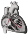
The heart is a muscular organ found in most animals. This organ pumps blood through the blood vessels of the circulatory system. The pumped blood carries oxygen and nutrients to the body, while carrying metabolic waste such as carbon dioxide to the lungs. In humans, the heart is approximately the size of a closed fist and is located between the lungs, in the middle compartment of the chest, called the mediastinum.

<span class="mw-page-title-main">Cardiac pacemaker</span> Network of cells that facilitate rhythmic heart contraction

The contraction of cardiac muscle in all animals is initiated by electrical impulses known as action potentials that in the heart are known as cardiac action potentials. The rate at which these impulses fire controls the rate of cardiac contraction, that is, the heart rate. The cells that create these rhythmic impulses, setting the pace for blood pumping, are called pacemaker cells, and they directly control the heart rate. They make up the cardiac pacemaker, that is, the natural pacemaker of the heart. In most humans, the highest concentration of pacemaker cells is in the sinoatrial (SA) node, the natural and primary pacemaker, and the resultant rhythm is a sinus rhythm.

<span class="mw-page-title-main">Systole</span> Part of the cardiac cycle when a heart chamber contracts

Systole is the part of the cardiac cycle during which some chambers of the heart contract after refilling with blood.

<span class="mw-page-title-main">Sinus node dysfunction</span> Medical condition

Sinus node dysfunction (SND), also known as sick sinus syndrome (SSS), is a group of abnormal heart rhythms (arrhythmias) usually caused by a malfunction of the sinus node, the heart's primary pacemaker. Tachycardia-bradycardia syndrome is a variant of sick sinus syndrome in which the arrhythmia alternates between fast and slow heart rates.

<span class="mw-page-title-main">Cardiac muscle</span> Muscular tissue of heart in vertebrates

Cardiac muscle is one of three types of vertebrate muscle tissues, with the other two being skeletal muscle and smooth muscle. It is an involuntary, striated muscle that constitutes the main tissue of the wall of the heart. The cardiac muscle (myocardium) forms a thick middle layer between the outer layer of the heart wall and the inner layer, with blood supplied via the coronary circulation. It is composed of individual cardiac muscle cells joined by intercalated discs, and encased by collagen fibers and other substances that form the extracellular matrix.

<span class="mw-page-title-main">Pacemaker potential</span>

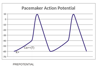
In the pacemaking cells of the heart (e.g., the sinoatrial node), the pacemaker potential (also called the pacemaker current) is the slow, positive increase in voltage across the cell's membrane (the membrane potential) that occurs between the end of one action potential and the beginning of the next action potential. This increase in membrane potential is what causes the cell membrane, which typically maintains a resting membrane potential around -65 mV, to reach the threshold potential and consequently fire the next action potential; thus, the pacemaker potential is what drives the self-generated rhythmic firing (automaticity) of pacemaker cells, and the rate of change (i.e., the slope) of the pacemaker potential is what determines the timing of the next action potential and thus the intrinsic firing rate of the cell. In a healthy sinoatrial node (SAN, a complex tissue within the right atrium containing pacemaker cells that normally determine the intrinsic firing rate for the entire heart), the pacemaker potential is the main determinant of the heart rate. Because the pacemaker potential represents the non-contracting time between heart beats (diastole), it is also called the diastolic depolarization. The amount of net inward current required to move the cell membrane potential during the pacemaker phase is extremely small, in the order of few pAs, but this net flux arises from time to time changing contribution of several currents that flow with different voltage and time dependence. Evidence in support of the active presence of K+, Ca2+, Na+ channels and Na+/K+ exchanger during the pacemaker phase have been variously reported in the literature, but several indications point to the “funny”(If) current as one of the most important.(see funny current). There is now substantial evidence that also sarcoplasmic reticulum (SR) Ca2+-transients participate to the generation of the diastolic depolarization via a process involving the Na–Ca exchanger.

<span class="mw-page-title-main">Cardiac conduction system</span> Aspect of heart function

The cardiac conduction system transmits the signals generated by the sinoatrial node – the heart's pacemaker, to cause the heart muscle to contract, and pump blood through the body's circulatory system. The pacemaking signal travels through the right atrium to the atrioventricular node, along the bundle of His, and through the bundle branches to Purkinje fibers in the walls of the ventricles. The Purkinje fibers transmit the signals more rapidly to stimulate contraction of the ventricles.

<span class="mw-page-title-main">Cardiac action potential</span> Biological process in the heart

Unlike the action potential in skeletal muscle cells, the cardiac action potential is not initiated by nervous activity. Instead, it arises from a group of specialized cells known as pacemaker cells, that have automatic action potential generation capability. In healthy hearts, these cells form the cardiac pacemaker and are found in the sinoatrial node in the right atrium. They produce roughly 60–100 action potentials every minute. The action potential passes along the cell membrane causing the cell to contract, therefore the activity of the sinoatrial node results in a resting heart rate of roughly 60–100 beats per minute. All cardiac muscle cells are electrically linked to one another, by intercalated discs which allow the action potential to pass from one cell to the next. This means that all atrial cells can contract together, and then all ventricular cells.

<span class="mw-page-title-main">Atrium (heart)</span> Part of the human heart

The atrium is one of the two upper chambers in the heart that receives blood from the circulatory system. The blood in the atria is pumped into the heart ventricles through the atrioventricular mitral and tricuspid heart valves.

<span class="mw-page-title-main">Cardiac cycle</span> Performance of the human heart

The cardiac cycle is the performance of the human heart from the beginning of one heartbeat to the beginning of the next. It consists of two periods: one during which the heart muscle relaxes and refills with blood, called diastole, following a period of robust contraction and pumping of blood, called systole. After emptying, the heart relaxes and expands to receive another influx of blood returning from the lungs and other systems of the body, before again contracting to pump blood to the lungs and those systems.

<span class="mw-page-title-main">Bachmann's bundle</span> Anatomical cardiac structure

In the heart's conduction system, Bachmann's bundle is a branch of the anterior internodal tract that resides on the inner wall of the left atrium. It is a broad band of cardiac muscle that passes from the right atrium, between the superior vena cava and the ascending aorta. Bachmann's bundle is, during normal sinus rhythm, the preferential path for electrical activation of the left atrium. It is therefore considered to be part of the "atrial conduction system" of the heart.

<span class="mw-page-title-main">Sinoatrial nodal artery</span> Artery of the heart which supplies the sinoatrial node

The sinoatrial nodal artery, sinuatrial nodal artery or sinoatrial artery is an artery of the heart which supplies the sinoatrial node, the natural pacemaker center of the heart. It is usually a branch of the right coronary artery. It passes between the right atrium, and the opening of the superior vena cava.

The pacemaker current is an electric current in the heart that flows through the HCN channel or pacemaker channel. Such channels are important parts of the electrical conduction system of the heart and form a component of the natural pacemaker.

The Bainbridge reflex or Bainbridge effect, also called the atrial reflex, is an increase in heart rate due to an increase in central venous pressure. Increased blood volume is detected by stretch receptors located in both sides of atria at the venoatrial junctions.

Sinoatrial arrest is a medical condition wherein the sinoatrial node of the heart transiently ceases to generate the electrical impulses that normally stimulate the myocardial tissues to contract and thus the heart to beat. It is defined as lasting from 2.0 seconds to several minutes. Since the heart contains multiple pacemakers, this interruption of the cardiac cycle generally lasts only a few seconds before another part of the heart, such as the atrio-ventricular junction or the ventricles, begins pacing and restores the heart action. This condition can be detected on an electrocardiogram (ECG) as a brief period of irregular length with no electrical activity before either the sinoatrial node resumes normal pacing, or another pacemaker begins pacing. If a pacemaker other than the sinoatrial node is pacing the heart, this condition is known as an escape rhythm. If no other pacemaker begins pacing during an episode of sinus arrest it becomes a cardiac arrest. This condition is sometimes confused with sinoatrial block, a condition in which the pacing impulse is generated, but fails to conduct through the myocardium. Differential diagnosis of the two conditions is possible by examining the exact length of the interruption of cardiac activity. If the next available pacemaker takes over, it is in the following order:

<span class="mw-page-title-main">Ectopic pacemaker</span> Cardiac condition

An ectopic pacemaker, also known as ectopic focus or ectopic foci, is an excitable group of cells that causes a premature heart beat outside the normally functioning SA node of the heart. It is thus a cardiac pacemaker that is ectopic, producing an ectopic beat. Acute occurrence is usually non-life-threatening, but chronic occurrence can progress into tachycardia, bradycardia or ventricular fibrillation. In a normal heart beat rhythm, the SA node usually suppresses the ectopic pacemaker activity due to the higher impulse rate of the SA node. However, in the instance of either a malfunctioning SA node or an ectopic focus bearing an intrinsic rate superior to SA node rate, ectopic pacemaker activity may take over the natural heart rhythm. This phenomenon is called an escape rhythm, the lower rhythm having escaped from the dominance of the upper rhythm. As a rule, premature ectopic beats indicate increased myocyte or conducting tissue excitability, whereas late ectopic beats indicate proximal pacemaker or conduction failure with an escape 'ectopic' beat.

Hyperpolarization-activated cyclic nucleotide–gated (HCN) channels are integral membrane proteins that serve as nonselective voltage-gated cation channels in the plasma membranes of heart and brain cells. HCN channels are sometimes referred to as pacemaker channels because they help to generate rhythmic activity within groups of heart and brain cells. HCN channels are activated by membrane hyperpolarization, are permeable to Na + and K +, and are constitutively open at voltages near the resting membrane potential. HCN channels are encoded by four genes and are widely expressed throughout the heart and the central nervous system.

Cardiac physiology or heart function is the study of healthy, unimpaired function of the heart: involving blood flow; myocardium structure; the electrical conduction system of the heart; the cardiac cycle and cardiac output and how these interact and depend on one another.

Cardiac excitation-contraction coupling (CardiacEC coupling) describes the series of events, from the production of an electrical impulse (action potential) to the contraction of muscles in the heart. This process is of vital importance as it allows for the heart to beat in a controlled manner, without the need for conscious input. EC coupling results in the sequential contraction of the heart muscles that allows blood to be pumped, first to the lungs (pulmonary circulation) and then around the rest of the body (systemic circulation) at a rate between 60 and 100 beats every minute, when the body is at rest. This rate can be altered, however, by nerves that work to either increase heart rate (sympathetic nerves) or decrease it (parasympathetic nerves), as the body's oxygen demands change. Ultimately, muscle contraction revolves around a charged atom (ion), calcium (Ca2+), which is responsible for converting the electrical energy of the action potential into mechanical energy (contraction) of the muscle. This is achieved in a region of the muscle cell, called the transverse tubule during a process known as calcium induced calcium release.

References

  1. 1 2 Hall, John E. (2011). Guyton and Hall textbook of medical physiology (Twelfth ed.). Philadelphia, Pa. p. 115. ISBN   9781416045748.{{cite book}}: CS1 maint: location missing publisher (link)
  2. 1 2 3 4 5 6 7 Monfredi, O.; Dobrzynski, H.; Mondal, T.; Boyett, M.R.; Morris, G.M. (2010). "The anatomy and physiology of the Sinoatrial Node-A contemporary review". Pacing and Clinical Electrophysiology. 33 (11): 1392–1406. doi:10.1111/j.1540-8159.2010.02838.x. PMID   20946278. S2CID   22207608.
  3. Csepe, Thomas A.; Zhao, Jichao; Hansen, Brian J.; Li, Ning; Sul, Lidiya V.; Lim, Praise; Wang, Yufeng; Simonetti, Orlando P.; Kilic, Ahmet (1 March 2017). "Human Sinoatrial Node Structure: 3D Microanatomy of Sinoatrial Conduction Pathways". Progress in Biophysics and Molecular Biology. 120 (1–3): 164–178. doi:10.1016/j.pbiomolbio.2015.12.011. ISSN   0079-6107. PMC   4808362 . PMID   26743207.
  4. Chandler, N.; Aslanidi, O.; Buckley, D.; Inada, S.; Birchall, S.; Atkinson, A.; Kirk, D.; Monfredi, O.; Molenaar, P.; Anderson, R.; Sharma, V.; Sigg, D.; Zhang, H.; Boyett, M.; Dobrzynski, H. (2011). "Computer three-dimensional anatomical recon-struction of the human sinus node and a novel paranodal area". Anatomical Record. 294 (6): 970–9. doi:10.1002/ar.21379. PMID   21538926. S2CID   45464625.
  5. Elsevier, Dorland's Illustrated Medical Dictionary, Elsevier.
  6. Chandler, NJ; Greener, ID; Tellez, JO; Inada, S; Musa, H; Molenaar, P; Difrancesco, D; et al. (2009). "Molecular architecture of the human sinus node: Insights into the function of the cardiac pacemaker". Circulation. 119 (12): 1562–1575. doi: 10.1161/circulationaha.108.804369 . PMID   19289639.
  7. Honjo, H.; Boyett, M.R.; Kodama, I.; Toyama, J. (1996). "Correlation between electrical activity and the size of rabbit sino-atrial node cells". The Journal of Physiology. 496 (3): 795–808. doi:10.1113/jphysiol.1996.sp021728. PMC   1160865 . PMID   8930845.
  8. 1 2 Boyett, Honjo; Kodama, I. (2000). "The sinoatrial node, a heterogeneous pace-maker structure". Cardiovascular Research. 47 (4): 658–87. doi: 10.1016/s0008-6363(00)00135-8 . PMID   10974216.
  9. Vikse, J.; Henry, B.M.; Roy, J.; Ramakrishnan, P.K.; Hsieh, W.C.; Walocha, J.A.; Tomaszewski, K.A. (2016b). "Anatomical variations in the Sinoatrial Nodal artery: A Meta-Analysis and clinical considerations". PLOS ONE. 11 (2): e0148331. Bibcode:2016PLoSO..1148331V. doi: 10.1371/journal.pone.0148331 . PMC   4743947 . PMID   26849441.
  10. Anderson, K.R.; Ho, S.Y.; Anderson, R.H. (1979). "Location and vascular supply of sinus node in human heart". Heart. 41 (1): 28–32. doi:10.1136/hrt.41.1.28. PMC   514694 . PMID   426954.
  11. Tsien, R. W.; Carpenter, D. O. (1 June 1978). "Ionic mechanisms of pacemaker activity in cardiac Purkinje fibers". Federation Proceedings. 37 (8): 2127–2131. ISSN   0014-9446. PMID   350631.
  12. Vassalle, M. (1977). "The relationship among cardiac pacemakers: Overdrive suppression". Circulation Research . 41 (3): 269–77. doi: 10.1161/01.res.41.3.269 . PMID   330018.
  13. Irisawa, H; Brown, HF; Giles, W (1993). "Cardiac pacemaking in the sinoatrial node". Physiol Rev. 73 (1): 197–227. doi:10.1152/physrev.1993.73.1.197. PMID   8380502.
  14. DiFrancesco, D (2010). "The role of the funny current in pacemaker activity". Circulation Research. 106 (3): 434–46. doi: 10.1161/circresaha.109.208041 . PMID   20167941.
  15. Joung, B.; Chen, P.; Lin, S. (2011). "The role of the calcium and the voltage clocks in sinoatrial node dysfunction". Yonsei Medical Journal. 52 (2): 211–9. doi:10.3349/ymj.2011.52.2.211. PMC   3051220 . PMID   21319337.
  16. Verkerk, A., Borren, van, Peters, R., Broekhuis, E., Lam, K., Coronel, R., Bakker, de, Tan, H. and Wilders, R. (2007) 'Single cells isolated from human sinoatrial node: Action potentials and numerical reconstruction of pacemaker current', Conference proceedings : ... Annual International Conference of the IEEE Engineering in Medicine and Biology Society. IEEE Engineering in Medicine and Biology Society. Annual Conference., 2007, pp. 904–7.
  17. Clark, R.B.; Mangoni, M.E.; Lueger, A.; Couette, B.; Nargeot, J.; Giles, W.R. (2004). "A rapidly activating delayed rectifier K+ current reg-ulates pacemaker activity in adult mouse sinoatrial node cells". American Journal of Physiology. Heart and Circulatory Physiology. 286 (5): 1757–1766. doi:10.1152/ajpheart.00753.2003. PMID   14693686. S2CID   476037.
  18. Gordan, R.; Gwathmey, J.K.; Xie, L.-H. (2015). "Autonomic and endocrine control of cardiovascular function". World Journal of Cardiology. 7 (4): 204–14. doi: 10.4330/wjc.v7.i4.204 . PMC   4404375 . PMID   25914789.
  19. Larsson, P.H. (2010) 'How is the heart rate regulated in the sinoatrial node? Another piece to the puzzle', 136(3).
  20. Osterrieder W., Noma A., Trautwein W. (1980) On the kinetics of the potassium current activated by acetylcholine in the SA node of the rabbit heart. Pflügers Arch. 386:101–109.
  21. Sinus node dysfunction Mount Sinai Hospital, New York
  22. Junctional Rhythm at eMedicine
  23. Silverman, M.E.; Hollman, A. (1 October 2007). "Discovery of the sinus node by Keith and Flack: on the centennial of their 1907 publication". Heart . 93 (10): 1184–1187. doi:10.1136/hrt.2006.105049. PMC   2000948 . PMID   17890694.
  24. Boyett MR, Dobrzynski H (June 2007). "The sinoatrial node is still setting the pace 100 years after its discovery". Circ. Res. 100 (11): 1543–5. doi:10.1161/CIRCRESAHA.107.101101. PMID   17556667. S2CID   17882001.