Pericardium

Last updated
Pericardium
Blausen 0470 HeartWall.png
Walls of the heart, showing pericardium at right.
Blausen 0724 PericardialSac.png
Cutaway illustration of pericardial sac
Details
LocationA sac around the heart
Artery Pericardiacophrenic artery
Nerve Phrenic nerve
Identifiers
Latin pericardium
Greek περίκάρδιον
MeSH D010496
TA98 A12.1.08.001
A12.1.08.002
A12.1.08.005
TA2 3341
FMA 9869
Anatomical terminology

The pericardium (PL: pericardia), also called pericardial sac, is a double-walled sac containing the heart and the roots of the great vessels. [1] It has two layers, an outer layer made of strong inelastic connective tissue (fibrous pericardium), and an inner layer made of serous membrane (serous pericardium). [2] [3] It encloses the pericardial cavity, which contains pericardial fluid, [2] and defines the middle mediastinum. It separates the heart from interference of other structures, protects it against infection and blunt trauma, and lubricates the heart's movements.

Contents

The English name originates from the Ancient Greek prefix peri- (περί) 'around' and the suffix -cardion (κάρδιον) 'heart'.

Anatomy

The pericardium is a tough fibroelastic sac which covers the heart from all sides except at the cardiac root (where the great vessels join the heart) and the bottom (where only the serous pericardium exists to cover the upper surface of the central tendon of diaphragm). [4] The fibrous pericardium is semi-rigid, while the serous pericardium is quite pliable.

The same mesothelium that constitutes the serous pericardium also covers the heart as the epicardium, resulting in a continuous serous membrane invaginated onto itself as two opposing surfaces (over the fibrous pericardium and over the heart). This creates a pouch-like potential space around the heart enclosed between the two opposing serosal surfaces, known as the pericardial space or pericardial cavity, which is filled with a small amount of serous fluid to lubricate the heart's movements and cushions it from any external jerk or shock.

Fibrous

The fibrous pericardium is the outside layer of the pericardium, made up of dense and loose connective tissue. [5] While capable of some change in shape, it is largely non-pliable, which acts to protect the heart against blunt forces and sudden pressure change from the outside. It is continuous with the outer adventitial layer of the neighboring great blood vessels, fused with the central fibrous area of the diaphragm on its posterior aspect [6] and attached to the posterior surface of the sternum by the sternopericardial ligaments. [7]

Serous

The serous pericardium, in turn, is divided into two parts:

Both of these layers function in lubricating the heart to prevent friction during heart activity.

The visceral serous pericardium extends to the root of the great vessels and joins the parietal serous pericardium at the anatomical base of the heart. This junction occurs at two areas: the ventricular outflow tracts where the aorta and pulmonary trunk leave the heart, and the inflow tracts where the superior/inferior vena cava and pulmonary veins enter the heart. [8] The root of the great vessels and the associated reflections of the serous pericardium creates various smaller sacs and tunnels known as pericardial sinuses , as well as radiographically significant pericardial recesses, [9] where pericardial fluid can pool and mimic mediastinal lymphadenopathy. [9]

Anatomical relationships

A transverse section of the thorax, showing the contents of the middle and the posterior mediastinum. The pleural cavity and the pericardial cavity are exaggerated since normally there is no space between the pleurae or between the pericardium and heart. Pericardium is also known as cardiac epidermis. Gray968.png
A transverse section of the thorax, showing the contents of the middle and the posterior mediastinum. The pleural cavity and the pericardial cavity are exaggerated since normally there is no space between the pleurae or between the pericardium and heart. Pericardium is also known as cardiac epidermis.

Function

The pericardium sets the heart in mediastinum and limits its motion, protects it from infection, lubricates it and prevents excessive dilation in cases of acute volume overload.

Clinical significance

Illustration of the pericardial sac and the sac when inflamed Pericarditis.jpg
Illustration of the pericardial sac and the sac when inflamed

Inflammation of the pericardium is called pericarditis. This condition typically causes chest pain that spreads to the back and is made worse by lying flat. In patients suffering with pericarditis, a pericardial friction rub can often be heard when listening to the heart with a stethoscope. Pericarditis is often caused by a viral infection (glandular fever, cytomegalovirus, or coxsackievirus), or more rarely with a bacterial infection, but may also occur following a myocardial infarction. Pericarditis is usually a short-lived condition that can be successfully treated with painkillers, anti-inflammatories, and colchicine. In some cases, pericarditis can become a long-term condition causing scarring of the pericardium which restricts the heart's movement, known as constrictive pericarditis. Constrictive pericarditis is sometimes treated by surgically removing the pericardium in a procedure called a pericardiectomy. [10]

Fluid can build up within the pericardial space, referred to as a pericardial effusion. Pericardial effusions often occur secondary to pericarditis, kidney failure, or tumours and frequently do not cause any symptoms. Large effusions or effusions that accumulate rapidly can compress the heart and restrict diastolic ventricular filling in a condition known as cardiac tamponade, causing pulsus paradoxus and potentially fatal circulatory failure. Fluid can be removed from the pericardial space for diagnosis or to relieve tamponade using a syringe in a procedure called pericardiocentesis. [11] For cases of recurrent pericardial effusion, an operation to create a hole between the pericardial and pleural spaces can be performed, known as a pericardial window or pericardiostomy.

The congenital absence of pericardium is rare. When it happens, it usually occurs on the left side. Those affected usually do not have any symptoms and they are usually discovered incidentally. About 30 to 50 percent of the affected people have other heart abnormalities such as atrial septal defect, patent ductus arteriosus, bicuspid aortic valve, and lung abnormalities. On chest X–ray, the heart looks posteriorly rotated. Another feature is the sharp delineation of pulmonary artery and transverse aorta due to lung deposition between these two structures. If there is partial absence of pericardium, there will be bulge of the left atrial appendage. On CT and MRI scans, similar findings as chest X–ray can be shown. The left sided partial pericardium defect is difficult to see because even a normal pericardium is difficult to be seen on CT and MRI. A complete pericardial defect will show the heart displaced to the left with part of the lungs squeezed between inferior border of heart and diaphragm. [12]

Additional images

Related Research Articles

<span class="mw-page-title-main">Pleural cavity</span> Thin fluid-filled space between the two pulmonary pleurae (visceral and parietal) of each lung

The pleural cavity, pleural space, or interpleural space is the potential space between the pleurae of the pleural sac that surrounds each lung. A small amount of serous pleural fluid is maintained in the pleural cavity to enable lubrication between the membranes, and also to create a pressure gradient.

<span class="mw-page-title-main">Cardiac tamponade</span> Buildup of fluid around the heart

Cardiac tamponade, also known as pericardial tamponade, is a compression of the heart due to pericardial effusion. Onset may be rapid or gradual. Symptoms typically include those of obstructive shock including shortness of breath, weakness, lightheadedness, and cough. Other symptoms may relate to the underlying cause.

<span class="mw-page-title-main">Constrictive pericarditis</span> Medical condition

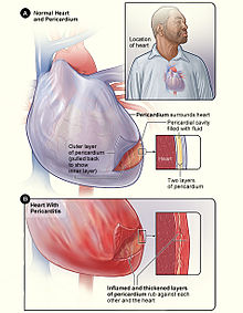
Constrictive pericarditis is a medical condition characterized by a thickened, fibrotic pericardium, limiting the heart's ability to function normally. In many cases, the condition continues to be difficult to diagnose and therefore benefits from a good understanding of the underlying cause.

<span class="mw-page-title-main">Pericarditis</span> Medical condition

Pericarditis is inflammation of the pericardium, the fibrous sac surrounding the heart. Symptoms typically include sudden onset of sharp chest pain, which may also be felt in the shoulders, neck, or back. The pain is typically less severe when sitting up and more severe when lying down or breathing deeply. Other symptoms of pericarditis can include fever, weakness, palpitations, and shortness of breath. The onset of symptoms can occasionally be gradual rather than sudden.

<span class="mw-page-title-main">Pericardiocentesis</span> Procedure where fluid is aspirated from the pericardium

Pericardiocentesis (PCC), also called pericardial tap, is a medical procedure where fluid is aspirated from the pericardium.

Kussmaul's sign is a paradoxical rise in jugular venous pressure (JVP) on inspiration, or a failure in the appropriate fall of the JVP with inspiration. It can be seen in some forms of heart disease and is usually indicative of limited right ventricular filling due to right heart dysfunction.

Dressler syndrome is a secondary form of pericarditis that occurs in the setting of injury to the heart or the pericardium. It consists of fever, pleuritic pain, pericarditis and/or pericardial effusion.

<span class="mw-page-title-main">Mediastinum</span> Central part of the thoracic cavity

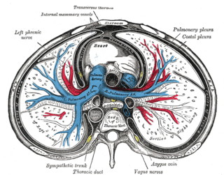
The mediastinum is the central compartment of the thoracic cavity. Surrounded by loose connective tissue, it is an undelineated region that contains a group of structures within the thorax, namely the heart and its vessels, the esophagus, the trachea, the phrenic and cardiac nerves, the thoracic duct, the thymus and the lymph nodes of the central chest.

<span class="mw-page-title-main">Serous membrane</span> Smooth coating lining contents & inner walls of body cavities

The serous membrane is a smooth tissue membrane of mesothelium lining the contents and inner walls of body cavities, which secrete serous fluid to allow lubricated sliding movements between opposing surfaces. The serous membrane that covers internal organs is called a visceral membrane; while the one that covers the cavity wall is called the parietal membrane. Between the two opposing serosal surfaces is often a potential space, mostly empty except for the small amount of serous fluid.

Pulsus paradoxus, also paradoxic pulse or paradoxical pulse, is an abnormally large decrease in stroke volume, systolic blood pressure and pulse wave amplitude during inspiration. Pulsus paradoxus is not related to pulse rate or heart rate, and it is not a paradoxical rise in systolic pressure. Normally, blood pressure drops less precipitously than 10 mmHg during inhalation. Pulsus paradoxus is a sign that is indicative of several conditions most commonly pericardial effusion.

<span class="mw-page-title-main">Pericardial effusion</span> Medical condition

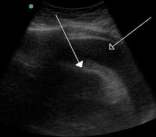
A pericardial effusion is an abnormal accumulation of fluid in the pericardial cavity. The pericardium is a two-part membrane surrounding the heart: the outer fibrous connective membrane and an inner two-layered serous membrane. The two layers of the serous membrane enclose the pericardial cavity between them. This pericardial space contains a small amount of pericardial fluid, normally 15-50 mL in volume. The pericardium, specifically the pericardial fluid provides lubrication, maintains the anatomic position of the heart in the chest, and also serves as a barrier to protect the heart from infection and inflammation in adjacent tissues and organs.

<span class="mw-page-title-main">Pericardial fluid</span>

Pericardial fluid is the serous fluid secreted by the serous layer of the pericardium into the pericardial cavity. The pericardium consists of two layers, an outer fibrous layer and the inner serous layer. This serous layer has two membranes which enclose the pericardial cavity into which is secreted the pericardial fluid. The fluid is similar to the cerebrospinal fluid of the brain which also serves to cushion and allow some movement of the organ.

<span class="mw-page-title-main">Acute pericarditis</span> Medical condition

Acute pericarditis is a type of pericarditis usually lasting less than 6 weeks. It is the most common condition affecting the pericardium.

<span class="mw-page-title-main">Electrical alternans</span> Medical condition

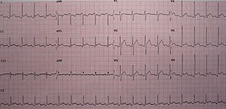
Electrical alternans is an electrocardiographic phenomenon of alternation of QRS complex amplitude or axis between beats and a possible wandering base-line. It is seen in cardiac tamponade and severe pericardial effusion and is thought to be related to changes in the ventricular electrical axis due to fluid in the pericardium, as the heart essentially wobbles in the fluid filled pericardial sac.

Pericardiectomy is the surgical removal of part or most of the pericardium. This operation is most commonly used to relieve constrictive pericarditis, or to remove a pericardium that is calcified and fibrous. It may also be used for severe or recurrent cases of pericardial effusion. Post-operative outcomes and mortality are significantly impacted by the disease it is used to treat.

The following outline is provided as an overview of and topical guide to cardiology, the branch of medicine dealing with disorders of the human heart. The field includes medical diagnosis and treatment of congenital heart defects, coronary artery disease, heart failure, valvular heart disease and electrophysiology. Physicians who specialize in cardiology are called cardiologists.

Obstructive shock is one of the four types of shock, caused by a physical obstruction in the flow of blood. Obstruction can occur at the level of the great vessels or the heart itself. Causes include pulmonary embolism, cardiac tamponade, and tension pneumothorax. These are all life-threatening. Symptoms may include shortness of breath, weakness, or altered mental status. Low blood pressure and tachycardia are often seen in shock. Other symptoms depend on the underlying cause.

<span class="mw-page-title-main">Hemopericardium</span> Medical condition

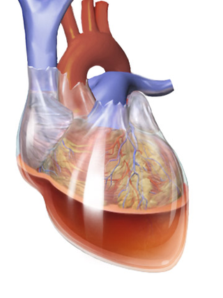
Hemopericardium refers to blood in the pericardial sac of the heart. It is clinically similar to a pericardial effusion, and, depending on the volume and rapidity with which it develops, may cause cardiac tamponade.

<span class="mw-page-title-main">Postpericardiotomy syndrome</span> Medical condition

Postpericardiotomy syndrome (PPS) is a medical syndrome referring to an immune phenomenon that occurs days to months after surgical incision of the pericardium. PPS can also be caused after a trauma, a puncture of the cardiac or pleural structures, after percutaneous coronary intervention, or due to pacemaker or pacemaker wire placement.

<span class="mw-page-title-main">Pulmonary pleurae</span> Serous membrane that lines the wall of the thoracic cavity and the surface of the lung

The pulmonary pleurae are the two opposing layers of serous membrane overlying the lungs and the inside of the surrounding chest walls.

References

  1. "Pericardiectomy". Johns Hopkins Medicine. 19 November 2019. Retrieved 20 Sep 2020.
  2. 1 2 Hegde, Sheila M. (2019-01-01), Solomon, Scott D.; Wu, Justina C.; Gillam, Linda D. (eds.), "33 - Pericardial Disease", Essential Echocardiography, Elsevier, pp. 347–353.e1, doi:10.1016/b978-0-323-39226-6.00033-3, ISBN   978-0-323-39226-6 , retrieved 2020-11-17
  3. Mou, Steven S.; McCrory, Michael C. (2019-01-01), Ungerleider, Ross M.; Meliones, Jon N.; Nelson McMillan, Kristen; Cooper, David S. (eds.), "28 - Inflammatory Heart Disease: Pericardial Effusion and Tamponade, Pericarditis, and Myocarditis", Critical Heart Disease in Infants and Children (Third Edition), Philadelphia: Elsevier, pp. 351–364.e5, doi:10.1016/b978-1-4557-0760-7.00028-0, ISBN   978-1-4557-0760-7, S2CID   80814860 , retrieved 2020-11-17
  4. Dorland's (2012). Dorland's Illustrated Medical Dictionary (32nd ed.). Elsevier Saunders. p. 1412. ISBN   978-1-4160-6257-8.
  5. Tortora, Gerard J.; Nielsen, Mark T. (2009). Principles of Human Anatomy (11th ed.). John Wiley & Sons. pp.  84–5. ISBN   978-0-471-78931-4.
  6. Rehman, Ibraheem; Nassereddin, Ali; Rehman, Afzal (2021), "Anatomy, Thorax, Pericardium", StatPearls, Treasure Island (FL): StatPearls Publishing, PMID   29489245 , retrieved 2021-09-12
  7. Winieski, Lawrence E. Snell's Clinical Anatomy By Regions. Wolters Kluwer. p. 241.
  8. 1 2 Laizzo, P.A. (2009). Handbook of Cardiac Anatomy, Physiology, and Devices (2nd ed.). Humana Press. pp. 125–8. ISBN   978-1-60327-371-8.
  9. 1 2 Dixon, Andrew; Hacking, Craig. "Pericardial recesses". Radiopaedia . Retrieved 2019-07-08.
  10. Byrne, John G; Karavas, Alexandros N; Colson, Yolonda L; Bueno, Raphael; Richards, William G; Sugarbaker, David J; Goldhaber, Samuel Z (2002). "Cardiac Decortication (Epicardiectomy) for Occult Constrictive Cardiac Physiology After Left Extrapleural Pneumonectomy". Chest. 122 (6): 2256–9. doi:10.1378/chest.122.6.2256. PMID   12475875.
  11. Davidson's 2010, pp. 638–639.
  12. Kligerman, Seth (January 2019). "Imaging of Pericardial Disease". Radiologic Clinics of North America. 57 (1): 179–199. doi:10.1016/j.rcl.2018.09.001. PMID   30454812. S2CID   53872634.
  13. Image by Mikael Häggström, MD. Sources for mentioned features:
    - "Mesothelial cytopathology". Libre Pathology. Retrieved 2022-10-18.
    - Shidham VB, Layfield LJ (2021). "Introduction to the second edition of 'Diagnostic Cytopathology of Serous Fluids' as CytoJournal Monograph (CMAS) in Open Access". CytoJournal. 18: 30. doi:10.25259/CMAS_02_01_2021. PMC   8813611 . PMID   35126608.