Pulmonary artery

Last updated
Pulmonary artery
Diagram of the human heart (cropped).svg
Anterior (frontal) view of the opened heart. White arrows indicate normal blood flow. (Pulmonary artery labelled at upper right.)
Details
Precursor Truncus arteriosus
System Cardiovascular, respiratory
Source Right ventricle
Identifiers
Latin arteria pulmonalis
MeSH D011651
TA98 A12.2.01.101
A12.2.01.201
TA2 4077, 4091
FMA 66326
Anatomical terminology

A pulmonary artery is an artery in the pulmonary circulation that carries deoxygenated blood from the right side of the heart to the lungs. The largest pulmonary artery is the main pulmonary artery or pulmonary trunk from the heart, and the smallest ones are the arterioles, which lead to the capillaries that surround the pulmonary alveoli.

Contents

Structure

The pulmonary arteries are blood vessels that carry systemic venous blood from the right ventricle of the heart to the microcirculation of the lungs. Unlike in other organs where arteries supply oxygenated blood, the blood carried by the pulmonary arteries is deoxygenated, as it is venous blood returning to the heart. The main pulmonary arteries emerge from the right side of the heart and then split into smaller arteries that progressively divide and become arterioles, eventually narrowing into the capillary microcirculation of the lungs where gas exchange occurs.[ citation needed ]

Pulmonary trunk

Volume rendering of a high resolution CT scan of the thorax. The anterior thoracic wall, the airways and the pulmonary vessels anterior to the root of the lung have been digitally removed to visualize the different levels of the pulmonary circulation. 3D CT of thorax, annotated.jpg
Volume rendering of a high resolution CT scan of the thorax. The anterior thoracic wall, the airways and the pulmonary vessels anterior to the root of the lung have been digitally removed to visualize the different levels of the pulmonary circulation.

In order of blood flow, the pulmonary arteries start as the pulmonary trunk that leaves the fibrous pericardium (parietal pericardium) of the ventricular outflow tract of right ventricle (also known as infundibulum or conus arteriosus. [1] The outflow track runs superiorly and to the left, posterior to the pulmonary valve. [1] The pulmonary trunk bifurcates into right and left pulmonary arteries below the arch of aorta and in front of the left main bronchus. [1] Pulmonary trunk is short and wide approximately 5 centimetres (2.0 in) in length [2] and 2 centimetres (0.79 in)-3 centimetres (1.2 in) in diameter. [3] [4]

The pulmonary trunk splits into the right and the left main pulmonary artery. [5] The left main pulmonary artery is shorter than the right, [1] passes behind and downwards the descending aorta and above the left main bronchus to the root of the left lung. Above, the left main pulmonary artery is connected to the concavity of the proximal descending aorta by the ligamentum arteriosum. [2] The right pulmonary artery pass across the midline of the body, below the carina of trachea, and comes in front of the right main bronchus. [1]

Branches

At the far end, pulmonary arteries (labelled at the bottom) become capillaries at the pulmonary alveoli. Alveolus diagram.svg
At the far end, pulmonary arteries (labelled at the bottom) become capillaries at the pulmonary alveoli.

The left main pulmonary artery then divides into two lobar arteries, one for each lobe of the left lung. [6]

At the right root of the lung, it bifurcates into artery that supplies the right upper lobe of the lung, in front of the right upper lobe bronchus, and interlobar artery that supplies the right middle and inferior lobes of the lung, running together with bronchus intermedius. [1]

The right and left main pulmonary (lungs) arteries give off branches that supplies the corresponding lung lobes. In such cases it is termed lobar arteries. [7] The lobar arteries branch into segmental arteries (roughly 1 for each segment). Segmental arteries run together with segmental bronchi, at the posterolateral surfaces of the bronchi. [7] These in turn branch into subsegmental pulmonary arteries. [7] These eventually form intralobular arteries. [8] The pulmonary arteries supply the alveoli of the lungs. In contrast, bronchial arteries, that has different origins, supply the bronchi of the lungs. [1]

Development

The pulmonary arteries originate from the truncus arteriosus and the sixth pharyngeal arch. The truncus arteriosus is a structure that forms during the development of the heart as a successor to the conus arteriosus. [9] :157

By the third week of development, the endocardial tubes have developed a swelling in the part closest to the heart. The swelling is known as the bulbus cordis and the upper part of this swelling develops into the truncus arteriosus. [9] :159–160 The structure is ultimately mesodermal in origin. [9] :157 During development of the heart, the heart tissues undergo folding, and the truncus arteriosus is exposed to what will eventually be both the left and right ventricles. As a septum develops between the two ventricles of the heart, two bulges form on either side of the truncus arteriosus. These progressively enlarge until the trunk splits into the aorta and pulmonary arteries. [9] :176–179

During early development, the ductus arteriosus connects the pulmonary trunk and the aortic arch, allowing blood to bypass the lungs. [10] :791

Function

The pulmonary artery carries deoxygenated blood from the right ventricle to the lungs. [11] The blood here passes through capillaries adjacent to alveoli and becomes oxygenated as part of the process of respiration. [12]

In contrast to the pulmonary arteries, the bronchial arteries supply nutrition to the lungs themselves. [10] :790

Pressure

The pulmonary artery pressure (PA pressure) is a measure of the blood pressure found in the main pulmonary artery. This is measured by inserting a catheter into the main pulmonary artery. [13] :190–191 The mean pressure is typically 9–18 mmHg, [14] and the wedge pressure measured in the left atrium may be 6–12 mmHg. The wedge pressure may be elevated in left heart failure, [13] :190–191 mitral valve stenosis, and other conditions, such as sickle cell disease. [15]

Clinical significance

The pulmonary artery is relevant in a number of clinical states. Pulmonary hypertension is used to describe an increase in the pressure of the pulmonary artery, and may be defined as a mean pulmonary artery pressure of greater than 25 mmHg. [13] :720 A pulmonary artery diameter of more than 29 mm (measured on a CT scan) is often used as an indicator for pulmonary hypertension. [16] In chest X-rays, a diameter of more than 16 mm for the right descending pulmonary artery is also an indicator for pulmonary hypertension. [17] This may occur as a result of heart problems such as heart failure, lung or airway disease such as COPD or scleroderma, or thromboembolic disease such as pulmonary embolism or emboli seen in sickle cell anaemia. [13] :720–721 Most recently, computational fluid based tools (non-invasive) have been proposed to be at par with the current clinical tests (invasive) of pulmonary hypertension. [18]

Pulmonary embolism refers to an embolus that lodges in the pulmonary circulation. This may arise from a deep venous thrombosis, especially after a period of immobility. A pulmonary embolus is a common cause of death in patients with cancer and stroke. [13] :720–721 A large pulmonary embolus that becomes lodged in the bifurcation of the pulmonary trunk with extensions into both the left and right main pulmonary arteries is called a saddle embolus. [19]

Several animal models have been utilized for investigating pulmonary artery related pathologies. Porcine model of pulmonary artery is the most frequently used and it was recently found that their mechanical properties vary with every subsequent branching. [20]

Additional images

See also

Related Research Articles

<span class="mw-page-title-main">Aorta</span> Largest artery in the human body

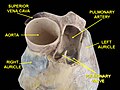
The aorta is the main and largest artery in the human body, originating from the left ventricle of the heart, branching upwards immediately after, and extending down to the abdomen, where it splits at the aortic bifurcation into two smaller arteries. The aorta distributes oxygenated blood to all parts of the body through the systemic circulation.

<span class="mw-page-title-main">Circulatory system</span> Organ system for circulating blood in animals

The circulatory system is a system of organs that includes the heart, blood vessels, and blood which is circulated throughout the entire body of a human or other vertebrate. It includes the cardiovascular system, or vascular system, that consists of the heart and blood vessels. The circulatory system has two divisions, a systemic circulation or circuit, and a pulmonary circulation or circuit. Some sources use the terms cardiovascular system and vascular system interchangeably with circulatory system.

<span class="mw-page-title-main">Coronary circulation</span> Circulation of blood in the blood vessels of the heart muscle (myocardium)

Coronary circulation is the circulation of blood in the arteries and veins that supply the heart muscle (myocardium). Coronary arteries supply oxygenated blood to the heart muscle. Cardiac veins then drain away the blood after it has been deoxygenated. Because the rest of the body, and most especially the brain, needs a steady supply of oxygenated blood that is free of all but the slightest interruptions, the heart is required to function continuously. Therefore its circulation is of major importance not only to its own tissues but to the entire body and even the level of consciousness of the brain from moment to moment. Interruptions of coronary circulation quickly cause heart attacks, in which the heart muscle is damaged by oxygen starvation. Such interruptions are usually caused by coronary ischemia linked to coronary artery disease, and sometimes to embolism from other causes like obstruction in blood flow through vessels.

<span class="mw-page-title-main">Heart valve</span> A flap of tissue that prevent backflow of blood around the heart

A heart valve is a biological one-way valve that allows blood to flow in one direction through the chambers of the heart. Four valves are usually present in a mammalian heart and together they determine the pathway of blood flow through the heart. A heart valve opens or closes according to differential blood pressure on each side.

<span class="mw-page-title-main">Ventricle (heart)</span> Chamber of the heart

A ventricle is one of two large chambers located toward the bottom of the heart that collect and expel blood towards the peripheral beds within the body and lungs. The blood pumped by a ventricle is supplied by an atrium, an adjacent chamber in the upper heart that is smaller than a ventricle. Interventricular means between the ventricles, while intraventricular means within one ventricle.

<span class="mw-page-title-main">Bronchus</span> Airway in the respiratory tract

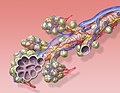
A bronchus is a passage or airway in the lower respiratory tract that conducts air into the lungs. The first or primary bronchi to branch from the trachea at the carina are the right main bronchus and the left main bronchus. These are the widest bronchi, and enter the right lung, and the left lung at each hilum. The main bronchi branch into narrower secondary bronchi or lobar bronchi, and these branch into narrower tertiary bronchi or segmental bronchi. Further divisions of the segmental bronchi are known as 4th order, 5th order, and 6th order segmental bronchi, or grouped together as subsegmental bronchi. The bronchi, when too narrow to be supported by cartilage, are known as bronchioles. No gas exchange takes place in the bronchi.

<span class="mw-page-title-main">Pulmonary circulation</span> Part of the circulatory system which carries blood from heart to lungs and back to the heart

The pulmonary circulation is a division of the circulatory system in all vertebrates. The circuit begins with deoxygenated blood returned from the body to the right atrium of the heart where it is pumped out from the right ventricle to the lungs. In the lungs the blood is oxygenated and returned to the left atrium to complete the circuit.

<span class="mw-page-title-main">Pulmonary vein</span> Veins that transfer oxygenated blood from the lungs to the heart

The pulmonary veins are the veins that transfer oxygenated blood from the lungs to the heart. The largest pulmonary veins are the four main pulmonary veins, two from each lung that drain into the left atrium of the heart. The pulmonary veins are part of the pulmonary circulation.

<span class="mw-page-title-main">Ductus arteriosus</span> Blood vessel connecting the pulmonary artery to the proximal descending aorta

The ductus arteriosus, also called the ductus Botalli, named after the Italian physiologist Leonardo Botallo, is a blood vessel in the developing fetus connecting the trunk of the pulmonary artery to the proximal descending aorta. It allows most of the blood from the right ventricle to bypass the fetus's fluid-filled non-functioning lungs. Upon closure at birth, it becomes the ligamentum arteriosum.

<span class="mw-page-title-main">Transposition of the great vessels</span> Group of congenital heart defects

Transposition of the great vessels (TGV) is a group of congenital heart defects involving an abnormal spatial arrangement of any of the great vessels: superior and/or inferior venae cavae, pulmonary artery, pulmonary veins, and aorta. Congenital heart diseases involving only the primary arteries belong to a sub-group called transposition of the great arteries (TGA), which is considered the most common congenital heart lesion that presents in neonates.

<span class="mw-page-title-main">Bronchial artery</span> Blood vessels supplying the lungs

In human anatomy, the bronchial arteries supply the lungs with oxygenated blood, and nutrition. Although there is much variation, there are usually two bronchial arteries that run to the left lung, and one to the right lung, and are a vital part of the respiratory system.

<span class="mw-page-title-main">Persistent truncus arteriosus</span> Medical condition

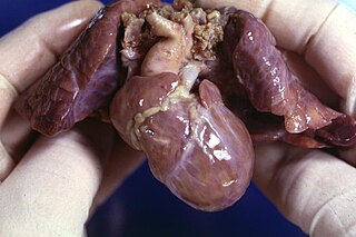
Persistent truncus arteriosus (PTA), often referred to simply as truncus arteriosus, is a rare form of congenital heart disease that presents at birth. In this condition, the embryological structure known as the truncus arteriosus fails to properly divide into the pulmonary trunk and aorta. This results in one arterial trunk arising from the heart and providing mixed blood to the coronary arteries, pulmonary arteries, and systemic circulation. For the International Classification of Diseases (ICD-11), the International Paediatric and Congenital Cardiac Code (IPCCC) was developed to standardize the nomenclature of congenital heart disease. Under this system, English is now the official language, and persistent truncus arteriosus should properly be termed common arterial trunk.

<span class="mw-page-title-main">Fetal circulation</span> Circulatory system of fetuses

In humans, the circulatory system is different before and after birth. The fetal circulation is composed of the placenta, umbilical blood vessels encapsulated by the umbilical cord, heart and systemic blood vessels. A major difference between the fetal circulation and postnatal circulation is that the lungs are not used during the fetal stage resulting in the presence of shunts to move oxygenated blood and nutrients from the placenta to the fetal tissue. At birth, the start of breathing and the severance of the umbilical cord prompt various changes that quickly transform fetal circulation into postnatal circulation.

<span class="mw-page-title-main">Thoracic aorta</span> Part of the aorta located in the thorax

The thoracic aorta is a part of the aorta located in the thorax. It is a continuation of the aortic arch. It is located within the posterior mediastinal cavity, but frequently bulges into the left pleural cavity. The descending thoracic aorta begins at the lower border of the fourth thoracic vertebra and ends in front of the lower border of the twelfth thoracic vertebra, at the aortic hiatus in the diaphragm where it becomes the abdominal aorta.

<span class="mw-page-title-main">Aortic arch</span> Part of the aorta

The aortic arch, arch of the aorta, or transverse aortic arch is the part of the aorta between the ascending and descending aorta. The arch travels backward, so that it ultimately runs to the left of the trachea.

A right-to-left shunt is a cardiac shunt which allows blood to flow from the right heart to the left heart. This terminology is used both for the abnormal state in humans and for normal physiological shunts in reptiles.

<span class="mw-page-title-main">Truncus arteriosus</span> Structure during embryonic development

The truncus arteriosus is a structure that is present during embryonic development. It is an arterial trunk that originates from both ventricles of the heart that later divides into the aorta and the pulmonary trunk.

<span class="mw-page-title-main">Root of the lung</span> Anatomical structure

The root of the lung is a group of structures that emerge at the hilum of each lung, just above the middle of the mediastinal surface and behind the cardiac impression of the lung. It is nearer to the back than the front. The root of the lung is connected by the structures that form it to the heart and the trachea. The rib cage is separated from the lung by a two-layered membranous coating, the pleura. The hilum is the large triangular depression where the connection between the parietal pleura and the visceral pleura is made, and this marks the meeting point between the mediastinum and the pleural cavities.

<span class="mw-page-title-main">Hypoplastic right heart syndrome</span> Type of congenital heart disease

Hypoplastic right heart syndrome or HRHS is a congenital heart defect in which the structures on the right side of the heart, particularly the right ventricle, are underdeveloped. This defect causes inadequate blood flow to the lungs, and thus a cyanotic infant.

<span class="mw-page-title-main">Heart development</span> Prenatal development of the heart

Heart development, also known as cardiogenesis, refers to the prenatal development of the heart. This begins with the formation of two endocardial tubes which merge to form the tubular heart, also called the primitive heart tube. The heart is the first functional organ in vertebrate embryos.

References

  1. 1 2 3 4 5 6 7 Ryan, Stephanie (2011). "2". Anatomy for diagnostic imaging (Third ed.). Elsevier Ltd. pp. 126, 133. ISBN   9780702029714.
  2. 1 2 Cheitlin MD, Ursell PC (2011). "Cardiac Anatomy". In Chatterjee K (ed.). Cardiology: An Illustrated Textbook. JP Medical Ltd. p. 6. ISBN   9789350252758.
  3. Edwards, P D; Bull, R K; Coulden, R (1998-10-01). "CT measurement of main pulmonary artery diameter". The British Journal of Radiology. 71 (850): 1018–1020. doi:10.1259/bjr.71.850.10211060. ISSN   0007-1285. PMID   10211060.
  4. Truong, Quynh A.; Massaro, Joseph M.; Rogers, Ian S.; Mahabadi, Amir A.; Kriegel, Matthias F.; Fox, Caroline S.; O'Donnell, Christopher J.; Hoffmann, Udo (2012-01-01). "Reference Values for Normal Pulmonary Artery Dimensions by Noncontrast Cardiac Computed Tomography". Circulation: Cardiovascular Imaging. 5 (1): 147–154. doi:10.1161/CIRCIMAGING.111.968610. PMC   3275437 . PMID   22178898.
  5. "Pulmonary Vasculature". University of Virginia School of Medicine . 2013. Retrieved 2017-06-24.
  6. Kandathil, Asha; Chamarthy, Murthy (June 2018). "Pulmonary vascular anatomy & anatomical variants". Cardiovascular Diagnosis and Therapy. 8 (3): 201–207. doi: 10.21037/cdt.2018.01.04 . ISSN   2223-3652. PMC   6039811 . PMID   30057869.
  7. 1 2 3 "Pulmonary Artery Anatomy". University of Virginia School of Medicine . 2013. Retrieved 2017-06-24.
  8. Takahashi M, Fukuoka J, Nitta N, Takazakura R, Nagatani Y, Murakami Y, et al. (2008). "Imaging of pulmonary emphysema: a pictorial review". International Journal of Chronic Obstructive Pulmonary Disease. 3 (2): 193–204. doi: 10.2147/COPD.S2639 . PMC   2629965 . PMID   18686729.
  9. 1 2 3 4 Schoenwolf GC, Larsen MJ, Bleyl SR, Brauer PR, Francis-West PH (2009). Larsen's human embryology (4th ed., Thoroughly rev. and updated. ed.). Philadelphia: Churchill Livingstone/Elsevier. pp. Development of the Urogenital system. ISBN   9780443068119.
  10. 1 2 Braunwald E (1992). Heart Disease: A Textbook of Cardiovascular Medicine (Fourth ed.). Philadelphia: W.B. Sanders.
  11. "22.4 Gas Exchange – Anatomy and Physiology". opentextbc.ca. Archived from the original on 2020-10-19. Retrieved 2019-05-22.
  12. "Exchanging Oxygen and Carbon Dioxide – Lung and Airway Disorders". MSD Manual Consumer Version. Retrieved 2019-05-22.
  13. 1 2 3 4 5 Colledge NR, Walker BR, Ralston SH, Britton R, eds. (2010). Davidson's Principles and Practice of Medicine (21st ed.). Edinburgh: Churchill Livingstone/Elsevier. ISBN   978-0-7020-3084-0.
  14. "Normal Hemodynamic Parameters – Adult" (PDF). Edwards Lifesciences LLC. Archived from the original (PDF) on 2010-11-10.
  15. Pashankar FD, Carbonella J, Bazzy-Asaad A, Friedman A (April 2008). "Prevalence and risk factors of elevated pulmonary artery pressures in children with sickle cell disease". Pediatrics. 121 (4): 777–782. doi:10.1542/peds.2007-0730. PMID   18381543. S2CID   26693444.
  16. Marini TJ, He K, Hobbs SK, Kaproth-Joslin K (December 2018). "Pictorial review of the pulmonary vasculature: from arteries to veins". Insights into Imaging. 9 (6): 971–987. doi:10.1007/s13244-018-0659-5. PMC   6269336 . PMID   30382495.
  17. Chang CH (December 1965). "The normal roentgenographic measurement of the right descending pulmonary artery in 1,085 cases and its clinical application. II. Clinical application of the measurement of the right descending pulmonary artery in the radiological diagnosis of pulmonary hypertensions from various causes" (PDF). Nagoya Journal of Medical Science. 28 (1): 67–80. PMID   5865788 . Retrieved 15 January 2022.
  18. Piskin S, Patnaik SS, Han D, Bordones AD, Murali S, Finol EA (March 2020). "A canonical correlation analysis of the relationship between clinical attributes and patient-specific hemodynamic indices in adult pulmonary hypertension". Medical Engineering & Physics. 77: 1–9. doi:10.1016/j.medengphy.2020.01.006. PMC   7069525 . PMID   32007361.
  19. Jones J, et al. "Saddle pulmonary embolism". Radiopaedia . Retrieved 2017-10-08.
  20. Pillalamarri NR, Patnaik SS, Piskin S, Gueldner P, Finol EA (January 2021). "Ex Vivo Regional Mechanical Characterization of Porcine Pulmonary Arteries". Experimental Mechanics. 61 (1): 285–303. doi:10.1007/s11340-020-00678-2. PMC   8011683 . PMID   33814554.