Related Research Articles

The trachea, also known as the windpipe, is a cartilaginous tube that connects the larynx to the bronchi of the lungs, allowing the passage of air, and so is present in almost all air-breathing animals with lungs. The trachea extends from the larynx and branches into the two primary bronchi. At the top of the trachea the cricoid cartilage attaches it to the larynx. The trachea is formed by a number of horseshoe-shaped rings, joined together vertically by overlying ligaments, and by the trachealis muscle at their ends. The epiglottis closes the opening to the larynx during swallowing.

Tetralogy of Fallot (TOF) is a congenital heart defect. Symptoms at birth may vary from none to severe. Later, there are typically episodes of bluish color to the skin known as cyanosis. When affected babies cry or have a bowel movement, they may develop a "tet spell" where they turn very blue, have difficulty breathing, become limp, and occasionally lose consciousness. Other symptoms may include a heart murmur, finger clubbing, and easy tiring upon breastfeeding.

A bronchus is a passage or airway in the respiratory system that conducts air into the lungs. The first bronchi to branch from the trachea are the right main bronchus and the left main bronchus, also known as the primary bronchi. These are the widest and enter the lungs at each hilum, where they branch into narrower secondary bronchi or lobar bronchi, and these branch into narrower tertiary bronchi or segmental bronchi. Further divisions of the segmental bronchi are known as 4th order, 5th order, and 6th order segmental bronchi, or grouped together as subsegmental bronchi. The bronchi when too narrow to be supported by cartilage are known as bronchioles. No gas exchange takes place in the bronchi.

Patent ductus arteriosus (PDA) is a medical condition in which the ductus arteriosus fails to close after birth: this allows a portion of oxygenated blood from the left heart to flow back to the lungs by flowing from the aorta, which has a higher pressure, to the pulmonary artery. Symptoms are uncommon at birth and shortly thereafter, but later in the first year of life there is often the onset of an increased work of breathing and failure to gain weight at a normal rate. With time, an uncorrected PDA usually leads to pulmonary hypertension followed by right-sided heart failure.

The Blalock–Thomas–Taussig shunt is a surgical procedure used to increase blood flow to the lungs in some forms of congenital heart disease. These conditions, in which a child is born with an abnormal heart include pulmonary atresia and Tetralogy of Fallot and are common causes of blue baby syndrome. The procedure involves connecting a branch of the subclavian artery or carotid artery to the pulmonary artery. In modern practice, this procedure is temporarily used to direct blood flow to the lungs and relieve cyanosis while the infant is waiting for corrective or definitive surgery. The Blalock–Thomas-Taussig shunt is used in the first step of the three stage palliation.

Cardiac catheterization is the insertion of a catheter into a chamber or vessel of the heart. This is done both for diagnostic and interventional purposes.

Transposition of the great vessels (TGV) is a group of congenital heart defects involving an abnormal spatial arrangement of any of the great vessels: superior and/or inferior venae cavae, pulmonary artery, pulmonary veins, and aorta. Congenital heart diseases involving only the primary arteries belong to a sub-group called transposition of the great arteries (TGA), which is considered the most common congenital heart lesion that presents in neonates.

Pulmonary atresia is a congenital malformation of the pulmonary valve in which the valve orifice fails to develop. The valve is completely closed thereby obstructing the outflow of blood from the heart to the lungs. The pulmonary valve is located on the right side of the heart between the right ventricle and pulmonary artery. In a normal functioning heart, the opening to the pulmonary valve has three flaps that open and close
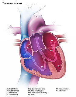
Persistent truncus arteriosus (PTA), often referred to simply as Truncus Arteriosus, is a rare form of congenital heart disease that presents at birth. In this condition, the embryological structure known as the truncus arteriosus fails to properly divide into the pulmonary trunk and aorta. This results in one arterial trunk arising from the heart and providing mixed blood to the coronary arteries, pulmonary arteries, and systemic circulation. For the International Classification of Diseases (ICD-11), the International Paediatric and Congenital Cardiac Code (IPCCC) was developed to standardize the nomenclature of congenital heart disease. Under this system, English is now the official language, and persistent truncus arteriosus should properly be termed Common arterial trunk.

Carotid endarterectomy (CEA) is a surgical procedure used to reduce the risk of stroke from carotid artery stenosis.

Carotid artery stenosis is a narrowing or constriction of any part of the carotid arteries, usually caused by atherosclerosis.
Levo-Transposition of the great arteries is an acyanotic congenital heart defect in which the primary arteries are transposed, with the aorta anterior and to the left of the pulmonary artery; the morphological left and right ventricles with their corresponding atrioventricular valves are also transposed.

The Norwood procedure is the first surgery of three staged heart surgeries to create a new functional systemic circuit in patients with hypoplastic left heart syndrome or other complex heart defects with single ventricle physiology. The Norwood procedure involves atrial septectomy and transection and ligation of the distal main pulmonary artery. The proximal pulmonary artery is then connected to the hypoplastic aortic arch, while the coarcted segment of the aorta is repaired. An aortopulmonary shunt is created to connect the aorta to the main pulmonary artery to provide pulmonary blood flow. The second surgery is the separation of the systemic and pulmonary circulation once pulmonary vascular resistance has fallen, by removing the aortopulmonary shunt followed by the creation of a bidirectional SVC-pulmonary shunt, also known as a modified Glenn procedure or Hemi-Fontan. The third surgery is the Fontan procedure, in which the inferior vena cava is connected to the branch pulmonary arteries. After this surgery is completed, all the venous blood returning from the body flows directly to the lungs.

Aberrant subclavian artery, or aberrant subclavian artery syndrome, is a rare anatomical variant of the origin of the right or left subclavian artery. This abnormality is the most common congenital vascular anomaly of the aortic arch, occurring in approximately 1% of individuals.
A vascular ring is a congenital defect in which there is an abnormal formation of the aorta and/or its surrounding blood vessels. The trachea and esophagus are completely encircled and sometimes compressed by a "ring" formed by these vessels, which can lead to breathing and digestive difficulties.

Lutembacher's syndrome is a very rare form of congenital heart disease that affects one of the chambers of the heart as well as a valve. It is commonly known as both congenital atrial septal defect (ASD) and acquired mitral stenosis (MS). Congenital atrial septal defect refers to a hole being in the septum or wall that separates the two atria; this condition is usually seen in fetuses and infants. Mitral stenosis refers to mitral valve leaflets sticking to each other making the opening for blood to pass from the atrium to the ventricles very small. With the valve being so small, blood has difficulty passing from the left atrium into the left ventricle. Septal defects that may occur with Lutembacher's syndrome include: Ostium primum atrial septal defect or ostium secundum which is more prevalent.
Hypoplastic right heart syndrome is a congenital heart defect in which the right atrium and right ventricle are underdeveloped. This defect causes inadequate blood flow to the lungs and thus, a blue or cyanotic infant.
Double aortic arch is a relatively rare congenital cardiovascular malformation. DAA is an anomaly of the aortic arch in which two aortic arches form a complete vascular ring that can compress the trachea and/or esophagus. Most commonly there is a larger (dominant) right arch behind and a smaller (hypoplastic) left aortic arch in front of the trachea/esophagus. The two arches join to form the descending aorta which is usually on the left side. In some cases the end of the smaller left aortic arch closes and the vascular tissue becomes a fibrous cord. Although in these cases a complete ring of two patent aortic arches is not present, the term ‘vascular ring’ is the accepted generic term even in these anomalies.

Anomalous aortic origin of a coronary artery (AAOCA) is a rare congenital heart defect in which a coronary artery inappropriately arises from the aorta, usually from the incorrect sinus of Valsalva. This anomalous coronary artery often takes an interarterial, intraconal, or intramural course, and is associated with an increased risk of sudden death in children.

Right-sided aortic arch is a rare anatomical variant in which the aortic arch is on the right side rather than on the left. During normal embryonic development, the aortic arch is formed by the left fourth aortic arch and the left dorsal aorta. In people with a right-sided aortic arch, instead the right dorsal aorta persists and the distal left aorta disappears.
References
- ↑ "Pulmonary Artery Sling: Background, Pathophysiology, Epidemiology". 7 January 2017.Cite journal requires
|journal=(help) - 1 2 Backer, Carl L. (2020). "Vascular Rings and Pulmonary Artery Sling". In Raja, Shahzad G. (ed.). Cardiac Surgery: A Complete Guide. Springer. p. 981. ISBN 978-3-030-24174-2. OCLC 1142507832.