Related Research Articles

In mammals, pregnancy is the period of reproduction during which a female carries one or more live offspring from implantation in the uterus through gestation. It begins when a fertilized zygote implants in the female's uterus, and ends once it leaves the uterus.

In all bilaterian animals, the mesoderm is one of the three primary germ layers in the very early embryo. The other two layers are the ectoderm and endoderm, with the mesoderm as the middle layer between them.

Embryology is the branch of biology that studies the prenatal development of gametes, fertilization, and development of embryos and fetuses. Additionally, embryology encompasses the study of congenital disorders that occur before birth, known as teratology.
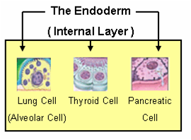
Endoderm is one of the three primary germ layers in the very early embryo. The other two layers are the ectoderm and mesoderm, with the endoderm being the innermost layer. Cells migrating inward along the archenteron form the inner layer of the gastrula, which develops into the endoderm.
The somites are a set of bilaterally paired blocks of paraxial mesoderm that form in the embryonic stage of somitogenesis, along the head-to-tail axis in segmented animals. In vertebrates, somites subdivide into the sclerotomes, myotomes, syndetomes and dermatomes that give rise to the vertebrae of the vertebral column, rib cage and part of the occipital bone; skeletal muscle, cartilage, tendons, and skin.
A germ layer is a primary layer of cells that forms during embryonic development. The three germ layers in vertebrates are particularly pronounced; however, all eumetazoans produce two or three primary germ layers. Some animals, like cnidarians, produce two germ layers making them diploblastic. Other animals such as chordates produce a third layer between these two layers, making them triploblastic. Germ layers eventually give rise to all of an animal’s tissues and organs through the process of organogenesis.

The gestational sac is the large cavity of fluid surrounding the embryo. During early embryogenesis it consists of the extraembryonic coelom, also called the chorionic cavity. The gestational sac is normally contained within the uterus. It is the only available structure that can be used to determine if an intrauterine pregnancy exists until the embryo is identified.

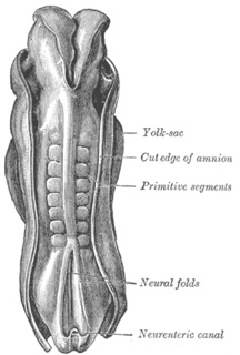
The yolk sac is a membranous sac attached to an embryo, formed by cells of the hypoblast adjacent to the embryonic disk. This is alternatively called the umbilical vesicle by the Terminologia Embryologica (TE), though yolk sac is far more widely used. In humans, the yolk sac is important in early embryonic blood supply, and much of it is incorporated into the primordial gut during the fourth week of development.

The neural plate is a key developmental structure that serves as the basis for the nervous system. Opposite the primitive streak in the embryo, ectodermal tissue thickens and flattens to become the neural plate. The region anterior to the primitive knot can be generally referred to as the neural plate. Cells take on a columnar appearance in the process as they continue to lengthen and narrow. The ends of the neural plate, known as the neural folds, push the ends of the plate up and together, folding into the neural tube, a structure critical to brain and spinal cord development. This process as a whole is termed primary neurulation.

A neurula is a vertebrate embryo at the early stage of development in which neurulation occurs. The neurula stage is preceded by the gastrula stage; consequentially, neurulation is preceded by gastrulation. Neurulation marks the beginning of the process of organogenesis.

In the embryonic development of vertebrates, pharyngeal pouches form on the endodermal side between the pharyngeal arches. The pharyngeal grooves form the lateral ectodermal surface of the neck region to separate the arches.

The vitelline veins are veins that drain blood from the yolk sac and the gut tube during gestation.

The dorsal aortae are paired embryological vessels which progress to form the descending aorta. The paired dorsal aortae arise from aortic arches that in turn arise from the aortic sac.
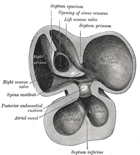
Endocardial cushions, or atrioventricular cushions, refer to a subset of cells in the development of the heart that play a vital role in the proper formation of the heart septa.

In amniote animal embryology, the epiblast is one of two distinct layers arising from the inner cell mass in the mammalian blastocyst or from the blastodisc in reptiles and birds. It derives the embryo proper through its differentiation into the three primary germ layers, ectoderm, mesoderm and endoderm, during gastrulation. The amnionic ectoderm and extraembryonic mesoderm also originate from the epiblast.
Bilaminar blastocyst or bilaminar disc refers to the epiblast and the hypoblast, evolved from the embryoblast. These two layers are sandwiched between two balloons: the primitive yolk sac and the amniotic cavity.
The heart is the first functional organ in a vertebrate embryo. There are 5 stages to heart development.

Human embryonic development, or human embryogenesis, refers to the development and formation of the human embryo. It is characterised by the processes of cell division and cellular differentiation of the embryo that occurs during the early stages of development. In biological terms, the development of the human body entails growth from a one-celled zygote to an adult human being. Fertilisation occurs when the sperm cell successfully enters and fuses with an egg cell (ovum). The genetic material of the sperm and egg then combine to form a single cell called a zygote and the germinal stage of development commences. Embryonic development in the human, covers the first eight weeks of development; at the beginning of the ninth week the embryo is termed a fetus. Human embryology is the study of this development during the first eight weeks after fertilisation. The normal period of gestation (pregnancy) is about nine months or 40 weeks.
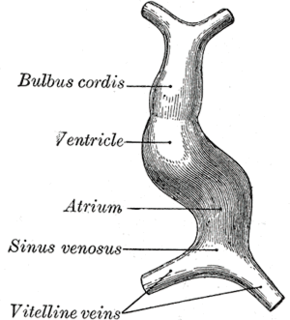
The tubular heart or primitive heart tube is the earliest stage of heart development.

Heart development refers to the prenatal development of the heart. This begins with the formation of two endocardial tubes which merge to form the tubular heart, also called the primitive heart tube. The heart is the first functional organ in vertebrate embryos, and in the human, beats spontaneously by week 4 of development.
References
- ↑ Kirby, Margaret L. (2007). Cardiac development. Oxford University Press. p. 119. ISBN 0-19-517819-X . Retrieved 20 April 2011.