Related Research Articles

The lymphatic system, or lymphoid system, is an organ system in vertebrates that is part of the immune system, and complementary to the circulatory system. It consists of a large network of lymphatic vessels, lymph nodes, lymphatic or lymphoid organs, and lymphoid tissues. The vessels carry a clear fluid called lymph back towards the heart, for re-circulation.

In human anatomy, the thoracic duct is the larger of the two lymph ducts of the lymphatic system. The thoracic duct usually begins from the upper aspect of the cisterna chyli, passing out of the abdomen through the aortic hiatus into first the posterior mediastinum and then the superior mediastinum, extending as high up as the root of the neck before descending to drain into the systemic (blood) circulation at the venous angle.
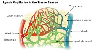
The lymphatic vessels are thin-walled vessels (tubes), structured like blood vessels, that carry lymph. As part of the lymphatic system, lymph vessels are complementary to the cardiovascular system. Lymph vessels are lined by endothelial cells, and have a thin layer of smooth muscle, and adventitia that binds the lymph vessels to the surrounding tissue. Lymph vessels are devoted to the propulsion of the lymph from the lymph capillaries, which are mainly concerned with the absorption of interstitial fluid from the tissues. Lymph capillaries are slightly bigger than their counterpart capillaries of the vascular system. Lymph vessels that carry lymph to a lymph node are called afferent lymph vessels, and those that carry it from a lymph node are called efferent lymph vessels, from where the lymph may travel to another lymph node, may be returned to a vein, or may travel to a larger lymph duct. Lymph ducts drain the lymph into one of the subclavian veins and thus return it to general circulation.

The subclavian vein is a paired large vein, one on either side of the body, that is responsible for draining blood from the upper extremities, allowing this blood to return to the heart. The left subclavian vein plays a key role in the absorption of lipids, by allowing products that have been carried by lymph in the thoracic duct to enter the bloodstream. The diameter of the subclavian veins is approximately 1–2 cm, depending on the individual.
The cisterna chyli or receptaculum chyli is a dilated sac at the lower end of the thoracic duct in most mammals into which lymph from the intestinal trunk and two lumbar lymphatic trunks flow. It receives fatty chyle from the intestines and thus acts as a conduit for the lipid products of digestion. It is the most common drainage trunk of most of the body's lymphatics. The cisterna chyli is a retroperitoneal structure.

Schlemm's canal is a circular lymphatic-like vessel in the eye. It collects aqueous humor from the anterior chamber and delivers it into the episcleral blood vessels. Canaloplasty may be used to widen it.

The midgut is the portion of the embryo from which most of the intestines develop. After it bends around the superior mesenteric artery, it is called the "midgut loop". It comprises the portion of the alimentary canal from the end of the foregut at the opening of the bile duct to the hindgut, about two-thirds of the way through the transverse colon.
Endothelial stem cells (ESCs) are one of three types of stem cells found in bone marrow. They are multipotent, which describes the ability to give rise to many cell types, whereas a pluripotent stem cell can give rise to all types. ESCs have the characteristic properties of a stem cell: self-renewal and differentiation. These parent stem cells, ESCs, give rise to progenitor cells, which are intermediate stem cells that lose potency. Progenitor stem cells are committed to differentiating along a particular cell developmental pathway. ESCs will eventually produce endothelial cells (ECs), which create the thin-walled endothelium that lines the inner surface of blood vessels and lymphatic vessels. The lymphatic vessels include things such as arteries and veins. Endothelial cells can be found throughout the whole vascular system and they also play a vital role in the movement of white blood cells

The right lymphatic duct is an important lymphatic vessel that drains the right upper quadrant of the body. It forms various combinations with the right subclavian vein and right internal jugular vein.

The jugular trunk is a lymphatic vessel in the neck. It is formed by vessels that emerge from the superior deep cervical lymph nodes and unite to efferents of the inferior deep cervical lymph nodes.

Human embryonic development, or human embryogenesis, is the development and formation of the human embryo. It is characterised by the processes of cell division and cellular differentiation of the embryo that occurs during the early stages of development. In biological terms, the development of the human body entails growth from a one-celled zygote to an adult human being. Fertilization occurs when the sperm cell successfully enters and fuses with an egg cell (ovum). The genetic material of the sperm and egg then combine to form the single cell zygote and the germinal stage of development commences. Embryonic development in the human, covers the first eight weeks of development; at the beginning of the ninth week the embryo is termed a fetus. The eight weeks has 23 stages.

C-fos-induced growth factor (FIGF) is a vascular endothelial growth factor that in humans is encoded by the FIGF gene.

Prospero homeobox protein 1 is a protein that in humans is encoded by the PROX1 gene. The Prox1 gene is critical for the development of multiple tissues. Prox1 activity is necessary and sufficient to specify a lymphatic endothelial cell fate in endothelial progenitors located in the embryonic veins.
The lymphatic endothelium is a specialised form of epithelium, distinct from but similar to vascular endothelium. A lymph capillary endothelial cell is distinct from other endothelial cells in that collagen fibers are directly attached to its plasma membrane.
A lymph duct is a great lymphatic vessel that empties lymph into one of the subclavian veins. There are two lymph ducts in the body—the right lymphatic duct and the thoracic duct. The right lymphatic duct drains lymph from the right upper limb, right side of thorax and right halves of head and neck. The thoracic duct drains lymph into the circulatory system at the left brachiocephalic vein between the left subclavian and left internal jugular veins.

Vascular remodelling is a process which occurs when an immature heart begins contracting, pushing fluid through the early vasculature. The process typically begins at day 22, and continues to the tenth week of human embryogenesis. This first passage of fluid initiates a signal cascade and cell movement based on physical cues including shear stress and circumferential stress, which is necessary for the remodelling of the vascular network, arterial-venous identity, angiogenesis, and the regulation of genes through mechanotransduction. This embryonic process is necessary for the future stability of the mature vascular network.
Lymph node stromal cells are essential to the structure and function of the lymph node whose functions include: creating an internal tissue scaffold for the support of hematopoietic cells; the release of small molecule chemical messengers that facilitate interactions between hematopoietic cells; the facilitation of the migration of hematopoietic cells; the presentation of antigens to immune cells at the initiation of the adaptive immune system; and the homeostasis of lymphocyte numbers. Stromal cells originate from multipotent mesenchymal stem cells.

The meningeal lymphatic vessels are a network of conventional lymphatic vessels located parallel to the dural venous sinuses and middle meningeal arteries of the mammalian central nervous system (CNS). As a part of the lymphatic system, the meningeal lymphatics are responsible for draining immune cells, small molecules, and excess fluid from the CNS into the deep cervical lymph nodes. Cerebrospinal fluid, and interstitial fluid are exchanged, and drained by the meningeal lymphatic vessels.
Guillermo Oliver is a Uruguayan-American research scientist. He is currently the Thomas D. Spies Professor of Lymphatic Metabolism at Northwestern University, and director of the Center for Vascular and Developmental Biology at the Feinberg Cardiovascular Research Institute. Oliver is an elected member of both the American Association for the Advancement of Science and the Academia de Ciencias de América Latina.
References
- 1 2 3 4 5 6 7 8 9 10 11 12 13 14 15 16 17 Schoenwolf, Gary C. (2009). Larsen's Human Embryology. Philadelphia, PA: Churchill Livingstone, Elsevier. pp. 425–428. ISBN 978-0443-06811-9.
- ↑ Dieter Marmé; Norbert Fusenig (6 December 2007). Tumor angiogenesis: basic mechanisms and cancer therapy. Springer. pp. 327–. ISBN 978-3-540-33176-6 . Retrieved 22 May 2011.
- 1 2 van der Putte, S.C.J. (1975). The Development of the Lymphatic System in Man. Germany: Springer-Verlag Berlin-Heidelberg. pp. 54–55. ISBN 3-540-07204-7.
- 1 2 3 4 Françoisa, Mathias; Kieran Shortb; Genevieve A. Seckerc; Alexander Combesa; Quenten Schwarzd; Tara-Lynne Davidsona; Ian Smythb; Young-Kwon Honge; Natasha L. Harveyc; Peter Koopmana (12 April 2012). "Segmental territories along the cardinal veins generate lymph sacs via a ballooning mechanism during embryonic lymphangiogenesis in mice". Developmental Biology. 364 (12): 89–98. doi: 10.1016/j.ydbio.2011.12.032 . PMID 22230615.