Superior epigastric artery

Last updated

Superior epigastric artery
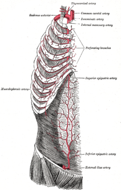
Gray522.png
Superior epigastric artery, internal thoracic artery and inferior epigastric artery. (Superior epigastric artery is labeled at right center.)
Details
Source Internal thoracic
Vein Superior epigastric vein
Identifiers
Latin arteria epigastrica superior
TA98 A12.2.08.041
TA2 4588
FMA 10646
Anatomical terminology

In human anatomy, the superior epigastric artery is a terminal [1] branch of the internal thoracic artery that provides arterial supply to the abdominal wall, and upper rectus abdominis muscle. It enters the rectus sheath to descend upon the inner surface of the rectus abdominis muscle. It ends by anastomosing with the inferior epigastric artery.

Contents

Structure

Origin

The superior epigastric artery arises from the internal thoracic artery (referred to as the internal mammary artery in the accompanying diagram). [2] [3]

Course and relations

The superior epigastric artery pierces the diaphragm [1] to enter the rectus sheath and descend upon the deep surface of the rectus abdominis. [4]

Along its course, it is accompanied by a similarly named vein, the superior epigastric vein.[ citation needed ]

Anastomoses

It anastomoses with the inferior epigastric artery [4] [2] within the rectus abdominis muscle [4] [1] at the umbilicus. [2]

Distribution

Where it anastomoses, the superior epigastric artery supplies the anterior part of the abdominal wall, [5] [6] upper rectus abdominis muscle, [5] and some of the diaphragm.[ citation needed ]

Collateralization in disease

Vascular disease

The superior epigastric arteries, inferior epigastric arteries, internal thoracic arteries and left subclavian artery and right subclavian artery / brachiocephalic are collateral vessels to the thoracic aorta and abdominal aorta. If the abdominal aorta develops a significant stenosis and/or blockage (as may be caused by atherosclerosis), this collateral pathway may develop sufficiently, over time, to supply blood to the lower limbs. [7]

Coarctation of the aorta

A congenitally narrowed aorta, due to coarctation, is often associated with a significant enlargement of the internal thoracic and epigastric arteries. [8]

See also

Related Research Articles

<span class="mw-page-title-main">Aorta</span> Largest artery in the human body

The aorta is the main and largest artery in the human body, originating from the left ventricle of the heart, branching upwards immediately after, and extending down to the abdomen, where it splits at the aortic bifurcation into two smaller arteries. The aorta distributes oxygenated blood to all parts of the body through the systemic circulation.

<span class="mw-page-title-main">Subclavian artery</span> Major arteries of the upper thorax, below the clavicle

In human anatomy, the subclavian arteries are paired major arteries of the upper thorax, below the clavicle. They receive blood from the aortic arch. The left subclavian artery supplies blood to the left arm and the right subclavian artery supplies blood to the right arm, with some branches supplying the head and thorax. On the left side of the body, the subclavian comes directly off the aortic arch, while on the right side it arises from the relatively short brachiocephalic artery when it bifurcates into the subclavian and the right common carotid artery.

<span class="mw-page-title-main">Abdominal aorta</span> Largest artery in the abdomen

In human anatomy, the abdominal aorta is the largest artery in the abdominal cavity. As part of the aorta, it is a direct continuation of the descending aorta.
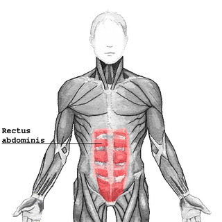
<span class="mw-page-title-main">Rectus abdominis muscle</span> Paired straight muscle

The rectus abdominis muscle, also known as the "abdominal muscle" or simply the "abs", is a pair of segmented skeletal muscle on the ventral aspect of a person's abdomen. The paired muscle is separated at the midline by a band of dense connective tissue called the linea alba, and the connective tissue defining each lateral margin of the rectus abdominus is the linea semilunaris. The muscle extends from the pubic symphysis, pubic crest and pubic tubercle inferiorly, to the xiphoid process and costal cartilages of the 5th–7th ribs superiorly.

<span class="mw-page-title-main">Superior orbital fissure</span> Foramen in the skull allowing for passage of cranial nerves

The superior orbital fissure is a foramen or cleft of the skull between the lesser and greater wings of the sphenoid bone. It gives passage to multiple structures, including the oculomotor nerve, trochlear nerve, ophthalmic nerve, abducens nerve, ophthalmic veins, and sympathetic fibres from the cavernous plexus.

<span class="mw-page-title-main">Internal thoracic artery</span> Artery of the thorax

In human anatomy, the internal thoracic artery (ITA), also known as the internal mammary artery, is an artery that supplies the anterior chest wall and the breasts. It is a paired artery, with one running along each side of the sternum, to continue after its bifurcation as the superior epigastric and musculophrenic arteries.

<span class="mw-page-title-main">Inferior epigastric artery</span> Blood vessel

In human anatomy, the inferior epigastric artery is an artery that arises from the external iliac artery. It is accompanied by the inferior epigastric vein; inferiorly, these two inferior epigastric vessels together travel within the lateral umbilical fold The inferior epigastric artery then traverses the arcuate line of rectus sheath to enter the rectus sheath, then anastomoses with the superior epigastric artery within the rectus sheath.

<span class="mw-page-title-main">Superior epigastric vein</span> Blood vessel

In human anatomy, the superior epigastric veins are two or more venae comitantes which accompany either superior epigastric artery before emptying into the internal thoracic vein. They participate in the drainage of the superior surface of the diaphragm.

<span class="mw-page-title-main">Conjoint tendon</span> Medial part of the posterior wall of the inguinal canal

The conjoint tendon is a sheath of connective tissue formed from the lower part of the common aponeurosis of the abdominal internal oblique muscle and the transversus abdominis muscle, joining the muscle to the pelvis. It forms the medial part of the posterior wall of the inguinal canal.

<span class="mw-page-title-main">Thyrocervical trunk</span> Artery of the neck

The thyrocervical trunk is a short artery of the neck. It arises from the subclavian artery, then promptly divides into its branches: the inferior thyroid artery, suprascapular artery, and (sometimes) the transverse cervical artery.

<span class="mw-page-title-main">Inferior thyroid artery</span> Artery of the neck

The inferior thyroid artery is an artery in the neck. It arises from the thyrocervical trunk and passes upward, in front of the vertebral artery and longus colli muscle. It then turns medially behind the carotid sheath and its contents, and also behind the sympathetic trunk, the middle cervical ganglion resting upon the vessel.

<span class="mw-page-title-main">Lateral circumflex femoral artery</span>

The lateral circumflex femoral artery is an artery in the upper thigh. It is usually a branch of the profunda femoris artery, and produces three branches. It is mostly distributed to the muscles of the lateral thigh, supplying arterial blood to muscles of the knee extensor group.

<span class="mw-page-title-main">Lumbar arteries</span> Arteries within the lower back

The lumbar arteries are arteries located in the lower back or lumbar region. The lumbar arteries are in parallel with the intercostals.

The subcostal arteries, so named because they lie below the last ribs, constitute the lowest pair of branches derived from the thoracic aorta, and are in series with the intercostal arteries.

In anatomy, arterial tree is used to refer to all arteries and/or the branching pattern of the arteries. This article regards the human arterial tree. Starting from the aorta:

<span class="mw-page-title-main">Arcuate line of rectus sheath</span> Line of demarcation in the human abdomen

The arcuate line of rectus sheath is a line of demarcation corresponding to the free inferior margin of the posterior layer of the rectus sheath inferior to which only the anterior layer of the rectus sheath is present and the rectus abdominis muscle is therefore in direct contact with the transversalis fascia. The arcuate line is concave inferior-wards.

<span class="mw-page-title-main">Deep circumflex iliac artery</span> Artery in the pelvis

The deep circumflex iliac artery is an artery in the pelvis that travels along the iliac crest of the pelvic bone.

<span class="mw-page-title-main">Linea semilunaris</span>

The linea semilunaris is a curved tendinous intersection found on either side of the rectus abdominis muscle.

<span class="mw-page-title-main">Lumbar veins</span> Veins that drain the posterior abdominal wall

The lumbar veins are four pairs of veins running along the inside of the posterior abdominal wall, and drain venous blood from parts of the abdominal wall. Each lumbar vein accompanies a single lumbar artery. The lower two pairs of lumbar veins all drain directly into the inferior vena cava, whereas the fate of the upper two pairs is more variable.

<span class="mw-page-title-main">Outline of human anatomy</span> Overview of and topical guide to human anatomy

The following outline is provided as an overview of and topical guide to human anatomy:
References

  1. 1 2 3 Sinnatamby, Chummy (2011). Last's Anatomy (12th ed.). Elsevier Australia. p. 225. ISBN   978-0-7295-3752-0.
  2. 1 2 3 Castro Ferreira, Marcus; Henrique Ishida, Luis; Munhoz, Alexandre (January 1, 2009), Wei, Fu-Chan; Mardini, Samir (eds.), "CHAPTER 19 - Rectus flap", Flaps and Reconstructive Surgery, Edinburgh: W.B. Saunders, pp. 207–223, doi:10.1016/b978-0-7216-0519-7.00019-8, ISBN   978-0-7216-0519-7 , retrieved November 22, 2020
  3. Ahmed, Abdul (January 1, 2017), Brennan, Peter A.; Schliephake, Henning; Ghali, G. E.; Cascarini, Luke (eds.), "37 - Common Free Vascularized Flaps: The Rectus Abdominis", Maxillofacial Surgery (Third Edition), Churchill Livingstone, pp. 533–542, doi:10.1016/b978-0-7020-6056-4.00038-1, ISBN   978-0-7020-6056-4 , retrieved November 22, 2020
  4. 1 2 3 The Big Picture: Gross Anatomy, Medical Course & Step 1 Review. David A. Morton, K. Bo Foreman, Kurt H. Albertine (2nd ed.). New York. 2018. ISBN   978-1-259-86264-9. OCLC   1044772257.{{cite book}}: CS1 maint: location missing publisher (link) CS1 maint: others (link)
  5. 1 2 Shell, Dan H.; Vásconez, Luis O.; de la Torre, Jorge I.; Chin, Gloria; Weinzweig, Norman (January 1, 2010), Weinzweig, Jeffrey (ed.), "Chapter 91 - Abdominal Wall Reconstruction", Plastic Surgery Secrets Plus (Second Edition), Philadelphia: Mosby, pp. 594–604, doi:10.1016/b978-0-323-03470-8.00091-0, ISBN   978-0-323-03470-8 , retrieved November 22, 2020
  6. DiEdwardo, Christine A.; Caterson, Stephanie A.; Barrall, David T. (January 1, 2010), Weinzweig, Jeffrey (ed.), "Chapter 80 - Abdominoplasty", Plastic Surgery Secrets Plus (Second Edition), Philadelphia: Mosby, pp. 532–537, doi:10.1016/b978-0-323-03470-8.00080-6, ISBN   978-0-323-03470-8 , retrieved November 22, 2020
  7. Yurdakul M, Tola M, Ozdemir E, Bayazit M, Cumhur T (April 2006). "Internal thoracic artery-inferior epigastric artery as a collateral pathway in aortoiliac occlusive disease". J. Vasc. Surg. 43 (4): 707–13. doi: 10.1016/j.jvs.2005.12.042 . PMID   16616225.
  8. Huhmann W, Kunitsch G, Dalichau H (1976). "[Coarctation of the aorta on the plain chest x-ray (author's transl)]". Dtsch Med Wochenschr. 101 (41): 1477–81. doi:10.1055/s-0028-1104294. PMID   964150. S2CID   260093972.