
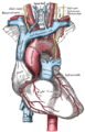
Coronary circulation is the circulation of blood in the arteries and veins that supply the heart muscle (myocardium). Coronary arteries supply oxygenated blood to the heart muscle. Cardiac veins then drain away the blood after it has been deoxygenated. Because the rest of the body, and most especially the brain, needs a steady supply of oxygenated blood that is free of all but the slightest interruptions, the heart is required to function continuously. Therefore its circulation is of major importance not only to its own tissues but to the entire body and even the level of consciousness of the brain from moment to moment. Interruptions of coronary circulation quickly cause heart attacks, in which the heart muscle is damaged by oxygen starvation. Such interruptions are usually caused by coronary ischemia linked to coronary artery disease, and sometimes to embolism from other causes like obstruction in blood flow through vessels.

A heart valve is a biological one-way valve that allows blood to flow in one direction through the chambers of the heart. Four valves are usually present in a mammalian heart and together they determine the pathway of blood flow through the heart. A heart valve opens or closes according to differential blood pressure on each side.

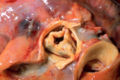
The papillary muscles are muscles located in the ventricles of the heart. They attach to the cusps of the atrioventricular valves via the chordae tendineae and contract to prevent inversion or prolapse of these valves on systole.

The coronary arteries are the arterial blood vessels of coronary circulation, which transport oxygenated blood to the heart muscle. The heart requires a continuous supply of oxygen to function and survive, much like any other tissue or organ of the body.

The atrioventricular node or AV node electrically connects the heart's atria and ventricles to coordinate beating in the top of the heart; it is part of the electrical conduction system of the heart. The AV node lies at the lower back section of the interatrial septum near the opening of the coronary sinus, and conducts the normal electrical impulse from the atria to the ventricles. The AV node is quite compact.

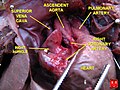
dextro-Transposition of the great arteries is a potentially life-threatening birth defect in the large arteries of the heart. The primary arteries are transposed.

The left coronary artery is a coronary artery that arises from the aorta above the left cusp of the aortic valve, and supplies blood to the left side of the heart muscle. The left coronary artery typically runs for 10–25 mm, then bifurcates into the left anterior descending artery, and the left circumflex artery.

The interventricular septum is the stout wall separating the ventricles, the lower chambers of the heart, from one another.

The coronary sinus is the largest vein of the heart. It drains over half of the deoxygenated blood from the heart muscle into the right atrium. It begins on the backside of the heart, in between the left atrium, and left ventricle; it begins at the junction of the great cardiac vein, and oblique vein of the left atrium. It receives multiple tributaries. It passes across the backside of the heart along a groove between left atrium and left ventricle, then drains into the right atrium at the orifice of the coronary sinus.

The coronary sulcus is a groove on the surface of the heart at the base of right auricle that separates the atria from the ventricles. The structure contains the trunks of the nutrient vessels of the heart, and is deficient in front, where it is crossed by the root of the pulmonary trunk. On the posterior surface of the heart, the coronary sulcus contains the coronary sinus. The right coronary artery, circumflex branch of left coronary artery, and small cardiac vein all travel along parts of the coronary sulcus.

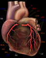
In the coronary circulation, the posterior descending artery (PDA), also called the posterior interventricular artery, is an artery running in the posterior interventricular sulcus to the apex of the heart where it meets with the left anterior descending artery also known as the anterior interventricular artery. The PDA supplies the posterior third of the interventricular septum. The remaining anterior two-thirds is supplied by the left anterior descending artery, which is a branch of left coronary artery.
The circumflex branch of left coronary artery is a branch of the left coronary artery. It winds around the left side of the heart along the atrioventricular groove. It supplies the posterolateral portion of the left ventricle.
The right marginal branch of right coronary artery is the largest marginal branch of the right coronary artery. It follows the acute margin of the heart. It supplies blood to both surfaces of the right ventricle.
Coronary artery anomalies are variations of the coronary circulation, affecting <1% of the general population. Symptoms include chest pain, shortness of breath and syncope, although cardiac arrest may be the first clinical presentation. Several varieties are identified, with a different potential to cause sudden cardiac death.
The left anterior descending artery is a branch of the left coronary artery. It supplies the anterior portion of the left ventricle. It provides about half of the arterial supply to the left ventricle and is thus considered the most important vessel supplying the left ventricle. Blockage of this artery is often called the widow-maker infarction due to a high risk of death.

The sinoatrial nodal artery, sinuatrial nodal artery or sinoatrial artery is an artery of the heart which supplies the sinoatrial node, the natural pacemaker center of the heart. It is usually a branch of the right coronary artery. It passes between the right atrium, and the opening of the superior vena cava.
The atrioventricular nodal branch is a coronary artery that supplies arterial blood to the atrioventricular node, which is responsible for initiating muscular contraction of the ventricles. The AV nodal branch is most often a branch of the right coronary artery.
The crux cordis or crux of the heart is the area on the lower back side of the heart where the coronary sulcus and the posterior interventricular sulcus meet. It is important surgically because the atrioventricular nodal artery, a small but vital vessel, passes in proximity to the crux of the heart. It is the anastomotic point of right and left coronary artery.

Heart development, also known as cardiogenesis, refers to the prenatal development of the heart. This begins with the formation of two endocardial tubes which merge to form the tubular heart, also called the primitive heart tube. The heart is the first functional organ in vertebrate embryos.
The heart is a muscular organ situated in the mediastinum. It consists of four chambers, four valves, two main arteries, and the conduction system. The left and right sides of the heart have different functions: the right side receives de-oxygenated blood through the superior and inferior venae cavae and pumps blood to the lungs through the pulmonary artery, and the left side receives saturated blood from the lungs.


