Internal thoracic artery

Last updated
Internal thoracic artery
Gray522.png
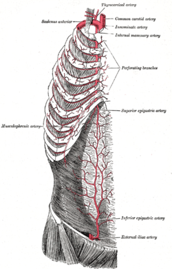
Right internal thoracic artery and its branches (labeled under its old name the Internal mammary artery, at upper right).
Details
Source Subclavian artery
Branches Pericardiocophrenic
Anterior intercostal branches
Musculophrenic
Superior epigastric
Perforating branches
Vein Internal thoracic vein
Identifiers
Latin arteria thoracica interna, arteria mammaria interna
MeSH D008323
TA98 A12.2.08.029
TA2 4576
FMA 3960
Anatomical terminology

In human anatomy, the internal thoracic artery (ITA), also known as the internal mammary artery, is an artery that supplies the anterior chest wall and the breasts. [1] It is a paired artery, with one running along each side of the sternum, to continue after its bifurcation as the superior epigastric and musculophrenic arteries.

Contents

Structure

The internal thoracic artery arises from the anterior surface of the subclavian artery near its origin. [2] [3] It has a width of between 1-2 mm. [4]

It travels downward on the inside of the rib cage, approximately 1 cm from the sides of the sternum, [3] and thus medial to the nipple. It is accompanied by the internal thoracic vein.

It runs deep to the abdominal external oblique muscle, but superficial to the vagus nerve.

In adults, the internal thoracic artery lies closest to the sternum at the first intercoastal space. The gap between the artery and lateral border of the sternum increases when going downwards, up to 1.1 cm to 1.3 cm at the sixth intercoastal space. In children, the gap ranges from 0.5 cm to 1.0 cm. [5]

Branches

After passing the sixth intercostal space, the internal thoracic artery splits into the following two terminal branches:

Function

The internal thoracic artery supplies the chest wall and the breasts. [1]

Clinical significance

Use in bypass grafts

The internal thoracic artery is the cardiac surgeon's blood vessel of choice for coronary artery bypass grafting. The left ITA has a superior long-term patency to saphenous vein grafts [6] [7] and other arterial grafts [8] (e.g. radial artery, gastroepiploic artery) when grafted to the left anterior descending coronary artery, generally the most important vessel, clinically, to revascularize. [1]

Plastic surgeons may use either the left or right internal thoracic arteries for autologous free flap reconstruction of the breast after mastectomy. Usually, a microvascular anastomosis is performed at the second intercostal space to the artery on which the free flap is based.

Additional images

Related Research Articles

<span class="mw-page-title-main">Coronary artery bypass surgery</span> Surgical procedure to restore normal blood flow to an obstructed coronary artery

Coronary artery bypass surgery, also known as coronary artery bypass graft, is a surgical procedure to treat coronary artery disease (CAD), the buildup of plaques in the arteries of the heart. It can relieve chest pain caused by CAD, slow the progression of CAD, and increase life expectancy. It aims to bypass narrowings in heart arteries by using arteries or veins harvested from other parts of the body, thus restoring adequate blood supply to the previously ischemic heart.
<span class="mw-page-title-main">Great saphenous vein</span> Large, subcutaneous, superficial vein of the leg

The great saphenous vein (GSV) or long saphenous vein is a large, subcutaneous, superficial vein of the leg. It is the longest vein in the body, running along the length of the lower limb, returning blood from the foot, leg and thigh to the deep femoral vein at the femoral triangle.

<span class="mw-page-title-main">Brachiocephalic vein</span> Vein

The left and right brachiocephalic veins are major veins in the upper chest, formed by the union of the ipsilateral internal jugular vein and subclavian vein behind the sternoclavicular joint. The left brachiocephalic vein is more than twice the length of the right brachiocephalic vein.

<span class="mw-page-title-main">Subclavian artery</span> Major arteries of the upper thorax, below the clavicle

In human anatomy, the subclavian arteries are paired major arteries of the upper thorax, below the clavicle. They receive blood from the aortic arch. The left subclavian artery supplies blood to the left arm and the right subclavian artery supplies blood to the right arm, with some branches supplying the head and thorax. On the left side of the body, the subclavian comes directly off the aortic arch, while on the right side it arises from the relatively short brachiocephalic artery when it bifurcates into the subclavian and the right common carotid artery.

<span class="mw-page-title-main">Stellate ganglion</span>

The stellate ganglion is a sympathetic ganglion formed by the fusion of the inferior cervical ganglion and the first thoracic ganglion, which is present in 80% of individuals. Sometimes, the second and the third thoracic ganglia are included in this fusion.

<span class="mw-page-title-main">Facial artery</span>

The facial artery is a branch of the external carotid artery that supplies structures of the superficial face.

<span class="mw-page-title-main">Intercostal nerves</span>

The intercostal nerves are part of the somatic nervous system, and arise from the anterior rami of the thoracic spinal nerves from T1 to T11. The intercostal nerves are distributed chiefly to the thoracic pleura and abdominal peritoneum, and differ from the anterior rami of the other spinal nerves in that each pursues an independent course without plexus formation.

<span class="mw-page-title-main">Inferior epigastric artery</span> Blood vessel

In human anatomy, the inferior epigastric artery is an artery that arises from the external iliac artery. It is accompanied by the inferior epigastric vein; inferiorly, these two inferior epigastric vessels together travel within the lateral umbilical fold The inferior epigastric artery then traverses the arcuate line of rectus sheath to enter the rectus sheath, then anastomoses with the superior epigastric artery within the rectus sheath.

<span class="mw-page-title-main">Superior epigastric artery</span> Blood vessel

In human anatomy, the superior epigastric artery is a terminal branch of the internal thoracic artery that provides arterial supply to the abdominal wall, and upper rectus abdominis muscle. It enters the rectus sheath to descend upon the inner surface of the rectus abdominis muscle. It ends by anastomosing with the inferior epigastric artery.

<span class="mw-page-title-main">Internal thoracic vein</span> Large blood vessel draining breasts and the chest wall

In human anatomy, the internal thoracic vein is the vein that drains the chest wall and breasts.

<span class="mw-page-title-main">Lateral pectoral nerve</span>

The lateral pectoral nerve arises from the lateral cord of the brachial plexus, and through it from the C5-7.

<span class="mw-page-title-main">Adductor canal</span> Aponeurotic tunnel in the middle third of the thigh

The adductor canal is an aponeurotic tunnel in the middle third of the thigh giving passage to parts of the femoral artery, vein, and nerve. It extends from the apex of the femoral triangle to the adductor hiatus.

<span class="mw-page-title-main">Intercostal space</span> Anatomic space between two ribs

The intercostal space (ICS) is the anatomic space between two ribs. Since there are 12 ribs on each side, there are 11 intercostal spaces, each numbered for the rib superior to it.

<span class="mw-page-title-main">Intercostal arteries</span> Arteries supplying the space between the ribs

The intercostal arteries are a group of arteries passing within an intercostal space. There are 9 anterior and 11 posterior intercostal arteries on each side of the body. The anterior intercostal arteries are branches of the internal thoracic artery and its terminal branch - the musculophrenic artery. The posterior intercostal arteries are branches of the supreme intercostal artery and thoracic aorta.

<span class="mw-page-title-main">Lumbar veins</span> Veins that drain the posterior abdominal wall

The lumbar veins are four pairs of veins running along the inside of the posterior abdominal wall, and drain venous blood from parts of the abdominal wall. Each lumbar vein accompanies a single lumbar artery. The lower two pairs of lumbar veins all drain directly into the inferior vena cava, whereas the fate of the upper two pairs is more variable.

<span class="mw-page-title-main">Outline of human anatomy</span> Overview of and topical guide to human anatomy

The following outline is provided as an overview of and topical guide to human anatomy:

Minimally invasive cardiac surgery, encompasses various aspects of cardiac surgical procedures that can be performed with minimally invasive approach either via mini-thoracotomy or mini-sternotomy. MICS CABG or the McGinn technique is heart surgery performed through several small incisions instead of the traditional open-heart surgery that requires a median sternotomy approach. MICS CABG is a beating-heart multi-vessel procedure performed under direct vision through an anterolateral mini-thoracotomy.

Vessel harvesting is a surgical technique that may be used in conjunction with a coronary artery bypass graft (CABG). For patients with coronary artery disease, a physician may recommend a bypass to reroute blood around blocked arteries to restore and improve blood flow and oxygen to the heart. To create the bypass graft, a surgeon will remove or "harvest" healthy blood vessels from another part of the body, often from the patient's leg or arm. This vessel becomes a graft, with one end attaching to a blood source above and the other end below the blocked area, creating a "conduit" channel or new blood flow connection across the heart.

In medicine, vein graft failure (VGF) is a condition in which vein grafts, which are used as alternative conduits in bypass surgeries, get occluded.
References

  1. 1 2 3 Kramer, R. S.; Morton, J. R.; Groom, R. C.; Robaczewski, D. L. (2018-01-01), "Coronary Artery Bypass Grafting", in Vasan, Ramachandran S.; Sawyer, Douglas B. (eds.), Encyclopedia of Cardiovascular Research and Medicine, Oxford: Elsevier, pp. 700–729, doi:10.1016/b978-0-12-809657-4.99754-0, ISBN   978-0-12-805154-2 , retrieved 2020-11-12
  2. Stewart, Charles E.; Urken, Mark L. (2009-01-01), Wei, Fu-Chan; Mardini, Samir (eds.), "Chapter 18 - Deltopectoral flap", Flaps and Reconstructive Surgery, Edinburgh: W.B. Saunders, pp. 193–205, doi:10.1016/b978-0-7216-0519-7.00018-6, ISBN   978-0-7216-0519-7 , retrieved 2020-11-12
  3. 1 2 Barral, Jean-Pierre; Croibier, Alain (2011-01-01), Barral, Jean-Pierre; Croibier, Alain (eds.), "13 - Vessels of the breast", Visceral Vascular Manipulations, Oxford: Churchill Livingstone, pp. 121–132, doi:10.1016/b978-0-7020-4351-2.00013-2, ISBN   978-0-7020-4351-2 , retrieved 2020-11-12
  4. Markiewicz, Michael R.; Ord, Robert; Fernandes, Rui P. (2017-01-01), Brennan, Peter A.; Schliephake, Henning; Ghali, G. E.; Cascarini, Luke (eds.), "43 - Local and Regional Flap Reconstruction of Maxillofacial Defects", Maxillofacial Surgery (Third Edition), Churchill Livingstone, pp. 616–635, doi:10.1016/b978-0-7020-6056-4.00044-7, ISBN   978-0-7020-6056-4 , retrieved 2020-11-12
  5. Jelicić N, Djordjević L, Stosić T (1996). "Unutrasnji grudni krvni sudovi (a. et vv. thoracicae internae) i njihov prakticni znacaj" [The internal thoracic blood vessels (internal thoracic arteries and veins) and their practical significance]. Srpski Arhiv Za Celokupno Lekarstvo (in Serbian). 124 (3–4): 58–61. PMID   9102819.
  6. Kitamura, S; Kawachi, K; Kawata, T; Kobayashi, S; Mizuguchi, K; Kameda, Y; Nishioka, H; Hamada, Y; Yoshida, Y (1996). "Ten-year survival and cardiac event-free rates in Japanese patients with the left anterior descending artery revascularized with internal thoracic artery or saphenous vein graft: a comparative study". Nippon Geka Gakkai Zasshi. 97 (3): 202–9. PMID   8649330.
  7. Arima, M; Kanoh, T; Suzuki, T; Kuremoto, K; Tanimoto, K; Oigawa, T; Matsuda, S (2005). "Serial angiographic follow-up beyond 10 years after coronary artery bypass grafting". Circulation Journal. 69 (8): 896–902. doi: 10.1253/circj.69.896 . PMID   16041156.
  8. Cohen, G; Tamariz, MG; Sever, JY; Liaghati, N; Guru, V; Christakis, GT; Bhatnagar, G; Cutrara, C; et al. (2001). "The radial artery versus the saphenous vein graft in contemporary CABG: a case-matched study". The Annals of Thoracic Surgery. 71 (1): 180–5, discussion 185–6. doi: 10.1016/S0003-4975(00)02285-2 . PMID   11216742.

Figures of ITA grafts