Thyroid ima artery

Last updated

Thyroid ima artery
Arterial supply of the thyroid gland.png
Location of the artery depicted in the image
Details
Source Brachiocephalic artery (most common)
Supplies Thyroid gland, trachea, parathyroid glands, thymus gland (as thymica accessoria)
Identifiers
Latin arteria thyroidea ima
TA98 A12.2.04.005
TA2 4180
FMA 3936
Anatomical terminology

The thyroid ima artery (thyroidea ima artery, arteria thyroidea ima, thyroid artery of Neubauer or the lowest thyroid artery) is an artery of the head and neck. It is an anatomical variant that, when present, supplies blood to the thyroid gland primarily, or the trachea, the parathyroid gland and the thymus gland (as thymica accessoria) in rare cases. It has also been reported to be a compensatory artery when one or both of the inferior thyroid arteries are absent, and in a few cases the only source of blood to the thyroid gland. Furthermore, it varies in origin, size, blood supply, and termination, and occurs in around 3.8% of the population and is 4.5 times more common in fetuses than in adults. [1] Because of the variations and rarity, it may lead to surgical complications, particularly during tracheostomy and other airway managements.

Contents

Structure

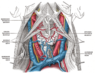
The veins of the thyroid gland (thyroid ima artery not labeled, but region of origin and route are visible) Gray560.png
The veins of the thyroid gland (thyroid ima artery not labeled, but region of origin and route are visible)

The thyroid ima artery is an embryonic artery and it occurs because of the failure of the vessel to close, remaining patent (open). [2]

The artery has a variable origin. It mostly arises from the brachiocephalic trunk, [3] but may also originate from the aortic arch, the right common carotid, the subclavian, the pericardiacophrenic artery, the thyrocervical trunk, the transverse scapular or the internal thoracic artery. [4] It ascends in front of the trachea in the superior mediastinum to the lower part of the thyroid gland. [2]

It differs in size and ranges from as small as accessory thyroid arteries to the size of primary thyroid vessels. The diameter of the lumen of the artery ranges from 3 to 5 millimetres (0.12 to 0.20 in). [5] The artery may be present as an accessory thyroid artery, but sometimes appears to compensate for incompetence or absence of one or more main thyroid vessels. [6] Since it begins from below the thyroid gland and ascends upwards, it is mostly associated with absence or reduced size of the inferior thyroid arteries. [7] In such cases, it is known as the accessory inferior thyroid artery. [5] [6] In rare cases, the artery has been seen to be compensating for absence of one or both superior thyroid arteries. [8]

In cases where the length of the thyroid ima artery is shorter, the artery ends by supplying the thymus gland and is known as thymica accessoria. [5]

Function

Thyroid ima marked on the image as TIA, parathyroid as PG Postoperative Hypoparathyroidism in Thyroid Surgery Fig4 Recorte.jpg
Thyroid ima marked on the image as TIA, parathyroid as PG

When present, the thyroid ima's chief supply is the thyroid gland, though it also supplies the trachea. The artery may extend and supply the parathyroid glands. An infrequently observed artery, it is more frequently reported in the context of enlarged parathyroid glands (parathyroid adenomas). The artery ends by supplying the thyroid gland, or the parathyroid glands, as a single unit or as multiple branches. [6] [9] The artery is also found to be the only supply of the parathyroid gland in rare cases. [6]

Clinical significance

The artery is only present in approximately 3–10% of the population. [10] [11] Thyroid ima artery is of surgical importance; due to its relatively small size and infrequent presence it can cause complications such as severe bleeding. [12] Knowledge of occurrence of the artery is especially important during tracheostomy, sternotomy and thyroidectomy. [5] Because the artery is smaller than the other thyroid vessels, and having an origin from one of the bigger vessels, a brisk cut while performing the surgery may cause complications such as severe hemorrhage and significant blood loss. [13] The artery, if dissected, may draw back into the mediastinum and further complicate the condition by causing hemorrhage and clots in the thoracic cavity. [14]

History

The thyroid ima artery was first defined by German anatomist Johann Ernst Neubauer  [ de ] in the year 1772. Hence, it was named the thyroid artery of Neubauer. [15] The artery originates lower than the inferior thyroid arteries, so it is also known as the lowest thyroid artery. [10] [11] Arteria thyroidea ima is the Latin name of the artery. [11] [3]

Other animals

The presence of thyroid ima artery is also observed in other higher primates. The artery has been reported in gorillas, gibbons, macaques and gray langurs. Variations in the origin were also seen; it was found to originate from the aorta in the thorax, or the carotid in the neck. [16]

Related Research Articles

<span class="mw-page-title-main">Trachea</span> Cartilaginous tube that connects the pharynx and larynx to the lungs

The trachea, also known as the windpipe, is a cartilaginous tube that connects the larynx to the bronchi of the lungs, allowing the passage of air, and so is present in almost all animals with lungs. The trachea extends from the larynx and branches into the two primary bronchi. At the top of the trachea the cricoid cartilage attaches it to the larynx. The trachea is formed by a number of horseshoe-shaped rings, joined together vertically by overlying ligaments, and by the trachealis muscle at their ends. The epiglottis closes the opening to the larynx during swallowing.

<span class="mw-page-title-main">Brachiocephalic artery</span> Artery of the mediastinum

The brachiocephalic artery, brachiocephalic trunk, or innominate artery is an artery of the mediastinum that supplies blood to the right arm, head, and neck.

<span class="mw-page-title-main">Parathyroid gland</span> Endocrine gland

Parathyroid glands are small endocrine glands in the neck of humans and other tetrapods. Humans usually have four parathyroid glands, located on the back of the thyroid gland in variable locations. The parathyroid gland produces and secretes parathyroid hormone in response to a low blood calcium, which plays a key role in regulating the amount of calcium in the blood and within the bones.

<span class="mw-page-title-main">Subclavian artery</span> Major arteries of the upper thorax, below the clavicle

In human anatomy, the subclavian arteries are paired major arteries of the upper thorax, below the clavicle. They receive blood from the aortic arch. The left subclavian artery supplies blood to the left arm and the right subclavian artery supplies blood to the right arm, with some branches supplying the head and thorax. On the left side of the body, the subclavian comes directly off the aortic arch, while on the right side it arises from the relatively short brachiocephalic artery when it bifurcates into the subclavian and the right common carotid artery.

<span class="mw-page-title-main">Thoracic duct</span> Lymphatic vessel

In human anatomy, the thoracic duct is the larger of the two lymph ducts of the lymphatic system. The thoracic duct usually begins from the upper aspect of the cisterna chyli, passing out of the abdomen through the aortic hiatus into first the posterior mediastinum and then the superior mediastinum, extending as high up as the root of the neck before descending to drain into the systemic (blood) circulation at the venous angle.

<span class="mw-page-title-main">External carotid artery</span> Major artery of the head and neck

The external carotid artery is a major artery of the head and neck. It arises from the common carotid artery when it splits into the external and internal carotid artery. The external carotid artery supplies blood to the face, brain and neck.
<span class="mw-page-title-main">Internal jugular vein</span> Blood vessel that drains the head

The internal jugular vein is a paired jugular vein that collects blood from the brain and the superficial parts of the face and neck. This vein runs in the carotid sheath with the common carotid artery and vagus nerve.

<span class="mw-page-title-main">Recurrent laryngeal nerve</span> Nerve in the human body

The recurrent laryngeal nerve (RLN) is a branch of the vagus nerve that supplies all the intrinsic muscles of the larynx, with the exception of the cricothyroid muscles. There are two recurrent laryngeal nerves, right and left. The right and left nerves are not symmetrical, with the left nerve looping under the aortic arch, and the right nerve looping under the right subclavian artery then traveling upwards. They both travel alongside the trachea. Additionally, the nerves are among the few nerves that follow a recurrent course, moving in the opposite direction to the nerve they branch from, a fact from which they gain their name.

<span class="mw-page-title-main">Vertebral artery</span> Major arteries of the neck

The vertebral arteries are major arteries of the neck. Typically, the vertebral arteries originate from the subclavian arteries. Each vessel courses superiorly along each side of the neck, merging within the skull to form the single, midline basilar artery. As the supplying component of the vertebrobasilar vascular system, the vertebral arteries supply blood to the upper spinal cord, brainstem, cerebellum, and posterior part of brain.

<span class="mw-page-title-main">Common carotid artery</span> One of the two arteries that supply the head and neck with blood

In anatomy, the left and right common carotid arteries (carotids) are arteries that supply the head and neck with oxygenated blood; they divide in the neck to form the external and internal carotid arteries.

<span class="mw-page-title-main">Superior thoracic aperture</span>

The superior thoracic aperture, also known as the thoracic outlet, or thoracic inlet refers to the opening at the top of the thoracic cavity. It is also clinically referred to as the thoracic outlet, in the case of thoracic outlet syndrome. A lower thoracic opening is the inferior thoracic aperture.

<span class="mw-page-title-main">Aortic arch</span> Part of the aorta

The aortic arch, arch of the aorta, or transverse aortic arch is the part of the aorta between the ascending and descending aorta. The arch travels backward, so that it ultimately runs to the left of the trachea.

<span class="mw-page-title-main">Aortic arches</span>

The aortic arches or pharyngeal arch arteries are a series of six paired embryological vascular structures which give rise to the great arteries of the neck and head. They are ventral to the dorsal aorta and arise from the aortic sac.

<span class="mw-page-title-main">Aberrant subclavian artery</span> Medical condition

Aberrant subclavian artery, or aberrant subclavian artery syndrome, is a rare anatomical variant of the origin of the right or left subclavian artery. This abnormality is the most common congenital vascular anomaly of the aortic arch, occurring in approximately 1% of individuals.

<span class="mw-page-title-main">Inferior thyroid artery</span> Artery of the neck

The inferior thyroid artery is an artery in the neck. It arises from the thyrocervical trunk and passes upward, in front of the vertebral artery and longus colli muscle. It then turns medially behind the carotid sheath and its contents, and also behind the sympathetic trunk, the middle cervical ganglion resting upon the vessel.

<span class="mw-page-title-main">Superior laryngeal nerve</span> Branch of the vagus nerve

The superior laryngeal nerve is a branch of the vagus nerve. It arises from the middle of the inferior ganglion of vagus nerve and additionally also receives a sympathetic branch from the superior cervical ganglion.
<span class="mw-page-title-main">Head and neck anatomy</span>

This article describes the anatomy of the head and neck of the human body, including the brain, bones, muscles, blood vessels, nerves, glands, nose, mouth, teeth, tongue, and throat.

<span class="mw-page-title-main">Pretracheal fascia</span>

The pretracheal fascia is a layer of the deep cervical fascia at the front of the neck. It attaches to the hyoid bone above, and - extending down into the thorax - blends with the fibrous pericardium below. It encloses the thyroid gland and parathyroid glands, trachea, and esophagus. It extends medially in front of the carotid vessels. It assists in forming the carotid sheath.
<span class="mw-page-title-main">Tracheoinnominate fistula</span> Medical condition

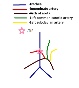
Tracheoinnominate fistula is an abnormal connection (fistula) between the innominate artery and the trachea. A TIF is a rare but life-threatening iatrogenic injury, usually the sequela of a tracheotomy.

In CT scan of the thyroid, focal and diffuse thyroid abnormalities are commonly encountered. These findings can often lead to a diagnostic dilemma, as the CT reflects nonspecific appearances. Ultrasound (US) examination has a superior spatial resolution and is considered the modality of choice for thyroid evaluation. Nevertheless, CT detects incidental thyroid nodules (ITNs) and plays an important role in the evaluation of thyroid cancer.

References

  1. Yurasakpong, Laphatrada; Nantasenamat, Chanin; Janta, Sirorat; Eiamratchanee, Pinthusorn; Coey, James; Chaiyamoon, Arada; Kruepunga, Nutmethee; Senarai, Thanyaporn; Langer, Martin Franz; Meemon, Krai; Suwannakhan, Athikhun (2022). "The decreasing prevalence of the thyroid ima artery: a systematic review and machine learning assisted meta-analysis". Annals of Anatomy - Anatomischer Anzeiger. 239: 151803. doi:10.1016/j.aanat.2021.151803. ISSN   0940-9602. PMID   34265384.
  2. 1 2 "Thyroidea ima artery (anatomy)". General Practice Notebook. Retrieved 21 January 2016.
  3. 1 2 Henry Gray (2015). Susan Standring (ed.). Gray's Anatomy: The Anatomical Basis of Clinical Practice (41st ed.). Elsevier Health Sciences. p. 1026. ISBN   978-0-7020-6851-5.
  4. Ronald A. Bergman; Adel K. Afifi; Ryosuke Miyauchi. "Thyroidea Ima (of Neubauer) Artery". Anatomy Atlases. Retrieved 21 January 2016.
  5. 1 2 3 4 G. Wyatt Pratt (1916). "The Thyroidea Ima Artery". Journal of Anatomy. 50 (Pt 3): 239–242.
  6. 1 2 3 4 Krudy A.G.; Doppman J.L.; Brennan M.F. (1980). "The significance of the thyroidea ima artery in arteriographic localization of parathyroid adenomas". Radiology. 136 (1): 45–51. doi:10.1148/radiology.136.1.7384523. PMID   7384523.
  7. Choi, Yeo-Kyu Youn, Kyu Eun Lee, June Young (2014). Color atlas of thyroid surgery : open, endoscopic and robotic procedures (Aufl. 2014 ed.). Berlin, Heidelberg: Springer Berlin Heidelberg. p. 3. ISBN   978-3-642-37262-9.{{cite book}}: CS1 maint: multiple names: authors list (link)
  8. Yilmaz E.; Celik H.H.; Durgun B.; Atasever A.; Ilgi S. (1993). "Arteria thyroidea ima arising from the brachiocephalic trunk with bilateral absence of inferior thyroid arteries: a case report". Surgical and Radiologic Anatomy. 15 (3): 197–199. doi:10.1007/bf01627705. PMID   8235962. S2CID   32274136.
  9. Beaumanoir (1882). "Preséntation de pièces anatomiques: artère thyroïdienne de Neubauer". Bulletins et Mémoires de la Société Anatomique (in French). 4: 316–317.
  10. 1 2 BD Chaurasia (2012). Garg, Krishna; Mittal, PS; Chandrupatla, Mrudula (eds.). BD Chaurasia's Human Anatomy: Head-Neck and Brain. Vol. 3 (6th ed.). CBS Publishers. p. 143. ISBN   978-81-239-2332-1.
  11. 1 2 3 Keith L. Moore; Arthur F. Dalley (1999). Clinically Oriented Anatomy . Lippincott Williams & Wilkins. p.  1027. ISBN   978-0-683-06141-3.
  12. Kamparoudi, Pagona; Paliouras, Dimitrios; Gogakos, Apostolos S.; Rallis, Thomas; Schizas, Nikolaos C.; Lazopoulos, Achilleas; Chatzinikolaou, Fotios; Sarafis, Pavlos; Serchan, Paschalitsa; Katsikogiannis, Nikolaos; Sarika, Eirini (2016). "Percutaneous tracheostomy—beware of the thyroidea-ima artery". Annals of Translational Medicine. 4 (22): 449. doi: 10.21037/atm.2016.11.04 . ISSN   2305-5839. PMC   5159410 . PMID   27999783.
  13. Vishram Singh (2012). P. Mahalingam (ed.). Clinical and Surgical Anatomy (2nd ed.). Elsevier Health Sciences. pp. 301–303. ISBN   978-81-312-0303-3.
  14. N. Anthony Moore; William A. Roy (2010). Rapid Review Gross and Developmental Anatomy. Elsevier Health Sciences. p. 220. ISBN   978-0-323-08048-4.
  15. C. Neubauer (1786). De arteria thyreoidea ima rariare arteriae innominate ramo. pp. 291–296, 309–314.
  16. Arthur Keith (1895). "The Modes of Origin of the Carotid and Subclavian Arteries from the Arch of the Aorta in Some of the Higher Primates". Journal of Anatomy and Physiology. 29 (Pt 3): 455.