
The aorta is the main and largest artery in the human body, originating from the left ventricle of the heart, branching upwards immediately after, and extending down to the abdomen, where it splits at the aortic bifurcation into two smaller arteries. The aorta distributes oxygenated blood to all parts of the body through the systemic circulation.

The rib cage or thoracic cage is an endoskeletal enclosure in the thorax of most vertebrates that comprises the ribs, vertebral column and sternum, which protect the vital organs of the thoracic cavity, such as the heart, lungs and great vessels and support the shoulder girdle to form the core part of the axial skeleton.

A spinal nerve is a mixed nerve, which carries motor, sensory, and autonomic signals between the spinal cord and the body. In the human body there are 31 pairs of spinal nerves, one on each side of the vertebral column. These are grouped into the corresponding cervical, thoracic, lumbar, sacral and coccygeal regions of the spine. There are eight pairs of cervical nerves, twelve pairs of thoracic nerves, five pairs of lumbar nerves, five pairs of sacral nerves, and one pair of coccygeal nerves. The spinal nerves are part of the peripheral nervous system.

In human anatomy, the subclavian arteries are paired major arteries of the upper thorax, below the clavicle. They receive blood from the aortic arch. The left subclavian artery supplies blood to the left arm and the right subclavian artery supplies blood to the right arm, with some branches supplying the head and thorax. On the left side of the body, the subclavian comes directly off the aortic arch, while on the right side it arises from the relatively short brachiocephalic artery when it bifurcates into the subclavian and the right common carotid artery.

The levator scapulae is a slender skeletal muscle situated at the back and side of the neck. It originates from the transverse processes of the four uppermost cervical vertebrae; it inserts onto the upper portion of the medial border of the scapula. It is innervated by the cervical nerves C3-C4, and frequently also by the dorsal scapular nerve. As the Latin name suggests, its main function is to lift the scapula.

In tetrapods, cervical vertebrae are the vertebrae of the neck, immediately below the skull. Truncal vertebrae lie caudal of cervical vertebrae. In sauropsid species, the cervical vertebrae bear cervical ribs. In lizards and saurischian dinosaurs, the cervical ribs are large; in birds, they are small and completely fused to the vertebrae. The vertebral transverse processes of mammals are homologous to the cervical ribs of other amniotes. Most mammals have seven cervical vertebrae, with the only three known exceptions being the manatee with six, the two-toed sloth with five or six, and the three-toed sloth with nine.
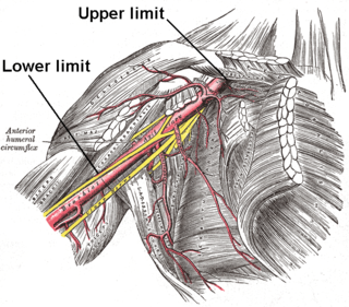
In human anatomy, the axillary artery is a large blood vessel that conveys oxygenated blood to the lateral aspect of the thorax, the axilla (armpit) and the upper limb. Its origin is at the lateral margin of the first rib, before which it is called the subclavian artery.

In human anatomy, the internal thoracic artery (ITA), also known as the internal mammary artery, is an artery that supplies the anterior chest wall and the breasts. It is a paired artery, with one running along each side of the sternum, to continue after its bifurcation as the superior epigastric and musculophrenic arteries.
In anatomy, the left and right common carotid arteries (carotids) are arteries that supply the head and neck with oxygenated blood; they divide in the neck to form the external and internal carotid arteries.

The scalene muscles are a group of three muscles on each side of the neck, identified as the anterior, the middle, and the posterior. They are innervated by the third to the eighth cervical spinal nerves (C3-C8).

A nerve plexus is a plexus of intersecting nerves. A nerve plexus is composed of afferent and efferent fibers that arise from the merging of the anterior rami of spinal nerves and blood vessels. There are five spinal nerve plexuses, except in the thoracic region, as well as other forms of autonomic plexuses, many of which are a part of the enteric nervous system. The nerves that arise from the plexuses have both sensory and motor functions. These functions include muscle contraction, the maintenance of body coordination and control, and the reaction to sensations such as heat, cold, pain, and pressure. There are several plexuses in the body, including:

The intercostal nerves are part of the somatic nervous system, and arise from the anterior rami of the thoracic spinal nerves from T1 to T11. The intercostal nerves are distributed chiefly to the thoracic pleura and abdominal peritoneum, and differ from the anterior rami of the other spinal nerves in that each pursues an independent course without plexus formation.

The inferior thyroid artery is an artery in the neck. It arises from the thyrocervical trunk and passes upward, in front of the vertebral artery and longus colli muscle. It then turns medially behind the carotid sheath and its contents, and also behind the sympathetic trunk, the middle cervical ganglion resting upon the vessel.

The deep cervical artery is an artery of the neck.
In anatomy, arterial tree is used to refer to all arteries and/or the branching pattern of the arteries. This article regards the human arterial tree. Starting from the aorta:

The carotid triangle is a portion of the anterior triangle of the neck.

The intercostal arteries are a group of arteries passing within an intercostal space. There are 9 anterior and 11 posterior intercostal arteries on each side of the body. The anterior intercostal arteries are branches of the internal thoracic artery and its terminal branch - the musculophrenic artery. The posterior intercostal arteries are branches of the supreme intercostal artery and thoracic aorta.

The inferior cervical ganglion is one of the three cervical sympathetic ganglia. It is situated between the base of the transverse process of the last cervical vertebra and the neck of the first rib, on the medial side of the costocervical artery.

The following outline is provided as an overview of and topical guide to human anatomy:
