
The aorta is the main and largest artery in the human body, originating from the left ventricle of the heart and extending down to the abdomen, where it splits into two smaller arteries. The aorta distributes oxygenated blood to all parts of the body through the systemic circulation.

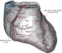
Coronary circulation is the circulation of blood in the blood vessels that supply the heart muscle (myocardium). Coronary arteries supply oxygenated blood to the heart muscle, and cardiac veins drain away the blood once it has been deoxygenated. Because the rest of the body, and most especially the brain, needs a steady supply of oxygenated blood that is free of all but the slightest interruptions, the heart is required to function continuously. Therefore its circulation is of major importance not only to its own tissues but to the entire body and even the level of consciousness of the brain from moment to moment. Interruptions of coronary circulation quickly cause heart attacks, in which the heart muscle is damaged by oxygen starvation. Such interruptions are usually caused by ischemic heart disease and sometimes by embolism from other causes like obstruction in blood flow through vessels.

The axillary nerve or the circumflex nerve is a nerve of the human body, that originates from the brachial plexus at the level of the axilla (armpit) and carries nerve fibers from C5 and C6. The axillary nerve travels through the quadrangular space with the posterior circumflex humeral artery and vein to innervate the deltoid and teres minor.
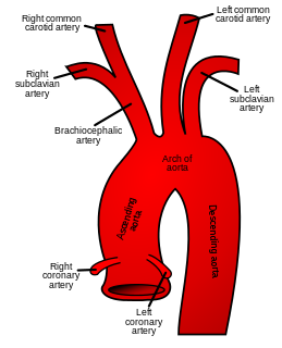
In human anatomy, the subclavian arteries are paired major arteries of the upper thorax, below the clavicle. They receive blood from the aortic arch. The left subclavian artery supplies blood to the left arm and the right subclavian artery supplies blood to the right arm, with some branches supplying the head and thorax. On the left side of the body, the subclavian comes directly off the aortic arch, while on the right side it arises from the relatively short brachiocephalic artery when it bifurcates into the subclavian and the right common carotid artery.

A pulmonary artery is an artery in the pulmonary circulation that carries deoxygenated blood from the right side of the heart to the lungs. The largest pulmonary artery is the main pulmonary artery or pulmonary trunk from the heart, and the smallest ones are the arterioles, which lead to the capillaries that surround the pulmonary alveoli.
The coronary arteries are the arterial blood vessels of coronary circulation, which transport oxygenated blood to the heart muscle. The heart requires a continuous supply of oxygen to function and survive, much like any other tissue or organ of the body.

The atrioventricular node or AV node is a part of the electrical conduction system of the heart that coordinates the top of the heart. It electrically connects the atria and ventricles. The AV node lies at the lower back section of the interatrial septum near the opening of the coronary sinus, and conducts the normal electrical impulse from the atria to the ventricles. The AV node is quite compact.
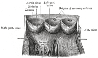
An aortic sinus, also known as a sinus of Valsalva, is one of the anatomic dilations of the ascending aorta, which occurs just above the aortic valve. These widenings are between the wall of the aorta and each of the three cusps of the aortic valve.

The subclavian vein is a paired large vein, one on either side of the body, that is responsible for draining blood from the upper extremities, allowing this blood to return to the heart. The left subclavian vein plays a key role in the absorption of lipids, by allowing products that have been carried by lymph to enter the bloodstream, where it can enter the hepatic portal vein. The diameter of the subclavian veins is approximately 1–2 cm, depending on the individual.

The stylohyoid muscle is a slender muscle, lying anterior and superior of the posterior belly of the digastric muscle. It is one of the suprahyoid muscles. It shares this muscle's innervation by the facial nerve, and functions to draw the hyoid bone backwards and elevate the tongue. Its origin is the styloid process of the temporal bone. It inserts on the body of the hyoid.

In human anatomy, the internal thoracic artery (ITA), previously commonly known as the internal mammary artery, is an artery that supplies the anterior chest wall and the breasts. It is a paired artery, with one running along each side of the sternum, to continue after its bifurcation as the superior epigastric and musculophrenic arteries.

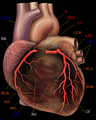
The left coronary artery is a coronary artery that arises from the aorta above the left cusp of the aortic valve, and feeds blood to the left side of the heart. It is also known as the left main coronary artery and the left main stem coronary artery.
In the blood supply of the heart, the right coronary artery (RCA) is an artery originating above the right cusp of the aortic valve, at the right aortic sinus in the heart. It travels down the right coronary sulcus, towards the crux of the heart. It supplies the right side of the heart, and the interventricular septum.

The thoracodorsal nerve is a nerve present in humans and other animals, also known as the middle subscapular nerve or the long subscapular nerve. It supplies the latissimus dorsi muscle.

The lingual artery arises from the external carotid artery between the superior thyroid artery and facial artery. It can be located easily in the tongue.

The cardiac plexus is a plexus of nerves situated at the base of the heart that innervates the heart.

The coronary sulcus is a groove on the surface of the heart that separates the atria from the ventricles. The structure contains the trunks of the nutrient vessels of the heart, and is deficient in front, where it is crossed by the root of the pulmonary trunk. On the posterior surface of the heart, the coronary sulcus contains the coronary sinus.

The "LCX", or left circumflex artery is an artery of the heart.
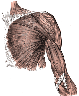
The bicipital aponeurosis is a broad aponeurosis of the biceps brachii, which is located in the cubital fossa of the elbow. It separates superficial from deep structures in much of the fossa.
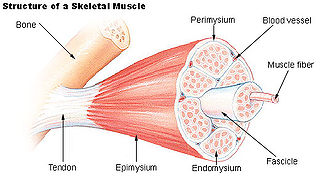
A muscle fascicle is a bundle of skeletal muscle fibers surrounded by perimysium, a type of connective tissue.

