Septin

Last updated
Cell division/GTP binding protein
Identifiers
SymbolCell_Div_GTP_bd
Pfam PF00735
Pfam clan CL0023
InterPro IPR000038
Available protein structures:
Pfam   structures / ECOD  
PDB RCSB PDB; PDBe; PDBj
PDBsum structure summary

Septins are a group of GTP-binding proteins expressed in all eukaryotic cells except plants. [1] [2] [3] Different septins form protein complexes with each other. These complexes can further assemble into filaments, rings and gauzes. Assembled as such, septins function in cells by localizing other proteins, either by providing a scaffold to which proteins can attach, or by forming a barrier preventing the diffusion of molecules from one compartment of the cell to another, [2] [3] [4] [5] or in the cell cortex as a barrier to the diffusion of membrane-bound proteins. [6]

Contents

Septins have been implicated in the localization of cellular processes at the site of cell division, and at the cell membrane at sites where specialized structures like cilia or flagella are attached to the cell body. [4] In yeast cells, they compartmentalize parts of the cell and build scaffolding to provide structural support during cell division at the septum, from which they derive their name. [3] Research in human cells suggests that septins build cages around pathogenic bacteria, that immobilize and prevent them from invading other cells. [7]

As filament forming proteins, septins can be considered part of the cytoskeleton. [4] Apart from forming non-polar filaments, septins associate with cell membranes, the cell cortex, actin filaments and microtubules. [4] [6]

Structure

schematic domain structure of septin polypeptide chain SeptinSequenceStructure v001.png
schematic domain structure of septin polypeptide chain
a) schematic of septin molecule with GTP binding domain to one side and the N and C termini of the polypeptide chain to the other
b) schematic of septin heterohexameric complex (of human septins), where different septins bind to each other via their GTP binding domains or via the N and C termini. Note the symmetry of the complex
c) schematic how septin complexes could align to form septin filaments Septin assembly.png
a) schematic of septin molecule with GTP binding domain to one side and the N and C termini of the polypeptide chain to the other
b) schematic of septin heterohexameric complex (of human septins), where different septins bind to each other via their GTP binding domains or via the N and C termini. Note the symmetry of the complex
c) schematic how septin complexes could align to form septin filaments

Septins are P-Loop-NTPase proteins that range in weight from 30-65 kDa. Septins are highly conserved between different eukaryotic species. They are composed of a variable-length proline rich N-terminus with a basic phosphoinositide binding motif important for membrane association, a GTP-binding domain, a highly conserved Septin Unique Element domain, and a C-terminal extension including a coiled coil domain of varying length. [4]

Septins interact either via their respective GTP-binding domains, or via both their N- and C-termini. Different organisms express a different number of septins, and from those symmetric oligomers are formed. For example, in yeast the octameric complex formed is Cdc11-Cdc12-Cdc3-Cdc10-Cdc10-Cdc3-Cdc12-Cdc11. [8] In humans, hexameric or octameric complexes are possible. Initially, it was indicated that the human complex was Sept7-Sept6-Sept2-Sept2-Sept6-Sept7; [9] but recently this order has been revised to Sept2-Sept6-Sept7-Sept7-Sept6-Sept2 [10] (or Sept2-Sept6-Sept7-Sept3-Sept3-Sept7-Sept6-Sept2 [11] in case of octameric hetero-oligomers). These complexes then associate to form non-polar filaments, filament bundles, cages or ring structures in cells. [4]

Occurrence

Septins are found in fungi, animals, and some eukaryotic algae but are not found in plants. [1]

In yeast

Septins in Saccharomyces cerevisiae (fluorescent micrograph)
* Green: septins (AgSEP7-GFP)
* Red: cell outline (phase contrast)
* Scale bar: 10 mm S cerevisiae septins.jpg
Septins in Saccharomyces cerevisiae (fluorescent micrograph)
• Green: septins (AgSEP7-GFP )
• Red: cell outline (phase contrast)
• Scale bar: 10 μm

There are seven different septins in Saccharomyces cerevisiae . Five of those are involved in mitosis, while two (Spr3 and Spr28) are specific to sporulation. [2] [3] Mitotic septins (Cdc3, Cdc10, Cdc11, Cdc12, Shs1) form a ring structure at the bud neck during cell division. [2] [4] They are involved in the selection of the bud-site, the positioning of the mitotic spindle, polarized growth, and cytokinesis. The sporulating septins (Spr3, Spr28) localize together with Cdc3 and Cdc11 to the edges of prospore membranes. [2]

Organization

Septins form a specialised region in the cell cortex known as the septin cortex. [12] The septin cortex undergoes several changes throughout the cell cycle: The first visible septin structure is a distinct ring which appears ~15 min before bud emergence. After bud emergence, the ring broadens to assume the shape of an hourglass around the mother-bud neck. During cytokinesis, the septin cortex splits into a double ring which eventually disappears. How can the septin cortex undergo such dramatic changes, although some of its functions may require it to be a stable structure? FRAP analysis has revealed that the turnover of septins at the neck undergoes multiple changes during the cell cycle. The predominant, functional conformation is characterized by a low turnover rate (frozen state), during which the septins are phosphorylated. Structural changes require a destabilization of the septin cortex (fluid state) induced by dephosphorylation prior to bud emergence, ring splitting and cell separation. [3]

The composition of the septin cortex does not only vary throughout the cell cycle but also along the mother-bud axis. This polarity of the septin network allows concentration of some proteins primarily to the mother side of the neck, some to the center and others to the bud site.

Functions

Scaffold

The septins act as a scaffold, recruiting many proteins. These protein complexes are involved in cytokinesis, chitin deposition, cell polarity, spore formation, in the morphogenesis checkpoint, spindle alignment checkpoint and bud site selection.

Cytokinesis

Budding yeast cytokinesis is driven through two septin dependent, redundant processes: recruitment and contraction of the actomyosin ring and formation of the septum by vesicle fusion with the plasma membrane. In contrast to septin mutants, disruption of one single pathway only leads to a delay in cytokinesis, not complete failure of cell division. Hence, the septins are predicted to act at the most upstream level of cytokinesis.

Cell polarity

After the isotropic-apical switch in budding yeast, cortical components, supposedly of the exocyst and polarisome, are delocalized from the apical pole to the entire plasma membrane of the bud, but not the mother cell. The septin ring at the neck serves as a cortical barrier that prevents membrane diffusion of these factors between the two compartments. This asymmetric distribution is abolished in septin mutants.

Some conditional septin mutants do not form buds at their normal axial location. Moreover, the typical localization of some bud-site-selection factors in a double ring at the neck is lost or disturbed in these mutants. This indicates that the septins may serve as anchoring site for such factors in axially budding cells.

In filamentous fungi

Since their discovery in S. cerevisiae, septin homologues have been found in other eukaryotic species, including filamentous fungi. Septins in filamentous fungi display a variety of different shapes within single cells, where they control aspects of filamentous morphology. [13] [14]

Candida albicans

The genome of C. albicans encodes homologues to all S. cerevisiae septins. Without Cdc3 and Cdc12 genes Candida albicans cannot proliferate, other septins affect morphology and chitin deposition, but are not essential. Candida albicans can display different morphologies of vegetative growth, which determines the appearance of septin structures. Newly forming hyphae form a septin ring at the base, Double rings form at sites of hyphal septation, and a septin cap forms at hyphal tips. Elongated septin-filaments encircle the spherical chlamydospores. Double rings of septins at the septation site also bear growth polarity, with the growing tip ring disassembling, while the basal ring remaining intact. [13]

Aspergillus nidulans

Five septins are found in A. nidulans (AnAspAp, AnAspBp, AnAspCp, AnAspDp, AnAspEp). AnAspBp forms single rings at septation sites that eventually split into double rings. Additionally, AnAspBp forms a ring at sites of branch emergence which broadens into a band as the branch grows. Like in C. albicans, double rings reflect polarity of the hypha. In the case of Aspergillus nidulans polarity is conveyed by disassembly of the more basal ring (the ring further away from the hyphal growth tip), leaving the apical ring intact, potentially as a growth guidance cue. [2] [13]

Ashbya gossypii

Septins in Ashbya gossypii (fluorescent micrograph) * Green: septins (AgSEP7-GFP)
* Red: cell outline (phase contrast)
* Inlay: 3D reconstruction of a discontinuous septin ring
* Scale bars: 10 mm A gossypii septins.jpg
Septins in Ashbya gossypii (fluorescent micrograph) • Green: septins (AgSEP7-GFP )
• Red: cell outline (phase contrast)
• Inlay: 3D reconstruction of a discontinuous septin ring
• Scale bars: 10 μm

The ascomycete A. gossypii possesses homologues to all S. cerevisiae septins, with one being duplicated (AgCDC3, AgCDC10, AgCDC11A, AgCDC11B, AgCDC12, AgSEP7). In vivo studies of AgSep7p-GFP have revealed that septins assemble into discontinuous hyphal rings close to growing tips and sites of branch formation, [2] and into asymmetric structures at the base of branching points. Rings are made of filaments which are long and diffuse close to growing tips and short and compact further away from the tip. During septum formation, the septin ring splits into two to form a double ring. Agcdc3Δ, Agcdc10Δ and Agcdc12Δ deletion mutants display aberrant morphology and are defective for actin-ring formation, chitin-ring formation, and sporulation. Due to the lack of septa, septin deletion mutants are highly sensitive, and damage of a single hypha can result in complete lysis of a young mycelium.

In animals

In contrast to septins in yeast, and in contrast to other cytoskeletal components of animals, septins do not form a continuous network in cells, but several dispersed ones in the cytoplasm of the cell cortex. These are integrated with actin bundles and microtubules. For example, the actin bundling protein anillin is required for correct spatial control of septin organization. [5] In the sperm cells of mammals, septins form a stable ring called annulus in the tail. In mice (and potentially in humans, too), defective annulus formation leads to male infertility. [4] [5]

Human

In humans, septins are involved in cytokinesis, cilium formation and neurogenesis through the capability to recruit other proteins or serve as a diffusion barrier. There are 13 different human genes coding for septins. The septin proteins produced by these genes are grouped into four subfamilies each named after its founding member: (i) SEPT2 (SEPT1, SEPT4, SEPT5), (ii) SEPT3 (SEPT9, SEPT12), (iii) SEPT6 (SEPT8, SEPT10, SEPT11, SEPT14), and (iv) SEPT7. Septin protein complexes are assembled to form either hetero-hexamers (incorporating monomers selected from three different groups and the monomer from each group is present in two copies; 3 x 2 = 6) or hetero-octamers (monomers from four different groups, each monomer present in two copies; 4 x 2 = 8). These hetero-oligomers in turn form higher-order structures such as filaments and rings. [4] [5] [1]

Septins form cage-like structures around bacterial pathogens, immobilizing harmful microbes and preventing them from invading healthy cells. This cellular defence system could potentially be exploited to create therapies for dysentery and other illnesses. For example, Shigella is a bacterium that causes lethal diarrhoea in humans. To propagate from cell to cell, Shigella bacteria develop actin-polymer 'tails', which propel the microbes and allow them to gain entry into neighbouring host cells. As part of the immune response, human cells produce a cell-signalling protein called TNF-α which trigger thick bundles of septin filaments to encircle the microbes within the infected host cell. [15] Microbes that become trapped in these septin cages are broken down by autophagy. [16] Disruptions in septins and mutations in the genes that code for them could be involved in causing leukaemia, colon cancer and neurodegenerative conditions such as Parkinson's disease and Alzheimer's disease. Potential therapies for these, as well as for bacterial conditions such as dysentery caused by Shigella, might bolster the body’s immune system with drugs that mimic the behaviour of TNF-α and allow the septin cages to proliferate. [7]

Caenorhabditis elegans

In the nematode worm Caenorhabditis elegans there are two genes coding for septins, and septin complexes contain the two different septins in a tetrameric UNC59-UNC61-UNC61-UNC59 complex. Septins in C.elegans concentrate at the cleavage furrow and the spindle midbody during cell division. Septins are also involved in cell migration and axon guidance in C.elegans. [2]

In mitochondria

The septin localized in the mitochondria is called mitochondrial septin (M-septin). It was identified as a CRMP/CRAM-interacting protein in the developing rat brain. [17]

History

The septins were discovered in 1970 by Leland H. Hartwell and colleagues in a screen for temperature-sensitive mutants affecting cell division (cdc mutants) in yeast ( Saccharomyces cerevisiae ). The screen revealed four mutants which prevented cytokinesis at restrictive temperature. The corresponding genes represent the four original septins, ScCDC3, ScCDC10, ScCDC11, and ScCDC12. [3] [4] Despite disrupted cytokinesis, the cells continued budding, DNA synthesis, and nuclear division, which resulted in large multinucleate cells with multiple, elongated buds. In 1976, analysis of electron micrographs revealed ~20 evenly spaced striations of 10-nm filaments around the mother-bud neck in wild-type but not in septin-mutant cells. [3] [4] [13] Immunofluorescence studies revealed that the septin proteins colocalize into a septin ring at the neck. [4] [13] The localization of all four septins is disrupted in conditional Sccdc3 and Sccdc12 mutants, indicating interdependence of the septin proteins. Strong support for this finding was provided by biochemical studies: The four original septins co-purified on affinity columns, together with a fifth septin protein, encoded by ScSEP7 or ScSHS1. Purified septins from budding yeast, Drosophila, Xenopus, and mammalian cells are able to self associate in vitro to form filaments. [13] How the septins interact in vitro to form hetero-oligomers that assemble into filaments was studied in detail in S. cerevisiae.

Micrographs of purified filaments raised the possibility that the septins are organized in parallel to the mother-bud axis. The 10-nm striations seen on electron micrographs may be the result of lateral interaction between the filaments. Mutant strains lacking factors important for septin organization support this view. Instead of continuous rings, the septins form bars oriented along the mother-bud axis in deletion mutants of ScGIN4, ScNAP1 and ScCLA4.

Related Research Articles

<span class="mw-page-title-main">Cytoskeleton</span> Network of filamentous proteins that forms the internal framework of cells

The cytoskeleton is a complex, dynamic network of interlinking protein filaments present in the cytoplasm of all cells, including those of bacteria and archaea. In eukaryotes, it extends from the cell nucleus to the cell membrane and is composed of similar proteins in the various organisms. It is composed of three main components: microfilaments, intermediate filaments, and microtubules, and these are all capable of rapid growth or disassembly depending on the cell's requirements.

<span class="mw-page-title-main">Cytokinesis</span> Part of the cell division process

Cytokinesis is the part of the cell division process and part of mitosis during which the cytoplasm of a single eukaryotic cell divides into two daughter cells. Cytoplasmic division begins during or after the late stages of nuclear division in mitosis and meiosis. During cytokinesis the spindle apparatus partitions and transports duplicated chromatids into the cytoplasm of the separating daughter cells. It thereby ensures that chromosome number and complement are maintained from one generation to the next and that, except in special cases, the daughter cells will be functional copies of the parent cell. After the completion of the telophase and cytokinesis, each daughter cell enters the interphase of the cell cycle.

<i>Saccharomyces cerevisiae</i> Species of yeast

Saccharomyces cerevisiae is a species of yeast. The species has been instrumental in winemaking, baking, and brewing since ancient times. It is believed to have been originally isolated from the skin of grapes. It is one of the most intensively studied eukaryotic model organisms in molecular and cell biology, much like Escherichia coli as the model bacterium. It is the microorganism behind the most common type of fermentation. S. cerevisiae cells are round to ovoid, 5–10 μm in diameter. It reproduces by budding.

<span class="mw-page-title-main">FtsZ</span> Protein encoded by the ftsZ gene

FtsZ is a protein encoded by the ftsZ gene that assembles into a ring at the future site of bacterial cell division. FtsZ is a prokaryotic homologue of the eukaryotic protein tubulin. The initials FtsZ mean "Filamenting temperature-sensitive mutant Z." The hypothesis was that cell division mutants of E. coli would grow as filaments due to the inability of the daughter cells to separate from one another. FtsZ is found in almost all bacteria, many archaea, all chloroplasts and some mitochondria, where it is essential for cell division. FtsZ assembles the cytoskeletal scaffold of the Z ring that, along with additional proteins, constricts to divide the cell in two.

The exocyst is an octameric protein complex involved in vesicle trafficking, specifically the tethering and spatial targeting of post-Golgi vesicles to the plasma membrane prior to vesicle fusion. It is implicated in a number of cell processes, including exocytosis, cell migration, and growth.

<span class="mw-page-title-main">Cell cortex</span> Layer on the inner face of a cell membrane

The cell cortex, also known as the actin cortex, cortical cytoskeleton or actomyosin cortex, is a specialized layer of cytoplasmic proteins on the inner face of the cell membrane. It functions as a modulator of membrane behavior and cell surface properties. In most eukaryotic cells lacking a cell wall, the cortex is an actin-rich network consisting of F-actin filaments, myosin motors, and actin-binding proteins. The actomyosin cortex is attached to the cell membrane via membrane-anchoring proteins called ERM proteins that plays a central role in cell shape control. The protein constituents of the cortex undergo rapid turnover, making the cortex both mechanically rigid and highly plastic, two properties essential to its function. In most cases, the cortex is in the range of 100 to 1000 nanometers thick.

<i>Eremothecium gossypii</i> Species of fungus

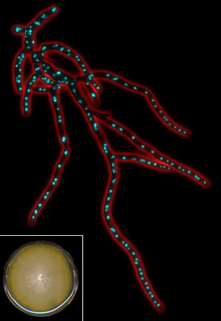
Eremothecium gossypii (also known as Ashbya gossypii) is a filamentous fungus or mold closely related to yeast, but growing exclusively in a filamentous way. It was originally isolated from cotton as a pathogen causing stigmatomycosis by Ashby and Nowell in 1926. This disease affects the development of hair cells in cotton bolls and can be transmitted to citrus fruits, which thereupon dry out and collapse (dry rot disease). In the first part of the 20th century, E. gossypii and two other fungi causing stigmatomycosis (Eremothecium coryli, Aureobasidium pullulans) made it virtually impossible to grow cotton in certain regions of the subtropics, causing severe economical losses. Control of the spore-transmitting insects - cotton stainer (Dysdercus suturellus) and Antestiopsis (antestia bugs) - permitted full eradication of infections. E. gossypii was recognized as a natural overproducer of riboflavin (vitamin B2), which protects its spores against ultraviolet light. This made it an interesting organism for industries, where genetically modified strains are still used to produce this vitamin.

The Rho family of GTPases is a family of small signaling G proteins, and is a subfamily of the Ras superfamily. The members of the Rho GTPase family have been shown to regulate many aspects of intracellular actin dynamics, and are found in all eukaryotic kingdoms, including yeasts and some plants. Three members of the family have been studied in detail: Cdc42, Rac1, and RhoA. All G proteins are "molecular switches", and Rho proteins play a role in organelle development, cytoskeletal dynamics, cell movement, and other common cellular functions.

<span class="mw-page-title-main">Prokaryotic cytoskeleton</span> Structural filaments in prokaryotes

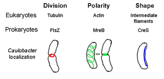
The prokaryotic cytoskeleton is the collective name for all structural filaments in prokaryotes. It was once thought that prokaryotic cells did not possess cytoskeletons, but advances in visualization technology and structure determination led to the discovery of filaments in these cells in the early 1990s. Not only have analogues for all major cytoskeletal proteins in eukaryotes been found in prokaryotes, cytoskeletal proteins with no known eukaryotic homologues have also been discovered. Cytoskeletal elements play essential roles in cell division, protection, shape determination, and polarity determination in various prokaryotes.

<span class="mw-page-title-main">SEPT2</span> Protein-coding gene in the species Homo sapiens

Septin 2, also known as SEPT2, is a protein which in humans is encoded by the SEPT2 gene.

<span class="mw-page-title-main">SEPT6</span> Protein-coding gene in the species Homo sapiens

Septin-6 is a protein that in humans is encoded by the SEPT6 gene.

<span class="mw-page-title-main">ANLN</span> Mammalian protein found in Homo sapiens

Anillin is a conserved protein implicated in cytoskeletal dynamics during cellularization and cytokinesis. The ANLN gene in humans and the scraps gene in Drosophila encode Anillin. In 1989, anillin was first isolated in embryos of Drosophila melanogaster. It was identified as an F-actin binding protein. Six years later, the anillin gene was cloned from cDNA originating from a Drosophila ovary. Staining with anti-anillin antibody showed the anillin localizes to the nucleus during interphase and to the contractile ring during cytokinesis. These observations agree with further research that found anillin in high concentrations near the cleavage furrow coinciding with RhoA, a key regulator of contractile ring formation.

<span class="mw-page-title-main">SEPT7</span> Protein-coding gene in the species Homo sapiens

Septin-7 is a protein that in humans is encoded by the SEPT7 gene.

The endosomal sorting complexes required for transport (ESCRT) machinery is made up of cytosolic protein complexes, known as ESCRT-0, ESCRT-I, ESCRT-II, and ESCRT-III. Together with a number of accessory proteins, these ESCRT complexes enable a unique mode of membrane remodeling that results in membranes bending/budding away from the cytoplasm. These ESCRT components have been isolated and studied in a number of organisms including yeast and humans. A eukaryotic signature protein, the machinery is found in all eukaryotes and some archaea.

<span class="mw-page-title-main">Cell polarity</span> Polar morphology of a cell, a specific orientation of the cell structure

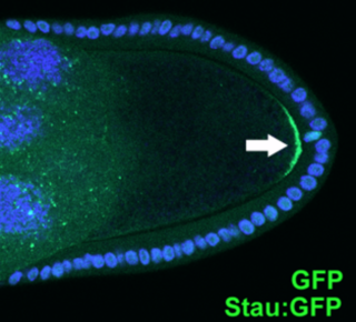
Cell polarity refers to spatial differences in shape, structure, and function within a cell. Almost all cell types exhibit some form of polarity, which enables them to carry out specialized functions. Classical examples of polarized cells are described below, including epithelial cells with apical-basal polarity, neurons in which signals propagate in one direction from dendrites to axons, and migrating cells. Furthermore, cell polarity is important during many types of asymmetric cell division to set up functional asymmetries between daughter cells.

Cdc14 and Cdc14 are a gene and its protein product respectively. Cdc14 is found in most of the eukaryotes. Cdc14 was defined by Hartwell in his famous screen for loci that control the cell cycle of Saccharomyces cerevisiae. Cdc14 was later shown to encode a protein phosphatase. Cdc14 is dual-specificity, which means it has serine/threonine and tyrosine-directed activity. A preference for serines next to proline is reported. Many early studies, especially in the budding yeast Saccharomyces cerevisiae, demonstrated that the protein plays a key role in regulating late mitotic processes. However, more recent work in a range of systems suggests that its cellular function is more complex.

<span class="mw-page-title-main">Cyclase-associated protein family</span>

In molecular biology, the cyclase-associated protein family (CAP) is a family of highly conserved actin-binding proteins present in a wide range of organisms including yeast, flies, plants, and mammals. CAPs are multifunctional proteins that contain several structural domains. CAP is involved in species-specific signalling pathways. In Drosophila, CAP functions in Hedgehog-mediated eye development and in establishing oocyte polarity. In Dictyostelium discoideum, CAP is involved in microfilament reorganisation near the plasma membrane in a PIP2-regulated manner and is required to perpetuate the cAMP relay signal to organise fruitbody formation. In plants, CAP is involved in plant signalling pathways required for co-ordinated organ expansion. In yeast, CAP is involved in adenylate cyclase activation, as well as in vesicle trafficking and endocytosis. In both yeast and mammals, CAPs appear to be involved in recycling G-actin monomers from ADF/cofilins for subsequent rounds of filament assembly. In mammals, there are two different CAPs that share 64% amino acid identity.

<span class="mw-page-title-main">Rong Li</span> American cell biologist (born 1967)

Rong Li is the Director of Mechanobiology Institute, a Singapore Research Center of Excellence, at the National University of Singapore. She is a Distinguished Professor at the National University of Singapore's Department of Biological Sciences and Bloomberg Distinguished Professor of Cell Biology and Chemical & Biomolecular Engineering at the Johns Hopkins School of Medicine and Whiting School of Engineering. She previously served as Director of Center for Cell Dynamics in the Johns Hopkins School of Medicine’s Institute for Basic Biomedical Sciences. She is a leader in understanding cellular asymmetry, division and evolution, and specifically, in how eukaryotic cells establish their distinct morphology and organization in order to carry out their specialized functions.

<span class="mw-page-title-main">Amy Gladfelter</span> American cell biologist (born 1974)

Amy S. Gladfelter is an American quantitative cell biologist who is interested in understanding fundamental mechanisms of cell organization. She is a Professor of Biology and the Associate Chair for Diversity Initiatives at the University of North Carolina at Chapel Hill, where she investigates cell cycle control and the septin cytoskeleton. She is also affiliated with the Lineberger Comprehensive Cancer Center and is a fellow of the Marine Biological Laboratory in Woods Hole, MA.

<span class="mw-page-title-main">Actomyosin ring</span> Cellular formation during cytokinesis

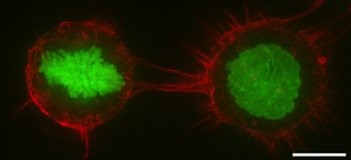
In molecular biology, an actomyosin ring or contractile ring, is a prominent structure during cytokinesis. It forms perpendicular to the axis of the spindle apparatus towards the end of telophase, in which sister chromatids are identically separated at the opposite sides of the spindle forming nuclei. The actomyosin ring follows an orderly sequence of events: identification of the active division site, formation of the ring, constriction of the ring, and disassembly of the ring. It is composed of actin and myosin II bundles, thus the term actomyosin. The actomyosin ring operates in contractile motion, although the mechanism on how or what triggers the constriction is still an evolving topic. Other cytoskeletal proteins are also involved in maintaining the stability of the ring and driving its constriction. Apart from cytokinesis, in which the ring constricts as the cells divide, actomyosin ring constriction has also been found to activate during wound closure. During this process, actin filaments are degraded, preserving the thickness of the ring. After cytokinesis is complete, one of the two daughter cells inherits a remnant known as the midbody ring.

References

  1. 1 2 3 Neubauer, K; Zieger, B (2017). "The Mammalian Septin Interactome". Frontiers in Cell and Developmental Biology. 5: 3. doi: 10.3389/fcell.2017.00003 . PMC   5293755 . PMID   28224124.
  2. 1 2 3 4 5 6 7 8 9 Weirich CS, Erzberger JP, Barral Y (2008). "The septin family of GTPases: architecture and dynamics". Nat. Rev. Mol. Cell Biol. 9 (6): 478–89. doi:10.1038/nrm2407. PMID   18478031. S2CID   2640351.
  3. 1 2 3 4 5 6 7 Douglas LM, Alvarez FJ, McCreary C, Konopka JB (2005). "Septin function in yeast model systems and pathogenic fungi". Eukaryotic Cell. 4 (9): 1503–12. doi:10.1128/EC.4.9.1503-1512.2005. PMC   1214204 . PMID   16151244.
  4. 1 2 3 4 5 6 7 8 9 10 11 12 13 Mostowy S, Cossart P (2012). "Septins: the fourth component of the cytoskeleton". Nat. Rev. Mol. Cell Biol. 13 (3): 183–94. doi:10.1038/nrm3284. PMID   22314400. S2CID   2418522.
  5. 1 2 3 4 Kinoshita M (2006). "Diversity of septin scaffolds". Curr. Opin. Cell Biol. 18 (1): 54–60. doi:10.1016/j.ceb.2005.12.005. PMID   16356703.
  6. 1 2 Bridges, AA; Gladfelter, AS (10 July 2015). "Septin Form and Function at the Cell Cortex". The Journal of Biological Chemistry. 290 (28): 17173–80. doi: 10.1074/jbc.R114.634444 . PMC   4498057 . PMID   25957401.
  7. 1 2 Mascarelli A (December 2011). "Septin proteins take bacterial prisoners: A cellular defence against microbial pathogens holds therapeutic potential". Nature. doi:10.1038/nature.2011.9540. S2CID   85080734.
  8. Bertin, A.; McMurray, M. A.; Grob, P.; Park, S.-S.; Garcia, G.; Patanwala, I.; Ng, H.-l.; Alber, T.; Thorner, J.; Nogales, E. (2008-06-12). "Saccharomyces cerevisiae septins: Supramolecular organization of heterooligomers and the mechanism of filament assembly". Proceedings of the National Academy of Sciences. 105 (24): 8274–8279. Bibcode:2008PNAS..105.8274B. doi: 10.1073/pnas.0803330105 . ISSN   0027-8424. PMC   2426963 . PMID   18550837.
  9. Sirajuddin, Minhajuddin; Farkasovsky, Marian; Hauer, Florian; Kühlmann, Dorothee; Macara, Ian G.; Weyand, Michael; Stark, Holger; Wittinghofer, Alfred (2007-07-18). "Structural insight into filament formation by mammalian septins". Nature. 449 (7160): 311–315. Bibcode:2007Natur.449..311S. doi:10.1038/nature06052. ISSN   0028-0836. PMID   17637674.
  10. Mendonça, Deborah C.; Macedo, Joci N.; Guimarães, Samuel L.; Barroso da Silva, Fernando L.; Cassago, Alexandre; Garratt, Richard C.; Portugal, Rodrigo V.; Araujo, Ana P. U. (September 2019). "A revised order of subunits in mammalian septin complexes". Cytoskeleton. 76 (9–10): 457–466. doi:10.1002/cm.21569. ISSN   1949-3584. PMID   31608568. S2CID   204536675.
  11. Soroor, Forooz; Kim, Moshe S.; Palander, Oliva; Balachandran, Yadu; Collins, Richard; Benlekbir, Samir; Rubinstein, John; Trimble, William S. (2019-03-07). "Revised subunit order of mammalian septin complexes explains their in vitro polymerization properties". bioRxiv: 569871. doi:10.1101/569871. hdl: 1807/109120 . S2CID   92158262 . Retrieved 2021-03-19.
  12. Gladfelter, AS; Pringle, JR; Lew, DJ (December 2001). "The septin cortex at the yeast mother-bud neck". Current Opinion in Microbiology. 4 (6): 681–9. doi:10.1016/s1369-5274(01)00269-7. PMID   11731320.
  13. 1 2 3 4 5 6 Gladfelter AS (2006). "Control of filamentous fungal cell shape by septins and formins". Nat. Rev. Microbiol. 4 (3): 223–9. doi:10.1038/nrmicro1345. PMID   16429163. S2CID   40080522.
  14. Harris, SD (2006). "Cell polarity in filamentous fungi: shaping the mold". International Review of Cytology. 251: 41–77. doi:10.1016/S0074-7696(06)51002-2. ISBN   9780123646552. PMID   16939777.
  15. Mostowy S, Bonazzi M, Hamon MA, Tham TN, Mallet A, Lelek M, Gouin E, Demangel C, Brosch R, Zimmer C, Sartori A, Kinoshita M, Lecuit M, Cossart P (2010). "Entrapment of intracytosolic bacteria by septin cage-like structures". Cell Host Microbe. 8 (5): 433–44. doi: 10.1016/j.chom.2010.10.009 . PMID   21075354.
  16. Mostowy S, Sancho-Shimizu V, Hamon MA, Simeone R, Brosch R, Johansen T, Cossart P (2011). "p62 and NDP52 proteins target intracytosolic Shigella and Listeria to different autophagy pathways". J. Biol. Chem. 286 (30): 26987–95. doi: 10.1074/jbc.M111.223610 . PMC   3143657 . PMID   21646350.
  17. Takahashi S, Inatome R, Yamamura H, Yanagi S (February 2003). "Isolation and expression of a novel mitochondrial septin that interacts with CRMP/CRAM in the developing neurones". Genes Cells. 8 (2): 81–93. doi: 10.1046/j.1365-2443.2003.00617.x . PMID   12581152.

Further reading