FtsA

Last updated
Cell division protein FtsA
FtsA-GFP.jpg
E. coli cells producing FtsA-GFP, which localizes to the cell division site.
Identifiers
Organism Escherichia coli (strain K12)
SymbolFtsA
Entrez 944778
RefSeq (Prot) NP_414636.1
UniProt P0ABH0
Other data
Chromosome Genome: 0.1 - 0.11 Mb
Search for
Structures Swiss-model
Domains InterPro
FtsA
Identifiers
SymbolFtsA
InterPro IPR020823
SHS2 "1C" domain inserted in FtsA
Identifiers
SymbolSHS2_FtsA
Pfam PF02491
InterPro IPR003494
SMART SM00842
Available protein structures:
Pfam   structures / ECOD  
PDB RCSB PDB; PDBe; PDBj
PDBsum structure summary

FtsA is a bacterial protein that is related to actin by overall structural similarity and in its ATP binding pocket. [1] [2] [3]

Contents

Along with other bacterial actin homologs such as MreB, ParM, and MamK, these proteins suggest that eukaryotic actin has a common ancestry. Like the other bacterial actins, FtsA binds ATP and can form actin-like filaments. [4] The FtsA-FtsA interface has been defined by structural as well as genetic analysis. [5] Although present in many diverse Gram-positive and Gram-negative species, FtsA is absent in actinobacteria and cyanobacteria. FtsA also is structurally similar to PilM, a type IV pilus ATPase. [6]

Function

FtsA is required for proper cytokinesis in bacteria such as Escherichia coli, Caulobacter crescentus, and Bacillus subtilis. Originally isolated in a screen for E. coli cells that could divide at 30˚C but not at 40˚C, [7] FtsA stands for "filamentous temperature sensitive A". Many thermosensitive alleles of E. coli ftsA exist, and all map in or near the ATP binding pocket. Suppressors that restore normal function map either to the binding pocket or to the FtsA-FtsA interface. [8]

FtsA localizes to the cytokinetic ring formed by FtsZ (Z ring). One of FtsA's functions in cytokinesis is to tether FtsZ polymers to the cytoplasmic membrane via a conserved C-terminal amphipathic helix, forming an "A ring" in the process. [9] Removal of this helix results in the formation of very long and stable polymer bundles of FtsA in the cell that do not function in cytokinesis. [5] Another essential division protein, ZipA, also tethers the Z ring to the membrane and exhibits overlapping function with FtsA. FtsZ, FtsA and ZipA together are called the proto-ring because they are involved in a specific initial phase of cytokinesis. [10] Another subdomain of FtsA (2B) is required for interactions with FtsZ, via the conserved C-terminus of FtsZ. [4] Other FtsZ regulators including MinC and ZipA bind to the same C terminus of FtsZ. Finally, subdomain 1C, which is in a unique position relative to MreB and actin, is required for FtsA to recruit downstream cell division proteins such as FtsN. [11] [12]

Although FtsA is essential for viability in E. coli, it can be deleted in B. subtilis. B. subtilis cells lacking FtsA divide poorly, but still survive. Another FtsZ-interacting protein, SepF (originally named YlmF; O31728 ), is able to replace FtsA in B. subtilis, suggesting that SepF and FtsA have overlapping functions. [13]

An allele of FtsA called FtsA* (R286W) is able to bypass the normal requirement for the ZipA in E. coli cytokinesis. [14] FtsA* also causes cells to divide at a shorter cell length than normal, suggesting that FtsA may normally receive signals from the septum synthesis machinery to regulate when cytokinesis can proceed. [15] Other FtsA*-like alleles have been found, and they mostly decrease FtsA-FtsA interactions. [5] Oligomeric state of FtsA is likely important for regulating its activity, its ability to recruit the later cell division proteins [5] and its ability to bind ATP. [8] Other cell division proteins of E. coli, including FtsN and the ABC transporter homologs FtsEX, seem to regulate septum constriction by signaling through FtsA, [16] [17] and the FtsQLB subcomplex is also involved in promoting FtsN-mediated septal constriction. [18] [19]

FtsA binds directly to the conserved C-terminal domain of FtsZ. [20] [4] This FtsA-FtsZ interaction is likely involved in regulating FtsZ polymer dynamics. In vitro, E. coli FtsA disassembles FtsZ polymers in the presence of ATP, both in solution, as FtsA* [21] and on supported lipid bilayers. [22] E. coli FtsA itself does not assemble into detectable structures except when on membranes, where it forms dodecameric minirings that often pack in clusters and bind to single FtsZ protofilaments. [23] In contrast, FtsA* forms arcs on lipid membranes but rarely closed minirings, supporting genetic evidence that this mutant has a weaker FtsA-FtsA interface. [5] When bound to the membrane, FtsA*-like mutants, which also can form double-stranded filaments, enhance close lateral interactions between FtsZ protofilaments, in contrast to FtsA, which keeps FtsZ protofilaments apart. [24] As FtsZ protofilament bundling may be important for promoting septum formation, a switch from an FtsA-like to an FtsA*-like conformation during cell cycle progression may serve to turn on septum synthesis enzymes (FtsWI) as well as condense FtsZ polymers, setting up a positive feedback loop. In support of this model, the cytoplasmic domain of FtsN, which activates FtsWI in E. coli and interacts directly with the 1C subdomain of FtsA, switches FtsA from the miniring form to the double stranded filament form on lipid surfaces in vitro. [25] These double filaments of E. coli FtsA are antiparallel, indicating that they themselves do not treadmill like FtsZ filaments.

Although E. coli FtsA has been the most extensively studied, more is becoming understood about FtsA proteins from other species. FtsA from Streptococcus pneumoniae forms helical filaments in the presence of ATP, [26] but no interactions with FtsZ in vitro have been reported yet. FtsA colocalizes with FtsZ in S. pneumoniae, but also is required for FtsZ ring localization, in contrast to E. coli where FtsZ rings remain localized upon inactivation of FtsA. FtsA from Staphylococcus aureus forms actin-like filaments similar to those of FtsA from Thermotoga maritima. [27] In addition, S. aureus FtsA enhances the GTPase activity of FtsZ. In a liposome system, FtsA* stimulates FtsZ to form rings that can divide liposomes, mimicking cytokinesis in vitro. [28]

Structure

Several crystal structures for FtsA are known, including a structure for E. coli FtsA. [29] Compared to MreB and eukaryotic actin, the subdomains are rearranged, and the 1B domain is swapped out for the SHS2 "1C" insert. [4] [30] [1] [31]

Related Research Articles

<span class="mw-page-title-main">Cytoskeleton</span> Network of filamentous proteins that forms the internal framework of cells

The cytoskeleton is a complex, dynamic network of interlinking protein filaments present in the cytoplasm of all cells, including those of bacteria and archaea. In eukaryotes, it extends from the cell nucleus to the cell membrane and is composed of similar proteins in the various organisms. It is composed of three main components, microfilaments, intermediate filaments and microtubules, and these are all capable of rapid growth or disassembly dependent on the cell's requirements.

<span class="mw-page-title-main">Cytokinesis</span> Part of the cell division process

Cytokinesis is the part of the cell division process during which the cytoplasm of a single eukaryotic cell divides into two daughter cells. Cytoplasmic division begins during or after the late stages of nuclear division in mitosis and meiosis. During cytokinesis the spindle apparatus partitions and transports duplicated chromatids into the cytoplasm of the separating daughter cells. It thereby ensures that chromosome number and complement are maintained from one generation to the next and that, except in special cases, the daughter cells will be functional copies of the parent cell. After the completion of the telophase and cytokinesis, each daughter cell enters the interphase of the cell cycle.

<span class="mw-page-title-main">MreB</span>

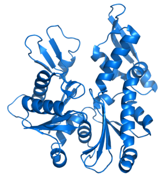
MreB is a protein found in bacteria that has been identified as a homologue of actin, as indicated by similarities in tertiary structure and conservation of active site peptide sequence. The conservation of protein structure suggests the common ancestry of the cytoskeletal elements formed by actin, found in eukaryotes, and MreB, found in prokaryotes. Indeed, recent studies have found that MreB proteins polymerize to form filaments that are similar to actin microfilaments. It has been shown to form multilayer sheets comprising diagonally interwoven filaments in the presence of ATP or GTP.

<span class="mw-page-title-main">FtsZ</span> Protein encoded by the ftsZ gene

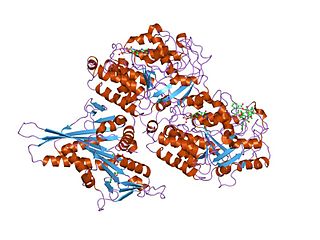
FtsZ is a protein encoded by the ftsZ gene that assembles into a ring at the future site of bacterial cell division. FtsZ is a prokaryotic homologue of the eukaryotic protein tubulin. The initials FtsZ mean "Filamenting temperature-sensitive mutant Z." The hypothesis was that cell division mutants of E. coli would grow as filaments due to the inability of the daughter cells to separate from one another. FtsZ is found in almost all bacteria, many archaea, all chloroplasts and some mitochondria, where it is essential for cell division. FtsZ assembles the cytoskeletal scaffold of the Z ring that, along with additional proteins, constricts to divide the cell in two.

<span class="mw-page-title-main">Tubulin</span> Superfamily of proteins that make up microtubules

Tubulin in molecular biology can refer either to the tubulin protein superfamily of globular proteins, or one of the member proteins of that superfamily. α- and β-tubulins polymerize into microtubules, a major component of the eukaryotic cytoskeleton. Microtubules function in many essential cellular processes, including mitosis. Tubulin-binding drugs kill cancerous cells by inhibiting microtubule dynamics, which are required for DNA segregation and therefore cell division.

<span class="mw-page-title-main">Filamentation</span>

Filamentation is the anomalous growth of certain bacteria, such as Escherichia coli, in which cells continue to elongate but do not divide. The cells that result from elongation without division have multiple chromosomal copies.

<span class="mw-page-title-main">Treadmilling</span> Simultaneous growth and breakdown on opposite ends of a protein filament

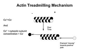
In molecular biology, treadmilling is a phenomenon observed within protein filaments of the cytoskeletons of many cells, especially in actin filaments and microtubules. It occurs when one end of a filament grows in length while the other end shrinks, resulting in a section of filament seemingly "moving" across a stratum or the cytosol. This is due to the constant removal of the protein subunits from these filaments at one end of the filament, while protein subunits are constantly added at the other end. Treadmilling was discovered by Wegner, who defined the thermodynamic and kinetic constraints. Wegner recognized that: “The equilibrium constant (K) for association of a monomer with a polymer is the same at both ends, since the addition of a monomer to each end leads to the same polymer.”; a simple reversible polymer can’t treadmill; ATP hydrolysis is required. GTP is hydrolyzed for microtubule treadmilling.

<span class="mw-page-title-main">DicF RNA</span> Non-coding RNA

DicF RNA is a non-coding RNA that is an antisense inhibitor of cell division gene ftsZ. DicF is bound by the Hfq protein which enhances its interaction with its targets. Pathogenic E. coli strains possess multiple copies of sRNA DicF in their genomes, while non-pathogenic strains do not. DicF and Hfq are both necessary to reduce FtsZ protein levels, leading to cell filamentation under anaerobic conditions.

<span class="mw-page-title-main">Prokaryotic cytoskeleton</span> Structural filaments in prokaryotes

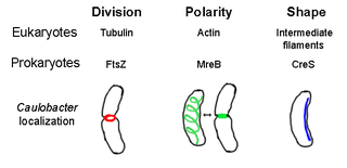
The prokaryotic cytoskeleton is the collective name for all structural filaments in prokaryotes. It was once thought that prokaryotic cells did not possess cytoskeletons, but advances in visualization technology and structure determination led to the discovery of filaments in these cells in the early 1990s. Not only have analogues for all major cytoskeletal proteins in eukaryotes been found in prokaryotes, cytoskeletal proteins with no known eukaryotic homologues have also been discovered. Cytoskeletal elements play essential roles in cell division, protection, shape determination, and polarity determination in various prokaryotes.

Fission, in biology, is the division of a single entity into two or more parts and the regeneration of those parts to separate entities resembling the original. The object experiencing fission is usually a cell, but the term may also refer to how organisms, bodies, populations, or species split into discrete parts. The fission may be binary fission, in which a single organism produces two parts, or multiple fission, in which a single entity produces multiple parts.

Bacterial morphological plasticity refers to changes in the shape and size that bacterial cells undergo when they encounter stressful environments. Although bacteria have evolved complex molecular strategies to maintain their shape, many are able to alter their shape as a survival strategy in response to protist predators, antibiotics, the immune response, and other threats.

<span class="mw-page-title-main">Min System</span> Mechanism used by E. coli in cell division

The Min System is a mechanism composed of three proteins MinC, MinD, and MinE used by E. coli as a means of properly localizing the septum prior to cell division. Each component participates in generating a dynamic oscillation of FtsZ protein inhibition between the two bacterial poles to precisely specify the mid-zone of the cell, allowing the cell to accurately divide in two. This system is known to function in conjunction with a second negative regulatory system, the nucleoid occlusion system (NO), to ensure proper spatial and temporal regulation of chromosomal segregation and division.

The MinC protein is one of three proteins in the Min system encoded by the minB operon and which is required to generate pole to pole oscillations prior to bacterial cell division as a means of specifying the midzone of the cell. This function is achieved by preventing the formation of the divisome Z-ring around the poles.

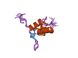
The MinE protein is one of three proteins of the Min system encoded by the minB operon required to generate pole to pole oscillations prior to bacterial cell division as a means of specifying the midzone of the cell, as seen in E.coli.

Joe Lutkenhaus is a professor at the University of Kansas Medical Center. He received a B.S. in organic chemistry from Iowa state University and then a PhD in biochemistry for the University of California, Los Angeles. Following his PhD, Lutkenhaus pursued his postdoctoral studies with William Donachie at the University of Edinburgh and then continued at the University of Connecticut Health Science center. In 2002, Lutkenhaus became a fellow of the American Academy of Microbiology.

The bacterial murein precursor exporter (MPE) family is a member of the cation diffusion facilitator (CDF) superfamily of membrane transporters. Members of the MPE family are found in a large variety of Gram-negative and Gram-positive bacteria and facilitate the translocation of lipid-linked murein precursors. A representative list of proteins belonging to the MPE family can be found in the Transporter Classification Database.

<span class="mw-page-title-main">Divisome</span> A protein complex in bacteria responsible for cell division

The divisome is a protein complex in bacteria that is responsible for cell division, constriction of inner and outer membranes during division, and peptidoglycan (PG) synthesis at the division site. The divisome is a membrane protein complex with proteins on both sides of the cytoplasmic membrane. In gram-negative cells it is located in the inner membrane. The divisome is nearly ubiquitous in bacteria although its composition may vary between species.

<span class="mw-page-title-main">Bacterial secretion system</span> Protein complexes present on the cell membranes of bacteria for secretion of substances

Bacterial secretion systems are protein complexes present on the cell membranes of bacteria for secretion of substances. Specifically, they are the cellular devices used by pathogenic bacteria to secrete their virulence factors to invade the host cells. They can be classified into different types based on their specific structure, composition and activity. Generally, proteins can be secreted through two different processes. One process is a one-step mechanism in which proteins from the cytoplasm of bacteria are transported and delivered directly through the cell membrane into the host cell. Another involves a two-step activity in which the proteins are first transported out of the inner cell membrane, then deposited in the periplasm, and finally through the outer cell membrane into the host cell.

<span class="mw-page-title-main">FtsK</span> Protein involved in bacterial cell division

FtsK, discovered in 1995 by the Donachie lab, is one of the largest proteins in E. coli at 1329 amino acids. It is involved in bacterial cell division and chromosome segregation. FtsK stands for "Filament temperature sensitive mutant K" because cells expressing a mutant ftsK allele called ftsK44, which encodes an FtsK variant containing an E58A residue change in the first periplasmic loop, fail to divide at high temperatures and form long filaments instead. FtsK, specifically its C-terminal domain, functions as a DNA translocase, interacts with other cell division proteins, and regulates Xer-mediated recombination. FtsK belongs to the AAA superfamily and is present in most bacteria.

In bacteriology, minicells are bacterial cells that are smaller than usual. The first minicells reported were from a strain of Escherichia coli that had a mutation in the Min System that lead to mis-localization of the septum during cell division and the production of cells of random sizes.

References

  1. 1 2 van den Ent F, Löwe J (Oct 2000). "Crystal structure of the cell division protein FtsA from Thermotoga maritima". The EMBO Journal. 19 (20): 5300–7. doi:10.1093/emboj/19.20.5300. PMC   313995 . PMID   11032797.
  2. Gunning PW, Ghoshdastider U, Whitaker S, Popp D, Robinson RC (Jun 2015). "The evolution of compositionally and functionally distinct actin filaments". Journal of Cell Science. 128 (11): 2009–19. doi: 10.1242/jcs.165563 . PMID   25788699.
  3. Ghoshdastider U, Jiang S, Popp D, Robinson RC (Jul 2015). "In search of the primordial actin filament". Proceedings of the National Academy of Sciences of the United States of America. 112 (30): 9150–1. doi: 10.1073/pnas.1511568112 . PMC   4522752 . PMID   26178194.
  4. 1 2 3 4 Szwedziak P, Wang Q, Freund SM, Löwe J (May 2012). "FtsA forms actin-like protofilaments". The EMBO Journal. 31 (10): 2249–60. doi:10.1038/emboj.2012.76. PMC   3364754 . PMID   22473211.
  5. 1 2 3 4 5 Pichoff S, Shen B, Sullivan B, Lutkenhaus J (Jan 2012). "FtsA mutants impaired for self-interaction bypass ZipA suggesting a model in which FtsA's self-interaction competes with its ability to recruit downstream division proteins". Molecular Microbiology. 83 (1): 151–67. doi:10.1111/j.1365-2958.2011.07923.x. PMC   3245357 . PMID   22111832.
  6. Karuppiah V, Derrick JP (Jul 2011). "Structure of the PilM-PilN inner membrane type IV pilus biogenesis complex from Thermus thermophilus". The Journal of Biological Chemistry. 286 (27): 24434–42. doi: 10.1074/jbc.M111.243535 . PMC   3129222 . PMID   21596754.
  7. Kohiyama M, Cousin D, Ryter A, Jacob F (April 1966). "Mutants thermosensibles d'Escherichia coli K12". Annales de l'Institute Pasteur. 110 (4): 465–86.
  8. 1 2 Herricks JR, Nguyen D, Margolin W (Nov 2014). "A thermosensitive defect in the ATP binding pocket of FtsA can be suppressed by allosteric changes in the dimer interface". Molecular Microbiology. 94 (3): 713–27. doi:10.1111/mmi.12790. PMC   4213309 . PMID   25213228.
  9. Pichoff S, Lutkenhaus J (Mar 2005). "Tethering the Z ring to the membrane through a conserved membrane targeting sequence in FtsA". Molecular Microbiology. 55 (6): 1722–34. doi: 10.1111/j.1365-2958.2005.04522.x . PMID   15752196.
  10. Rico AI, Krupka M, Vicente M (Jul 2013). "In the beginning, Escherichia coli assembled the proto-ring: an initial phase of division". The Journal of Biological Chemistry. 288 (29): 20830–6. doi: 10.1074/jbc.R113.479519 . PMC   3774354 . PMID   23740256.
  11. Rico AI, García-Ovalle M, Mingorance J, Vicente M (Sep 2004). "Role of two essential domains of Escherichia coli FtsA in localization and progression of the division ring". Molecular Microbiology. 53 (5): 1359–71. doi: 10.1111/j.1365-2958.2004.04245.x . PMID   15387815.
  12. Busiek KK, Eraso JM, Wang Y, Margolin W (Apr 2012). "The early divisome protein FtsA interacts directly through its 1c subdomain with the cytoplasmic domain of the late divisome protein FtsN". Journal of Bacteriology. 194 (8): 1989–2000. doi:10.1128/JB.06683-11. PMC   3318488 . PMID   22328664.
  13. Ishikawa S, Kawai Y, Hiramatsu K, Kuwano M, Ogasawara N (Jun 2006). "A new FtsZ-interacting protein, YlmF, complements the activity of FtsA during progression of cell division in Bacillus subtilis". Molecular Microbiology. 60 (6): 1364–80. doi:10.1111/j.1365-2958.2006.05184.x. PMID   16796675. S2CID   19570920.
  14. Geissler B, Elraheb D, Margolin W (Apr 2003). "A gain-of-function mutation in ftsA bypasses the requirement for the essential cell division gene zipA in Escherichia coli". Proceedings of the National Academy of Sciences of the United States of America. 100 (7): 4197–202. doi: 10.1073/pnas.0635003100 . PMC   153070 . PMID   12634424.
  15. Geissler B, Shiomi D, Margolin W (Mar 2007). "The ftsA* gain-of-function allele of Escherichia coli and its effects on the stability and dynamics of the Z ring". Microbiology. 153 (Pt 3): 814–25. doi:10.1099/mic.0.2006/001834-0. PMC   4757590 . PMID   17322202.
  16. Du S, Pichoff S, Lutkenhaus J (Aug 2016). "FtsEX acts on FtsA to regulate divisome assembly and activity". Proc Natl Acad Sci USA. 113 (34): 5052–5061. doi: 10.1073/pnas.1606656113 . PMC   5003251 . PMID   27503875.
  17. Pichoff S, Du S, Lutkenhaus J (Mar 2015). "The bypass of ZipA by overexpression of FtsN requires a previously unknown conserved FtsN motif essential for FtsA-FtsN interaction supporting a model in which FtsA monomers recruit late cell division proteins to the Z ring". Molecular Microbiology. 95 (6): 971–987. doi:10.1111/mmi.12907. PMC   4364298 . PMID   25496259.
  18. Tsang MJ, Bernhardt TG (Mar 2015). "A role for the FtsQLB complex in cytokinetic ring activation revealed by an ftsL allele that accelerates division". Molecular Microbiology. 95 (6): 924–944. doi:10.1111/mmi.12905. PMC   4414402 . PMID   25496050.
  19. Liu B, Persons L, Lee L, de Boer P (Mar 2015). "Roles for both FtsA and the FtsBLQ subcomplex in FtsN-stimulated cell constriction in Escherichia coli". Molecular Microbiology. 95 (6): 945–970. doi:10.1111/mmi.12906. PMC   4428282 . PMID   25496160.
  20. Pichoff S, Lutkenhaus J (2002). "Unique and overlapping roles for ZipA and FtsA in septal ring assembly in Escherichia coli". EMBO Journal. 21 (4): 685–93. doi:10.1093/emboj/21.4.685. PMC   125861 . PMID   11847116.
  21. Beuria TK, Mullapudi S, Mileykovskaya E, Sadasivam M, Dowhan W, Margolin W (May 2009). "Adenine nucleotide-dependent regulation of assembly of bacterial tubulin-like FtsZ by a hypermorph of bacterial actin-like FtsA". The Journal of Biological Chemistry. 284 (21): 14079–86. doi: 10.1074/jbc.M808872200 . PMC   2682856 . PMID   19297332.
  22. Loose M, Mitchison TJ (Jan 2014). "The bacterial cell division proteins FtsA and FtsZ self-organize into dynamic cytoskeletal patterns". Nature Cell Biology. 16 (1): 38–46. doi:10.1038/ncb2885. PMC   4019675 . PMID   24316672.
  23. Krupka M, Rowlett VW, Morado D, Vitrac H, Schoenemann K, Liu J, Margolin W (July 2017). "Escherichia coli FtsA forms lipid-bound minirings that antagonize lateral interactions between FtsZ protofilaments". Nature Communications. 8: 15957. doi:10.1038/ncomms15957. PMC   5508204 . PMID   28695917.
  24. Schoenemann KM, Krupka M, Rowlett VW, Distelhorst SL, Hu B, Margolin W (September 2018). "Gain-of-function variants of FtsA form diverse oligomeric structures on lipids and enhance FtsZ protofilament bundling". Molecular Microbiology. 109 (5): 676–693. doi:10.1111/mmi.14069. PMC   6181759 . PMID   29995995.
  25. Nierhaus T, McLaughlin SH, Bürmann F, Kureisaite-Ciziene D, Maslen SL, Skehel JM, Yu CW, Freund SM, Funke LF, Chin JW, Löwe J (September 2022). "Bacterial divisome protein FtsA forms curved antiparallel double filaments when binding to FtsN". Nature Microbiology. 7: 1686–1701. doi:10.1038/s41564-022-01206-9. PMID   36123441.
  26. Lara B, Rico AI, Petruzzelli S, Santona A, Dumas J, Biton J, Vicente M, Mingorance J, Massidda O (2005). "Cell division in cocci: localization and properties of the Streptococcus pneumoniae FtsA protein" (PDF). Molecular Microbiology. 55 (3): 699–711. doi:10.1111/j.1365-2958.2004.04432.x. hdl: 11572/187538 . PMID   15660997. S2CID   42834683.
  27. Mura A, Fadda D, Perez A, Danforth ML, Musu D, Rico AI, Krupka M, Denapaite D, Tsui HT, Branny P, Vicente M, Winkler ME, Margolin W, Massidda O (February 2017). "Roles of the essential protein FtsA in cell growth and division in Streptococcus pneumoniae". Journal of Bacteriology. 199 (3): e00608-16. doi: 10.1128/JB.00608-16 . PMC   5237122 . PMID   27872183.
  28. Osawa M, Erickson HP (2013). "Liposome division by a simple bacterial division machinery". Proceedings of the National Academy of Sciences of the United States of America. 110 (27): 11000–4. doi: 10.1073/pnas.1222254110 . PMC   3703997 . PMID   23776220.
  29. Nierhaus T, McLaughlin SH, Bürmann F, Kureisaite-Ciziene D, Maslen SL, Skehel JM, Yu CW, Freund SM, Funke LF, Chin JW, Löwe J (September 2022). "Bacterial divisome protein FtsA forms curved antiparallel double filaments when binding to FtsN". Nature Microbiology. 7: 1686–1701. doi:10.1038/s41564-022-01206-9. PMID   36123441.
  30. Fujita J, Maeda Y, Nagao C, Tsuchiya Y, Miyazaki Y, Hirose M, Mizohata E, Matsumoto Y, Inoue T, Mizuguchi K, Matsumura H (May 2014). "Crystal structure of FtsA from Staphylococcus aureus". FEBS Letters. 588 (10): 1879–85. doi: 10.1016/j.febslet.2014.04.008 . PMID   24746687.
  31. Anantharaman V, Aravind L (September 2004). "The SHS2 module is a common structural theme in functionally diverse protein groups, like Rpb7p, FtsA, GyrI, and MTH1598/TM1083 superfamilies". Proteins. 56 (4): 795–807. doi:10.1002/prot.20140. PMID   15281131. S2CID   9140384.