Pilus

Last updated
Schematic drawing of bacterial conjugation. 1- Donor cell produces pilus. 2- Pilus attaches to recipient cell, brings the two cells together. 3- The mobile plasmid is nicked and a single strand of DNA is then transferred to the recipient cell. 4- Both cells recircularize their plasmids, synthesize second strands, and reproduce pili; both cells are now viable donors. Conjugation.svg
Schematic drawing of bacterial conjugation. 1- Donor cell produces pilus. 2- Pilus attaches to recipient cell, brings the two cells together. 3- The mobile plasmid is nicked and a single strand of DNA is then transferred to the recipient cell. 4- Both cells recircularize their plasmids, synthesize second strands, and reproduce pili; both cells are now viable donors.

A pilus (Latin for 'hair'; pl.: pili) is a hair-like appendage found on the surface of many bacteria and archaea. [1] The terms pilus and fimbria (Latin for 'fringe'; plural: fimbriae) can be used interchangeably, although some researchers reserve the term pilus for the appendage required for bacterial conjugation. All conjugative pili are primarily composed of pilinfibrous proteins, which are oligomeric.

Contents

Dozens of these structures can exist on the bacterial and archaeal surface. Some bacteria, viruses or bacteriophages attach to receptors on pili at the start of their reproductive cycle.

Pili are antigenic. They are also fragile and constantly replaced, sometimes with pili of different composition, resulting in altered antigenicity. Specific host responses to old pili structures are not effective on the new structure. Recombination between genes of some (but not all) pili code for variable (V) and constant (C) regions of the pili (similar to immunoglobulin diversity). As the primary antigenic determinants, virulence factors and impunity factors on the cell surface of a number of species of Gram negative and some Gram positive bacteria, including Enterobacteriaceae, Pseudomonadaceae, and Neisseriaceae, there has been much interest in the study of pili as organelle of adhesion and as vaccine components. The first detailed study of pili was done by Brinton and co-workers who demonstrated the existence of two distinct phases within one bacterial strain: pileated (p+) and non-pileated) [2]

Types by function

A few names are given to different types of pili by their function. The classification does not always overlap with the structural or evolutionary-based types, as convergent evolution occurs. [3]

Conjugative pili

Conjugative pili allow for the transfer of DNA between bacteria, in the process of bacterial conjugation. They are sometimes called "sex pili", in analogy to sexual reproduction, because they allow for the exchange of genes via the formation of "mating pairs". Perhaps the most well-studied is the F-pilus of Escherichia coli , encoded by the F sex factor.

Escherichia coli undergoing conjugation. Bacteria produce long extracellular appendages called sex pili, which connect two neighbouring cells and serve as a physical conduit for transfer of DNA. Adapted from Bacterial conjugation.png
Escherichia coli undergoing conjugation. Bacteria produce long extracellular appendages called sex pili, which connect two neighbouring cells and serve as a physical conduit for transfer of DNA. Adapted from

A sex pilus is typically 6 to 7 nm in diameter. During conjugation, a pilus emerging from the donor bacterium ensnares the recipient bacterium, draws it in close, and eventually triggers the formation of a mating bridge, which establishes direct contact and the formation of a controlled pore that allows transfer of DNA from the donor to the recipient. Typically, the DNA transferred consists of the genes required to make and transfer pili (often encoded on a plasmid), and so is a kind of selfish DNA; however, other pieces of DNA are often co-transferred and this can result in dissemination of genetic traits throughout a bacterial population, such as antibiotic resistance. The connection established by the F-pilus is extremely mechanically and thermochemically resistant thanks to the robust properties of the F-pilus, which ensures successful gene transfer in a variety of environments. [5] Not all bacteria can make conjugative pili, but conjugation can occur between bacteria of different species.[ citation needed ]

Proposed conjugation mechanisms between donor and recipient cells in archaea (left) and bacteria (right). The schematic shows how ssDNA substrates are generated by the HerA-NurA machinery in the donor archaeal cells and by the plasmid-encoded relaxosome in bacteria. The figure is reproduced from Beltran et al Fig5.png
Proposed conjugation mechanisms between donor and recipient cells in archaea (left) and bacteria (right). The schematic shows how ssDNA substrates are generated by the HerA-NurA machinery in the donor archaeal cells and by the plasmid-encoded relaxosome in bacteria. The figure is reproduced from

Hyperthermophilic archaea encode pili structurally similar to the bacterial conjugative pili. [6] However, unlike in bacteria, where conjugation apparatus typically mediates the transfer of mobile genetic elements, such as plasmids or transposons, the conjugative machinery of hyperthermophilic archaea, called Ced (Crenarchaeal system for exchange of DNA) [7] and Ted (Thermoproteales system for exchange of DNA), [6] appears to be responsible for the transfer of cellular DNA between members of the same species. It has been suggested that in these archaea the conjugation machinery has been fully domesticated for promoting DNA repair through homologous recombination rather than spread of mobile genetic elements. [6]

Fimbriae

Escherichia coli. E. coli fimbriae.png
Escherichia coli .

Fimbria (Latin for 'fringe', pl.: fimbriae) is a term used for a short pilus that is used to attach the bacterium to a surface, sometimes also called an "attachment pilus" [8] or adhesive pilus. The term "fimbria" can refer to many different (structural) types of pilus, as many different types of pili have been used for adhesion, a case of convergent evolution. [3] The Gene Ontology system does not treat fimbriae as a distinct type of appendage, using the generic pilus (GO:0009289) type instead.

This appendage ranges from 3–10 nanometers in diameter and can be as much as several micrometers long. Fimbriae are used by bacteria to adhere to one another and to adhere to animal cells and some inanimate objects. A bacterium can have as many as 1,000 fimbriae. Fimbriae are only visible with the use of an electron microscope. They may be straight or flexible.

Fimbriae possess adhesins which attach them to some sort of substratum so that the bacteria can withstand shear forces and obtain nutrients. For example, E. coli uses them to attach to mannose receptors.

Some aerobic bacteria form a very thin layer at the surface of a broth culture. This layer, called a pellicle, consists of many aerobic bacteria that adhere to the surface by their fimbriae. Thus, fimbriae allow the aerobic bacteria to remain both on the broth, from which they take nutrients, and near the air.

Fimbriae are required for the formation of biofilm, as they attach bacteria to host surfaces for colonization during infection. Fimbriae are either located at the poles of a cell or are evenly spread over its entire surface.

This term was also used in a lax sense to refer to all pili, by those who use "pilus" to specifically refer to sex pili. [9]

Types by assembling system or structure

Transfer

The Tra (transfer) family includes all known sex pili (as of 2010). They are related to the type IV secretion system (T4SS). [3] They can be classified into the F-like type (after the F-pilus) and the P-like type. Like their secretion counterparts, the pilus injects material, DNA in this case, into another cell. [10]

Type IV pili

Type IV Pilus Twitching Motility
1. Pre-PilA is made in the cytoplasm and moves into the inner membrane. 2. Pre-PilA is inserted into the inner membrane. 3. PilD, a peptidase, removes a leader sequence, thus making the Pre-PilA shorter and into PilA, the main building-block protein of Pili. 4. PilF, a NTP-Binding protein that provides energy for Type IV Pili Assembly. 5. The secretin protein, PilQ, found on the outer membrane of the cell is necessary for the development/extension of the pilus. PilC is the first proteins to form the pilus and are responsible for overall attachment of the pilus. 6. Once the Type IV Pilus attaches or interacts with what it needs to, it begins to retract. This occurs with the PilT beginning to degrade the last parts of the PilA in the pilus. The mechanism of PilT is very similar to PilF. 7. Degradation of the pilus into the components to be utilized and synthesized into PilA again. Type IV Pilus Twitching Motility Steps.svg
Type IV Pilus Twitching Motility1. Pre-PilA is made in the cytoplasm and moves into the inner membrane. 2. Pre-PilA is inserted into the inner membrane. 3. PilD, a peptidase, removes a leader sequence, thus making the Pre-PilA shorter and into PilA, the main building-block protein of Pili. 4. PilF, a NTP-Binding protein that provides energy for Type IV Pili Assembly. 5. The secretin protein, PilQ, found on the outer membrane of the cell is necessary for the development/extension of the pilus. PilC is the first proteins to form the pilus and are responsible for overall attachment of the pilus. 6. Once the Type IV Pilus attaches or interacts with what it needs to, it begins to retract. This occurs with the PilT beginning to degrade the last parts of the PilA in the pilus. The mechanism of PilT is very similar to PilF. 7. Degradation of the pilus into the components to be utilized and synthesized into PilA again.
Type IVa pilus machine architectural model Type IVa pilus machine architectural model.pdf
Type IVa pilus machine architectural model

Some pili, called type IV pili (T4P), generate motile forces. [12] The external ends of the pili adhere to a solid substrate, either the surface to which the bacterium is attached or to other bacteria. Then, when the pili contract, they pull the bacterium forward like a grappling hook. Movement produced by type IV pili is typically jerky, so it is called twitching motility, as opposed to other forms of bacterial motility such as that produced by flagella. However, some bacteria, for example Myxococcus xanthus , exhibit gliding motility. Bacterial type IV pili are similar in structure to the component proteins of archaella (archaeal flagella), and both are related to the Type II secretion system (T2SS); [13] they are unified by the group of Type IV filament systems. Besides archaella, many archaea produce adhesive type 4 pili, which enable archaeal cells to adhere to different substrates. The N-terminal alpha-helical portions of the archaeal type 4 pilins and archaellins are homologous to the corresponding regions of bacterial T4P; however, the C-terminal beta-strand-rich domains appear to be unrelated in bacterial and archaeal pilins. [14]

Genetic transformation is the process by which a recipient bacterial cell takes up DNA from a neighboring cell and integrates this DNA into its genome by homologous recombination. In Neisseria meningitidis (also called meningococcus), DNA transformation requires the presence of short DNA uptake sequences (DUSs) which are 9-10 monomers residing in coding regions of the donor DNA. Specific recognition of DUSs is mediated by a type IV pilin. [15] Menningococcal type IV pili bind DNA through the minor pilin ComP via an electropositive stripe that is predicted to be exposed on the filament's surface. ComP displays an exquisite binding preference for selective DUSs. The distribution of DUSs within the N. meningitides genome favors certain genes, suggesting that there is a bias for genes involved in genomic maintenance and repair. [16] [17]

This family was originally identified as "type IV fimbriae" by their appearance under the microscope. This classification survived as it happens to correspond to a clade. [18]

Type 1 fimbriae

Another type are called type 1 fimbriae. [19] They contain FimH adhesins at the "tips". The chaperone-usher pathway is responsible for moving many types of fimbriae out of the cell, including type 1 fimbriae [20] and the P fimbriae. [21]

Curli

This figure depicts fimbriae adhesion. In this process the fimbriae of a bacterial cell (right) adhere to specific proteins, called receptors, found on the outer membrane of a host cell (left). They do this by a specific interaction between the receptors of the host cell and the perfectly matched adhesions found on the bacteria's fimbriae. This process of bacteria adhering to a host cell can result in the colonization of that host cell as more and more bacteria collect around it, and is integral to the continued survival of the bacteria, enabling them to infect tissues and entire organs. Fimbriae Adhesion to a Host Cell.jpg
This figure depicts fimbriae adhesion. In this process the fimbriae of a bacterial cell (right) adhere to specific proteins, called receptors, found on the outer membrane of a host cell (left). They do this by a specific interaction between the receptors of the host cell and the perfectly matched adhesions found on the bacteria's fimbriae. This process of bacteria adhering to a host cell can result in the colonization of that host cell as more and more bacteria collect around it, and is integral to the continued survival of the bacteria, enabling them to infect tissues and entire organs.

"Gram-negative bacteria assemble functional amyloid surface fibers called curli." [23] Curli are a type of fimbriae. [19] Curli are composed of proteins called curlins. [23] Some of the genes involved are CsgA, CsgB, CsgC, CsgD, CsgE, CsgF, and CsgG. [23]

Virulence

Pili are responsible for virulence in the pathogenic strains of many bacteria, including E. coli, Vibrio cholerae , and many strains of Streptococcus . [24] [25] This is because the presence of pili greatly enhances bacteria's ability to bind to body tissues, which then increases replication rates and ability to interact with the host organism. [24] If a species of bacteria has multiple strains but only some are pathogenic, it is likely that the pathogenic strains will have pili while the nonpathogenic strains do not. [26] [27]

The development of attachment pili may then result in the development of further virulence traits. Fimbriae are one of the primary mechanisms of virulence for E. coli , Bordetella pertussis , Staphylococcus and Streptococcus bacteria. Their presence greatly enhances the bacteria's ability to attach to the host and cause disease. [28] Nonpathogenic strains of V. cholerae first evolved pili, allowing them to bind to human tissues and form microcolonies. [24] [27] These pili then served as binding sites for the lysogenic bacteriophage that carries the disease-causing toxin. [24] [27] The gene for this toxin, once incorporated into the bacterium's genome, is expressed when the gene coding for the pilus is expressed (hence the name "toxin mediated pilus"). [24]

See also

Related Research Articles

<span class="mw-page-title-main">Bacterial conjugation</span> Method of bacterial gene transfer

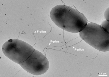
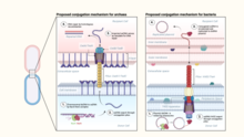
Bacterial conjugation is the transfer of genetic material between bacterial cells by direct cell-to-cell contact or by a bridge-like connection between two cells. This takes place through a pilus. It is a parasexual mode of reproduction in bacteria.

<i>Neisseria gonorrhoeae</i> Species of bacterium

Neisseria gonorrhoeae, also known as gonococcus (singular) or gonococci (plural), is a species of Gram-negative diplococci bacteria isolated by Albert Neisser in 1879. It causes the sexually transmitted genitourinary infection gonorrhea as well as other forms of gonococcal disease including disseminated gonococcemia, septic arthritis, and gonococcal ophthalmia neonatorum.

The evolution of flagella is of great interest to biologists because the three known varieties of flagella – each represent a sophisticated cellular structure that requires the interaction of many different systems.

<i>Neisseria</i> Genus of bacteria

Neisseria is a large genus of bacteria that colonize the mucosal surfaces of many animals. Of the 11 species that colonize humans, only two are pathogens, N. meningitidis and N. gonorrhoeae.

<span class="mw-page-title-main">Secretion</span> Controlled release of substances by cells or tissues

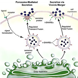
Secretion is the movement of material from one point to another, such as a secreted chemical substance from a cell or gland. In contrast, excretion is the removal of certain substances or waste products from a cell or organism. The classical mechanism of cell secretion is via secretory portals at the plasma membrane called porosomes. Porosomes are permanent cup-shaped lipoprotein structures embedded in the cell membrane, where secretory vesicles transiently dock and fuse to release intra-vesicular contents from the cell.

<i>Agrobacterium tumefaciens</i> Bacterium, genetic engineering tool

Agrobacterium tumefaciens is the causal agent of crown gall disease in over 140 species of eudicots. It is a rod-shaped, Gram-negative soil bacterium. Symptoms are caused by the insertion of a small segment of DNA, from a plasmid into the plant cell, which is incorporated at a semi-random location into the plant genome. Plant genomes can be engineered by use of Agrobacterium for the delivery of sequences hosted in T-DNA binary vectors.

<span class="mw-page-title-main">Appendage</span> External body part or natural prolongation, that protrudes from an organisms body

An appendage is an external body part, or natural prolongation, that protrudes from an organism's or microorganism's body.

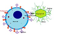
Adhesins are cell-surface components or appendages of bacteria that facilitate adhesion or adherence to other cells or to surfaces, usually in the host they are infecting or living in. Adhesins are a type of virulence factor.

The F-plasmid allows genes to be transferred from one bacterium carrying the factor to another bacterium lacking the factor by conjugation. The F factor was the first plasmid to be discovered. Unlike other plasmids, F factor is constitutive for transfer proteins due to a mutation in the gene finO. The F plasmid belongs to F-like plasmids, a class of conjugative plasmids that control sexual functions of bacteria with a fertility inhibition (Fin) system.

Pilin refers to a class of fibrous proteins that are found in pilus structures in bacteria. These structures can be used for the exchange of genetic material, or as a cell adhesion mechanism. Although not all bacteria have pili or fimbriae, bacterial pathogens often use their fimbriae to attach to host cells. In Gram-negative bacteria, where pili are more common, individual pilin molecules are linked by noncovalent protein-protein interactions, while Gram-positive bacteria often have polymerized LPXTG pilin.

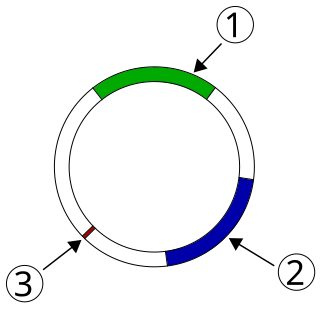
Transfer genes or tra genes, are some genes necessary for non-sexual transfer of genetic material in both gram-positive and gram-negative bacteria. The tra locus includes the pilin gene and regulatory genes, which together form pili on the cell surface, polymeric proteins that can attach themselves to the surface of F-bacteria and initiate the conjugation. The existence of the tra region of a plasmid genome was first discovered in 1979 by David H. Figurski and Donald R. Helinski In the course of their work, Figurski and Helinski also discovered a second key fact about the tra region – that it can act in trans to the mobilization marker which it affects.

In biology, phase variation is a method for dealing with rapidly varying environments without requiring random mutation. It involves the variation of protein expression, frequently in an on-off fashion, within different parts of a bacterial population. As such the phenotype can switch at frequencies that are much higher than classical mutation rates. Phase variation contributes to virulence by generating heterogeneity. Although it has been most commonly studied in the context of immune evasion, it is observed in many other areas as well and is employed by various types of bacteria, including Salmonella species.

<span class="mw-page-title-main">Bacterial motility</span> Ability of bacteria to move independently using metabolic energy

Bacterial motility is the ability of bacteria to move independently using metabolic energy. Most motility mechanisms that evolved among bacteria also evolved in parallel among the archaea. Most rod-shaped bacteria can move using their own power, which allows colonization of new environments and discovery of new resources for survival. Bacterial movement depends not only on the characteristics of the medium, but also on the use of different appendages to propel. Swarming and swimming movements are both powered by rotating flagella. Whereas swarming is a multicellular 2D movement over a surface and requires the presence of surfactants, swimming is movement of individual cells in liquid environments.

<span class="mw-page-title-main">Sortase</span> Group of prokaryotic enzymes

Sortase refers to a group of prokaryotic enzymes that modify surface proteins by recognizing and cleaving a carboxyl-terminal sorting signal. For most substrates of sortase enzymes, the recognition signal consists of the motif LPXTG (Leu-Pro-any-Thr-Gly), then a highly hydrophobic transmembrane sequence, followed by a cluster of basic residues such as arginine. Cleavage occurs between the Thr and Gly, with transient attachment through the Thr residue to the active site Cys residue, followed by transpeptidation that attaches the protein covalently to cell wall components. Sortases occur in almost all Gram-positive bacteria and the occasional Gram-negative bacterium or Archaea, where cell wall LPXTG-mediated decoration has not been reported. Although sortase A, the "housekeeping" sortase, typically acts on many protein targets, other forms of sortase recognize variant forms of the cleavage motif, or catalyze the assembly of pilins into pili.

<span class="mw-page-title-main">Plasmid-mediated resistance</span> Antibiotic resistance caused by a plasmid

Plasmid-mediated resistance is the transfer of antibiotic resistance genes which are carried on plasmids. Plasmids possess mechanisms that ensure their independent replication as well as those that regulate their replication number and guarantee stable inheritance during cell division. By the conjugation process, they can stimulate lateral transfer between bacteria from various genera and kingdoms. Numerous plasmids contain addiction-inducing systems that are typically based on toxin-antitoxin factors and capable of killing daughter cells that don't inherit the plasmid during cell division. Plasmids often carry multiple antibiotic resistance genes, contributing to the spread of multidrug-resistance (MDR). Antibiotic resistance mediated by MDR plasmids severely limits the treatment options for the infections caused by Gram-negative bacteria, especially family Enterobacteriaceae. The global spread of MDR plasmids has been enhanced by selective pressure from antimicrobial medications used in medical facilities and when raising animals for food.

Gabriel Waksman FMedSci, FRS, is Courtauld professor of biochemistry and molecular biology at University College London (UCL), and professor of structural and molecular biology at Birkbeck College, University of London. He is the director of the Institute of Structural and Molecular Biology (ISMB) at UCL and Birkbeck, head of the Department of Structural and Molecular Biology at UCL, and head of the Department of Biological Sciences at Birkbeck.

<span class="mw-page-title-main">Twitching motility</span> Form of crawling bacterial motility

Twitching motility is a form of crawling bacterial motility used to move over surfaces. Twitching is mediated by the activity of hair-like filaments called type IV pili which extend from the cell's exterior, bind to surrounding solid substrates, and retract, pulling the cell forwards in a manner similar to the action of a grappling hook. The name twitching motility is derived from the characteristic jerky and irregular motions of individual cells when viewed under the microscope. It has been observed in many bacterial species, but is most well studied in Pseudomonas aeruginosa, Neisseria gonorrhoeae and Myxococcus xanthus. Active movement mediated by the twitching system has been shown to be an important component of the pathogenic mechanisms of several species.

<span class="mw-page-title-main">Curli</span> A proteinaceous extracellular fiber produced by enteric bacteria

The Curli protein is a type of amyloid fiber produced by certain strains of enterobacteria. They are extracellular fibers located on bacteria such as E. coli and Salmonella spp. These fibers serve to promote cell community behavior through biofilm formation in the extracellular matrix. Amyloids are associated with several human neurodegenerative diseases such as Alzheimer's disease, Huntington's disease, Parkinson's disease, and prion diseases. The study of curli may help to understand human diseases thought to arise from improper amyloid fiber formation. The curli pili are generally assembled through the extracellular nucleation precipitation pathway.

P fimbriae are chaperone-usher type fimbrial appendages found on the surface of many Escherichia coli bacteria. The P fimbriae is considered to be one of the most important virulence factor in uropathogenic E. coli and plays an important role in upper urinary tract infections. P fimbriae mediate adherence to host cells, a key event in the pathogenesis of urinary tract infections.

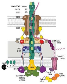
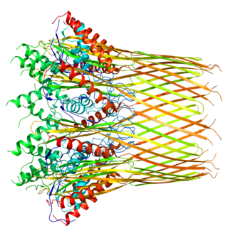
The bacterial type IV secretion system, also known as the type IV secretion system or the T4SS, is a secretion protein complex found in gram negative bacteria, gram positive bacteria, and archaea. It is able to transport proteins and DNA across the cell membrane. The type IV secretion system is just one of many bacterial secretion systems. Type IV secretion systems are related to conjugation machinery which generally involve a single-step secretion system and the use of a pilus. Type IV secretion systems are used for conjugation, DNA exchange with the extracellular space, and for delivering proteins to target cells. The type IV secretion system is divided into type IVA and type IVB based on genetic ancestry.

References

  1. "pilus" at Dorland's Medical Dictionary
  2. Brinton, Charles (1954). "Electrophoresis and phage susceptibility studies on a filament-producing variant of the E. coli bacterium". Biochimica et Biophysica Acta. 15 (4): 533–542. doi:10.1016/0006-3002(54)90011-6. PMID   13230101.
  3. 1 2 3 4 Chagnot, C; Zorgani, MA; Astruc, T; Desvaux, M (14 October 2013). "Proteinaceous determinants of surface colonization in bacteria: bacterial adhesion and biofilm formation from a protein secretion perspective". Frontiers in Microbiology. 4: 303. doi: 10.3389/fmicb.2013.00303 . PMC   3796261 . PMID   24133488.
  4. "Gut bacteria use super-polymers to dodge antibiotics | Imperial News | Imperial College London". Imperial News.
  5. Patkowski, Jonasz B.; Dahlberg, Tobias; Amin, Himani; Gahlot, Dharmender K.; Vijayrajratnam, Sukhithasri; Vogel, Joseph P.; Francis, Matthew S.; Baker, Joseph L.; Andersson, Magnus; Costa, Tiago R. D. (5 April 2023). "The F-pilus biomechanical adaptability accelerates conjugative dissemination of antimicrobial resistance and biofilm formation". Nature Communications. 14 (1): 1879. doi:10.1038/s41467-023-37600-y. PMC   10076315 . PMID   37019921.
  6. 1 2 3 4 Beltran, Leticia C.; Cvirkaite-Krupovic, Virginija; Miller, Jessalyn; Wang, Fengbin; Kreutzberger, Mark A. B.; Patkowski, Jonasz B.; Costa, Tiago R. D.; Schouten, Stefan; Levental, Ilya; Conticello, Vincent P.; Egelman, Edward H.; Krupovic, Mart (2023-02-07). "Archaeal DNA-import apparatus is homologous to bacterial conjugation machinery". Nature Communications. 14 (1): 666. doi:10.1038/s41467-023-36349-8. ISSN   2041-1723. PMC   9905601 . PMID   36750723.
  7. van Wolferen, Marleen; Wagner, Alexander; van der Does, Chris; Albers, Sonja-Verena (2016-03-01). "The archaeal Ced system imports DNA". Proceedings of the National Academy of Sciences of the United States of America. 113 (9): 2496–2501. Bibcode:2016PNAS..113.2496V. doi: 10.1073/pnas.1513740113 . ISSN   1091-6490. PMC   4780597 . PMID   26884154.
  8. Proft, T.; Baker, E. N. (February 2009). "Pili in Gram-negative and Gram-positive bacteria — structure, assembly and their role in disease". Cellular and Molecular Life Sciences. 66 (4): 613–635. doi: 10.1007/s00018-008-8477-4 . PMID   18953686. S2CID   860681.
  9. Ottow, JC (1975). "Ecology, physiology, and genetics of fimbriae and pili". Annual Review of Microbiology. 29: 79–108. doi:10.1146/annurev.mi.29.100175.000455. PMID   1180526.
  10. Filloux, A (July 2010). "A variety of bacterial pili involved in horizontal gene transfer". Journal of Bacteriology. 192 (13): 3243–5. doi:10.1128/JB.00424-10. PMC   2897649 . PMID   20418394.
  11. Joan, Slonczewski (2017). Microbiology : an evolving science. Foster, John Watkins (Fourth ed.). New York: W. W. Norton & Company. pp. 1000–1002. ISBN   9780393614039. OCLC   951925510.
  12. Mattick JS (2002). "Type IV pili and twitching motility". Annu. Rev. Microbiol. 56 (1): 289–314. doi:10.1146/annurev.micro.56.012302.160938. PMID   12142488.
  13. Jarrell; et al. (2009). "Archaeal Flagella and Pili". Pili and Flagella: Current Research and Future Trends. Caister Academic Press. ISBN   978-1-904455-48-6.
  14. Wang, F; Cvirkaite-Krupovic, V; Kreutzberger, MAB; Su, Z; de Oliveira, GAP; Osinski, T; Sherman, N; DiMaio, F; Wall, JS; Prangishvili, D; Krupovic, M; Egelman, EH (2019). "An extensively glycosylated archaeal pilus survives extreme conditions". Nature Microbiology. 4 (8): 1401–1410. doi:10.1038/s41564-019-0458-x. PMC   6656605 . PMID   31110358.
  15. Cehovin A, Simpson PJ, McDowell MA, Brown DR, Noschese R, Pallett M, Brady J, Baldwin GS, Lea SM, Matthews SJ, Pelicic V (2013). "Specific DNA recognition mediated by a type IV pilin". Proc. Natl. Acad. Sci. U.S.A. 110 (8): 3065–70. Bibcode:2013PNAS..110.3065C. doi: 10.1073/pnas.1218832110 . PMC   3581936 . PMID   23386723.
  16. Davidsen T, Rødland EA, Lagesen K, Seeberg E, Rognes T, Tønjum T (2004). "Biased distribution of DNA uptake sequences towards genome maintenance genes". Nucleic Acids Res. 32 (3): 1050–8. doi:10.1093/nar/gkh255. PMC   373393 . PMID   14960717.
  17. Caugant DA, Maiden MC (2009). "Meningococcal carriage and disease--population biology and evolution". Vaccine. 27 Suppl 2 (4): B64–70. doi:10.1016/j.vaccine.2009.04.061. PMC   2719693 . PMID   19464092.
  18. Nuccio SP, et al. (2007). "Evolution of the chaperone/usher assembly pathway: fimbrial classification goes Greek". Microbiology and Molecular Biology Reviews. 71 (4): 551–575. doi:10.1128/MMBR.00014-07. PMC   2168650 . PMID   18063717.
  19. 1 2 Cookson, AL; Cooley, WA; Woodward, MJ (2002), "The role of type 1 and curli fimbriae of Shiga toxin-producing Escherichia coli in adherence to abiotic surfaces", Int J Med Microbiol, 292 (3–4): 195–205, doi:10.1078/1438-4221-00203, PMID   12398210.
  20. Kolenda, Rafal; Ugorski, Maciej; Grzymajlo, Krzysztof (14 May 2019). "Everything You Always Wanted to Know About Salmonella Type 1 Fimbriae, but Were Afraid to Ask". Frontiers in Microbiology. 10: 1017. doi: 10.3389/fmicb.2019.01017 . PMC   6527747 . PMID   31139165.
  21. Rice JC, Peng T, Spence JS, Wang HQ, Goldblum RM, Corthésy B, Nowicki BJ (December 2005). "Pyelonephritic Escherichia coli expressing P fimbriae decrease immune response of the mouse kidney". Journal of the American Society of Nephrology. 16 (12): 3583–91. doi: 10.1681/ASN.2005030243 . PMID   16236807.
  22. WI, Kenneth Todar, Madison. "Colonization and Invasion by Bacterial Pathogens". www.textbookofbacteriology.net. Retrieved 2016-12-03.{{cite web}}: CS1 maint: multiple names: authors list (link)
  23. 1 2 3 Epstein, EA; Reizian, MA; Chapman, MR (2009), "Spatial clustering of the curlin secretion lipoprotein requires curli fiber assembly.", J Bacteriol, 191 (2): 608–615, doi:10.1128/JB.01244-08, PMC   2620823 , PMID   19011034.
  24. 1 2 3 4 5 Craig, Lisa; Taylor, Ronald (2014). "Chapter 1: The Vibrio cholerae Toxin Coregulated Pilus: Structure, Assembly, and Function with Implications for Vaccine Design". In Barocchi, Michèle; Telford, John (eds.). Bacterial Pili: Structure, Synthesis, and Role in Disease. C.A.B. International. pp. 1–16. ISBN   978-1-78064-255-0.
  25. Rinaudo, Daniela; Moschioni, Monica (2014). "Chapter 13: Pilus-based Vaccine Development in Streptococci: Variability, Diversity, and Immunological Resposes". In Barocchi, Michèle; Telford, John (eds.). Bacterial Pili: Structure, Synthesis, and Role in Disease. C.A.B. International. pp. 182–202. ISBN   978-1-78064-255-0.
  26. Todar, Kenneth. "Textbook of Bacteriology: Bacterial Structure in Relationship to Pathogenicity". Textbook of Bacteriology. Retrieved 24 November 2017.
  27. 1 2 3 Georgiadou, Michaella; Pelicic, Vladimir (2014). "Chapter 5: Type IV Pili: Functions & Biogenesis". In Barocchi, Michèle; Telford, John (eds.). Bacterial Pili: Structure, Synthesis, and Role in Disease. C.A.B. International. pp. 71–84. ISBN   978-1-78064-255-0.
  28. Connell I, Agace W, Klemm P, Schembri M, Mărild S, Svanborg C (September 1996). "Type 1 fimbrial expression enhances Escherichia coli virulence for the urinary tract". Proc. Natl. Acad. Sci. U.S.A. 93 (18): 9827–32. Bibcode:1996PNAS...93.9827C. doi: 10.1073/pnas.93.18.9827 . PMC   38514 . PMID   8790416.