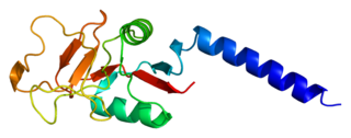
Mannose receptor

Last updated
Macrophage mannose receptor
Identifiers
SymbolMMR
Membranome 56
mannose receptor, C type 1
Identifiers
SymbolMRC1
Alt. symbolsCD206
NCBI gene 4360
HGNC 7228
OMIM 153618
RefSeq NM_002438
UniProt P22897
Other data
Locus Chr. 10 p13
Search for
Structures Swiss-model
Domains InterPro
mannose receptor, C type 2
Identifiers
SymbolMRC2
Alt. symbolsCD280
NCBI gene 9902
HGNC 16875
RefSeq NM_006039
UniProt Q9UBG0
Other data
Locus Chr. 17 q23
Search for
Structures Swiss-model
Domains InterPro

The mannose receptor (Cluster of Differentiation 206, CD206) is a C-type lectin primarily present on the surface of macrophages, immature dendritic cells and liver sinusoidal endothelial cells, but is also expressed on the surface of skin cells such as human dermal fibroblasts and keratinocytes. [1] [2] It is the first member of a family of endocytic receptors that includes Endo180 (CD280), M-type PLA2R, and DEC-205 (CD205). [3]

Contents

The receptor recognises terminal mannose, N-acetylglucosamine and fucose residues on glycans attached to proteins [4] found on the surface of some microorganisms, playing a role in both the innate and adaptive immune systems. Additional functions include clearance of glycoproteins from circulation, including sulphated glycoprotein hormones and glycoproteins released in response to pathological events. [5] The mannose receptor recycles continuously between the plasma membrane and endosomal compartments in a clathrin-dependent manner. [6]

Structure

Domain organisation

Domain organisation of the mannose receptor, adapted from Introduction to Glycobiology. Mannose receptor domain organisation.jpg
Domain organisation of the mannose receptor, adapted from Introduction to Glycobiology.

The mannose receptor is a type I transmembrane protein, with an extracellular N-terminus and an intracellular C-terminus. It is first synthesised as an inactive precursor, but is proteolytically cleaved to its active form in the Golgi apparatus. [8] In general, The extracellular portion of the receptor is composed of 8 consecutive C-type carbohydrate recognition domains (CRDs) closest to the plasma membrane, followed by a single fibronectin type II repeat domain and an N-terminal cysteine-rich domain. The cytoplasmic tail is not capable of signal transduction in isolation, since it lacks the appropriate signaling motifs. [9]

N-terminal cysteine-rich domain

The N-terminal cysteine-rich domain is homologous to the ricin B chain and binds to sulphated sugar moieties, with particularly high affinity for N-Acetylgalactosamine and galactose residues sulphated at positions 3 and 4 of their pyranose rings. [10]

Other ligands include chondroitin sulfates A and B, as well as sulphated Lewisx and Lewisa structures. [6] The mannose receptor is the only member of the family in which this domain is functional. [5]

The mannose receptor N-terminal cysteine-rich domain (pink) bound to its sulphated N-Acetylgalactosamine ligand (cyan). PBD ID: 1DQO Mannose receptor Cys-rich domain.png
The mannose receptor N-terminal cysteine-rich domain (pink) bound to its sulphated N-Acetylgalactosamine ligand (cyan). PBD ID: 1DQO

Fibronectin type II repeat domain

The fibronectin type II repeat domain is conserved amongst all members of the mannose receptor family. Collagens I-IV bind this region with high affinity, while collagen V binds only weakly. Through this domain, the mannose receptor internalises collagen in macrophages and liver sinusoidal cells, independent of the lectin activity of the receptor. [9] Along with the N-terminal cysteine-rich domain, this domain is the most highly conserved between mice and humans (92%). [8]

C-type carbohydrate recognition domains (CRDs)

The 8 tandem CRDs in the extracellular region of the mannose receptor share only 30% homology with each other. They each contain at least some of the amino acid residues necessary for Ca2+ and ligand binding, common to functional C-type CRDs. Only CRDs 4 and 5 contain all of the residues required for sugar binding, forming a protease-resistant ligand-binding core. The most common ligand is terminal mannose residues, but N-acetylglucosamine and fucose also bind. [8]

The main interaction between CRD-4 and its sugar ligand is through direct ligation to the conserved Ca2+ in the sugar-binding site, in a similar way to the binding mechanism of mannan-binding lectin (MBL). However, a quarter of the free energy of sugar-binding is associated with the hydrophobic stacking interactions formed between one face of the sugar ring and the side chain of a conserved tyrosine residue in the binding site, which is not seen in MBL. Despite the similarities in mannose-binding between the mannose receptor and MBL, these differences suggest that mannose-binding by the mannose receptor evolved separately to that of other C-type lectins. [11]

Individually, the CRDs bind mannose with only weak affinity. High affinity binding is thought to result from the clustering of multiple CRDs. This clustering allows for binding of multivalent, branched ligands such as high-mannose N-linked oligosaccharides. [12]

Conformation

It has been suggested that the mannose receptor can exist in at least two different structural conformations. The C-type CRDs are each separated by linker regions of 10-20 amino acids containing a number of proline residues, whose cyclic side chain is fairly rigid and favours a conformation in which the N-terminal cysteine-rich domain is extended as far away from the plasma membrane as possible. [13]

Alternatively, interactions between neighbouring CRDs may hold them in close proximity to one another and cause the extracellular region of the receptor to bend, bringing the N-terminal cysteine-rich domain into close contact with the CRDs. This would position CRDs 4 and 5 furthest from the membrane to maximise their interaction with potential ligands. The resistance to proteolysis shown by CRDs 4 and 5 suggests physical interactions between the two domains does occur, thereby supporting the existence of this U-shaped conformation. [13]

It is thought that transitions between these two conformations occur in a pH-dependent manner, regulating ligand selectivity and release during endocytosis. The lower, more acidic pH of early endosomes is thought to be responsible for ligand release. [13]

Proteolytic processing

A functional, soluble form of the mannose receptor is produced upon proteolytic cleavage of the membrane-bound form by metalloproteases found in the extracellular environment. [14] [15]

The soluble protein consists of the entire extracellular region of the receptor and it may be involved in transport of mannosylated proteins away from sites of inflammation. [9] Shedding of the mannose receptor from macrophages has been shown to be enhanced upon recognition of fungal pathogens such as Candida albicans and Aspergillus fumigatus , which suggests the soluble form may play a role in fungal pathogen recognition. In this way, the balance between membrane-bound and soluble mannose receptor could affect targeting of fungal pathogens during the course of infection. [16]

Glycosylation

The mannose receptor is heavily glycosylated and its N-linked glycosylation sites are highly conserved between mice and humans, indicating an important role for this post-translational modification. The presence of sialic acid residues on N-linked glycans of the mannose receptor is important for its role in binding both sulphated and mannosylated glycoproteins. Sialylation regulates multimerisation of the receptor, which is known to influence binding to sulphated glycoproteins. Terminal sialic acid residues are also known to be required for binding to mannosylated glycans. The absence of sialic acid reduces the receptors ability to bind and internalise mannosylated glycans, but does not affect its localisation to the plasma membrane or its endocytic activity. [9] [17]

Function

Phagocytosis of pathogens

A number of pathogenic microorganisms, including C. albicans, [15] [18] Pneumocystis carinii [19] [20] and Leishmania donovani [21] [22] display glycans on their surfaces with terminal mannose residues that are recognised by the C-type CRDs of the mannose receptor, thereby acting as a marker of non-self. Upon recognition, the receptor internalises the bound pathogen and transports it to lysosomes for degradation via the phagocytic pathway. In this way, the mannose receptor acts as a pattern recognition receptor. The presence of a di-aromatic FENTLY (Phe-Glu-Asn-Thr-Leu-Tyr) sequence motif in the cytoplasmic tail of the receptor is vital for its clathrin-mediated internalization. [6] This is supported by the evidence that Cos-1 cells transfected with the mannose receptor lacking its C-terminal tail are unable to endocytose C. albicans and P. carinii. [6]

Surprisingly, mannose receptor knockout mice do not show increased susceptibility to infection, which suggests that the receptor is not essential for phagocytosis. However, its involvement cannot be rejected since other mechanisms may compensate. For example, infection of knockout mice with P. carinii resulted in increased recruitment of macrophages to the site of infection. Furthermore, other receptors present on the surface of phagocytic cells, such as DC-SIGN, SIGNR1 and Endo180, exhibit similar ligand binding ability to the mannose receptor and so it is likely that in its absence, these proteins are able to compensate and induce phagocytosis. [6]

The ability of the mannose receptor to aid in pathogen internalisation is also thought to facilitate infection by Mycobacterium tuberculosis and Mycobacterium leprae . These bacteria reside and multiply in macrophages, preventing formation of the phagolysosome to avoid degradation. Hence, by mediating their entrance into the macrophage, blocking the mannose receptor helps these pathogens to infect and grow in their target cell. [6] [23]

Clathrin-mediated endocytosis

The CRD regions of the mannose receptor on liver sinusoidal endothelial cells remove a number of waste material ranging from soluble macromolecules to large particulate matter. [24] These include lysosomal enzymes, [25] collagen α-chains, [26] C-terminal propeptides of type I pro-collagens, [27] and tissue plasminogen activator. [28] Binding studies indicate that each liver sinusoidal endothelial cell expresses a surface pool of 20,000-25,000 mannose receptors. The mannose receptor on liver sinusoidal endothelial cell is a rapidly recycling receptor, with a Ke (endocytotic rate constant) of 4.12 min-1, which corresponds to a half-life of 10 s for the surface pool of receptor-ligand complexes. [29]

As opposed to macrophages that use the mannose receptors for phagocytosis of particulate matter >200 nm, the mannose receptor on liver sinusoidal endothelial cells mediates clathrin-mediated endocytosis of macromolecules and nanoparticles <200 nm. [24]

Antigen presentation

The mannose receptor may also play a role in antigen uptake and presentation by immature dendritic cells in the adaptive immune system. Upon binding to the receptor, mannosylated antigens are internalised and transported to endocytic compartments within the cell for loading onto Major Histocompatibility Complex (MHC) molecules or other related antigen-presentation molecules. An indirect example of this is the processing of the glycolipid antigen lipoarabinomannan, derived from Mycobacteria. Lipoarabinomannan (LAM) is presented to T cells in complex with CD1b, but is also able to bind to the mannose receptor. Since the presence of mannan, an alternative ligand, inhibits LAM-dependent T cell proliferation, it is suggested that the receptor binds extracellular LAM, internalises it and then transports it to endocytic vesicles to be loaded onto CD1b. [8]

Mature dendritic cells and macrophages use the mannose receptor for antigen presentation in a different way. The cleaved, soluble receptor binds to circulating antigens and directs them to effector cells in lymphoid organs via its cysteine-rich domain, thus activating the adaptive immune system. [8]

Intracellular signalling

The cytoplasmic tail of the mannose receptor does not contain any signalling motifs, yet the receptor has proven to be essential for production of both pro- and anti-inflammatory cytokines, indicating a more passive role for the receptor in phagocytosis of pathogens. [6] [8] This suggests that the mannose receptor is assisted by other cell surface receptors in order to trigger a signalling cascade. For example, it has been shown that HEK 293 cells co-transfected with human mannose receptor and human Toll-like receptor 2 cDNA are able to secrete IL-8 in response to P. carinii infection, whereas those transfected with either receptor alone did not. [30] It is possible that the two receptors form a complex on the cell surface that facilitates signal transduction upon pathogenic challenge.

Resolution of inflammation

Another key role of the mannose receptor is to regulate the levels of molecules released into the circulation during the inflammatory response. In response to pathological events, glycoproteins including lysosomal hydrolases, tissue plasminogen activator and neutrophil myeloperoxidase are released to help fight off any invading microorganisms. Once the threat has subsided, these glycoproteins can be damaging to host tissues so their levels in the circulation must be strictly controlled. [6]

High-mannose oligosaccharides present on the surface of these glycoproteins act to mark their transient nature, since they are eventually recognised by the mannose receptor and removed from the circulation. Mannose receptor knockout mice are less able to clear these proteins, and show increased concentrations of a number of lysosomal hydrolases in the blood. [5]

Consistent with this function, the mannose receptor is expressed at low levels during inflammation and at high levels during the resolution of inflammation, to ensure inflammatory agents are removed from the circulation only at the appropriate time. [5]

Clearance of glycoprotein hormones

The N-terminal cysteine-rich domain of the mannose receptor plays an important role in the recognition of sulphated glycoprotein hormones and their clearance from the circulation. [8]

Glycoprotein hormones such as lutropin, which triggers release of the egg during ovulation, must stimulate their receptors in pulses to avoid receptor desensitisation. Glycans on their surface are capped with sulphated N-Acetylgalactosamine (GalNAc), making them ligands for the cysteine-rich ricin homology domain of the mannose receptor. This tag ensures a cycle of release, stimulation, and removal from the circulation. [7]

Knockout mice lacking the enzyme required to add the sulphated GalNAc capping structure show longer half-lives for lutropin, which results in increased receptor activation and oestrogen production. Female knockout mice reach sexual maturity faster than their wild-type counterparts, have a longer oestrus cycle and produce more litters. Thus, the sulphated GalNAc tag is very important in regulating serum concentrations of certain glycoprotein hormones. [7]

Types

Humans express two types of mannose receptors, each encoded by its own gene:

GeneProteinAlternative names
MRC1Macrophage mannose receptor 1C-type mannose receptor 1,
C-type lectin domain family 13 member D (CLEC13D),
CD206, MMR
MRC2Macrophage mannose receptor 2C-type mannose receptor 2,
Urokinase-type plasminogen activator receptor-associated protein,
CD280

Applications in health and disease

The selective internalisation properties of the mannose receptor indicate a number of potential applications in health and disease. By manipulating the glycosylation of important bioactive proteins to a highly mannosylated state, their serum levels could be tightly regulated and they could be targeted specifically to cells expressing the mannose receptor. There is also potential for use of the mannose receptor as a target for improved macrophage activation and antigen presentation. [5] [8] [31]

MRC2/Endo180 [32] interacts with Basigin/CD147 via its fourth C-type lectin domain to form a molecular epithelial-mesenchymal transition suppressor complex that if disrupted results in the induction of invasive prostate epithelial cell behavior associated with poor prostate cancer survival. [33] Increased basement membrane stiffness due to its glycation can also trigger Endo180-dependent invasion of prostate epithelial cells and this bio-mechanical mechanism is associated with poor prostate cancer survival. [34] It has been suggested that stabilization of the Endo180-CD147 epithelial-mesenchymal transition suppressor complex and targeting of the non-complexed form of Endo180 in invasive cells could have therapeutic benefit in the prevention of cancer progression and metastasis. [35]

Related Research Articles

<span class="mw-page-title-main">Concanavalin A</span> Lectin (carbohydrate-binding protein) originally extracted from the jack-bean

Concanavalin A (ConA) is a lectin originally extracted from the jack-bean. It is a member of the legume lectin family. It binds specifically to certain structures found in various sugars, glycoproteins, and glycolipids, mainly internal and nonreducing terminal α-D-mannosyl and α-D-glucosyl groups. Its physiological function in plants, however, is still unknown. ConA is a plant mitogen, and is known for its ability to stimulate mouse T-cell subsets giving rise to four functionally distinct T cell populations, including precursors to regulatory T cells; a subset of human suppressor T-cells is also sensitive to ConA. ConA was the first lectin to be available on a commercial basis, and is widely used in biology and biochemistry to characterize glycoproteins and other sugar-containing entities on the surface of various cells. It is also used to purify glycosylated macromolecules in lectin affinity chromatography, as well as to study immune regulation by various immune cells.

Pattern recognition receptors (PRRs) play a crucial role in the proper function of the innate immune system. PRRs are germline-encoded host sensors, which detect molecules typical for the pathogens. They are proteins expressed mainly by cells of the innate immune system, such as dendritic cells, macrophages, monocytes, neutrophils, as well as by epithelial cells, to identify two classes of molecules: pathogen-associated molecular patterns (PAMPs), which are associated with microbial pathogens, and damage-associated molecular patterns (DAMPs), which are associated with components of host's cells that are released during cell damage or death. They are also called primitive pattern recognition receptors because they evolved before other parts of the immune system, particularly before adaptive immunity. PRRs also mediate the initiation of antigen-specific adaptive immune response and release of inflammatory cytokines.

Scavenger receptors are a large and diverse superfamily of cell surface receptors. Its properties were first recorded in 1970 by Drs. Brown and Goldstein, with the defining property being the ability to bind and remove modified low density lipoproteins (LDL). Today scavenger receptors are known to be involved in a wide range of processes, such as: homeostasis, apoptosis, inflammatory diseases and pathogen clearance. Scavenger receptors are mainly found on myeloid cells and other cells that bind to numerous ligands, primarily endogenous and modified host-molecules together with pathogen-associated molecular patterns(PAMPs), and remove them. The Kupffer cells in the liver are particularly rich in scavenger receptors, includes SR-A I, SR-A II, and MARCO.

<span class="mw-page-title-main">Selectin</span> Family of cell adhesion molecules

The selectins are a family of cell adhesion molecules. All selectins are single-chain transmembrane glycoproteins that share similar properties to C-type lectins due to a related amino terminus and calcium-dependent binding. Selectins bind to sugar moieties and so are considered to be a type of lectin, cell adhesion proteins that bind sugar polymers.

<span class="mw-page-title-main">DC-SIGN</span> Protein-coding gene in the species Homo sapiens

DC-SIGN also known as CD209 is a protein which in humans is encoded by the CD209 gene.

The asialoglycoprotein receptors (ASGPR) are lectins which bind asialoglycoprotein and glycoproteins from which a sialic acid has been removed to expose galactose residues. The receptors, which are integral membrane proteins and are located on mammalian hepatocytes, remove target glycoproteins from circulation. The asialoglycoprotein receptor has been demonstrated to have high expression on the surface of hepatocytes and several human carcinoma cell lines It is also weakly expressed by glandular cells of the gallbladder and the stomach. Lactobionic acid has been used as a targeting moiety for drug delivery to cells expressing asialoglycoprotein receptors.

<span class="mw-page-title-main">Endoglin</span> Protein-coding gene in the species Homo sapiens

Endoglin (ENG) is a type I membrane glycoprotein located on cell surfaces and is part of the TGF beta receptor complex. It is also commonly referred to as CD105, END, FLJ41744, HHT1, ORW and ORW1. It has a crucial role in angiogenesis, therefore, making it an important protein for tumor growth, survival and metastasis of cancer cells to other locations in the body.

Collectins (collagen-containing C-type lectins) are a part of the innate immune system. They form a family of collagenous Ca2+-dependent defense lectins, which are found in animals. Collectins are soluble pattern recognition receptors (PRRs). Their function is to bind to oligosaccharide structure or lipids that are on the surface of microorganisms. Like other PRRs they bind pathogen-associated molecular patterns (PAMPs) and danger-associated molecular patterns (DAMPs) of oligosaccharide origin. Binding of collectins to microorganisms may trigger elimination of microorganisms by aggregation, complement activation, opsonization, activation of phagocytosis, or inhibition of microbial growth. Other functions of collectins are modulation of inflammatory, allergic responses, adaptive immune system and clearance of apoptotic cells.

<span class="mw-page-title-main">Mannan-binding lectin</span> Mammalian protein found in Homo sapiens

Mannose-binding lectin (MBL), also called mannan-binding lectin or mannan-binding protein (MBP), is a lectin that is instrumental in innate immunity as an opsonin and via the lectin pathway.

Siglecs(Sialic acid-binding immunoglobulin-type lectins) are cell surface proteins that bind sialic acid. They are found primarily on the surface of immune cells and are a subset of the I-type lectins. There are 14 different mammalian Siglecs, providing an array of different functions based on cell surface receptor-ligand interactions.

<span class="mw-page-title-main">Sialoadhesin</span> Protein-coding gene in the species Homo sapiens

Sialoadhesin is a cell adhesion molecule found on the surface of macrophages. It is found in especially high amounts on macrophages of the spleen, liver, lymph node, bone marrow, colon, and lungs. Also, in patients with rheumatoid arthritis, the protein has been found in great amounts on macrophages of the affected tissues. It is defined as an I-type lectin, since it contains 17 immunoglobulin (Ig) domains, and thus also belongs to the immunoglobulin superfamily (IgSF). Sialoadhesin binds to certain molecules called sialic acids. During this binding process a salt bridge (protein) is formed between a highly conserved arginine residue and the carboxylate group of the sialic acid. Since sialoadhesin binds sialic acids with its N-terminal IgV-domain, it is also a member of the SIGLEC family. Alternate names for sialoadhesin include siglec-1 and CD169.

<span class="mw-page-title-main">Langerin</span>

Langerin (CD207) is a type II transmembrane protein which is encoded by the CD207 gene in humans. It was discovered by scientists Sem Saeland and Jenny Valladeau as a main part of Birbeck granules. Langerin is C-type lectin receptor on Langerhans cells (LCs) and in mice also on dermal interstitial CD103+ dendritic cells (DC) and on resident CD8+ DC in lymph nodes.

Visna-maedi virus from the genus Lentivirus and subfamily Orthoretrovirinae, is a retrovirus that causes encephalitis and chronic pneumonitis in sheep. It is known as visna when found in the brain, and maedi when infecting the lungs. Lifelong, persistent infections in sheep occur in the lungs, lymph nodes, spleen, joints, central nervous system, and mammary glands; The condition is sometimes known as ovine progressive pneumonia (OPP), particularly in the United States, or Montana sheep disease. White blood cells of the monocyte/macrophage lineage are the main target of the virus.

The mannose 6-phosphate receptors (MPRs) are transmembrane glycoproteins that target enzymes to lysosomes in vertebrates.

<span class="mw-page-title-main">OLR1</span> Protein-coding gene in the species Homo sapiens

Oxidized low-density lipoprotein receptor 1 also known as lectin-type oxidized LDL receptor 1 (LOX-1) is a protein that in humans is encoded by the OLR1 gene.

<span class="mw-page-title-main">CLEC4M</span> Protein-coding gene in the species Homo sapiens

C-type lectin domain family 4 member M is a protein that in humans is encoded by the CLEC4M gene. CLEC4M has also been designated as CD299.

<span class="mw-page-title-main">CLEC7A</span> Protein-coding gene in humans

C-type lectin domain family 7 member A or Dectin-1 is a protein that in humans is encoded by the CLEC7A gene. CLEC7A is a member of the C-type lectin/C-type lectin-like domain (CTL/CTLD) superfamily. The encoded glycoprotein is a small type II membrane receptor with an extracellular C-type lectin-like domain fold and a cytoplasmic domain with a partial immunoreceptor tyrosine-based activation motif. It functions as a pattern-recognition receptor for a variety of β-1,3-linked and β-1,6-linked glucans from fungi and plants, and in this way plays a role in innate immune response. Expression is found on myeloid dendritic cells, monocytes, macrophages and B cells. Alternate transcriptional splice variants, encoding different isoforms, have been characterized. This gene is closely linked to other CTL/CTLD superfamily members on chromosome 12p13 in the natural killer gene complex region.

<span class="mw-page-title-main">TNFRSF12A</span> Protein-coding gene in the species Homo sapiens

Tumor necrosis factor receptor superfamily member 12A also known as the TWEAK receptor (TWEAKR) is a protein that in humans is encoded by the TNFRSF12A gene.

Collagen receptors are membrane proteins that bind the extracellular matrix protein collagen, the most abundant protein in mammals. They control mainly cell proliferation, migration and adhesion, coagulation cascade activation and they affect ECM structure by regulation of MMP.

Liver sinusoidal endothelial cells (LSECs) form the lining of the smallest blood vessels in the liver, also called the hepatic sinusoids. LSECs are highly specialized endothelial cells with characteristic morphology and function. They constitute an important part of the reticuloendothelial system (RES).

References

  1. Szolnoky G, Bata-Csörgö Z, Kenderessy AS, Kiss M, Pivarcsi A, Novák Z, Nagy Newman K, Michel G, Ruzicka T, Maródi L, Dobozy A, Kemény L (August 2001). "A mannose-binding receptor is expressed on human keratinocytes and mediates killing of Candida albicans". Journal of Investigative Dermatology. 117 (2): 205–13. doi: 10.1046/j.1523-1747.2001.14071.x . PMID   11511295.
  2. Sheikh H, Yarwood H, Ashworth A, Isacke CM (March 2000). "Endo180, an endocytic recycling glycoprotein related to the macrophage mannose receptor is expressed on fibroblasts, endothelial cells and macrophages and functions as a lectin receptor". Journal of Cell Science. 113 (6): 1021–32. doi:10.1242/jcs.113.6.1021. PMID   10683150.
  3. East L, Isacke CM (2002). "The mannose receptor family". Biochimica et Biophysica Acta (BBA) - General Subjects. 1572 (2–3): 364–86. doi:10.1016/S0304-4165(02)00319-7. PMID   12223280.
  4. Schlesinger PH, Doebber TW, Mandell BF, White R, DeSchryver C, Rodman JS, Miller MJ, Stahl P (1978). "Plasma clearance of glycoproteins with terminal mannose and N-acetylglucosamine by liver non-parenchymal cells. Studies with beta-glucuronidase, N-acetyl-β-D-glucosaminidase, ribonuclease B and agalacto-orosomucoid". Biochemical Journal. 176 (1): 103–9. doi:10.1042/bj1760103. PMC   1186209 . PMID   728098.
  5. 1 2 3 4 5 Lee SJ, Evers S, Roeder D, Parlow AF, Risteli J, Risteli L, Lee YC, Feizi T, Langen H, Nussenzweig MC (2002). "Mannose receptor-mediated regulation of serum glycoprotein homeostasis". Science. 295 (5561): 1898–901. Bibcode:2002Sci...295.1898L. doi:10.1126/science.1069540. PMID   11884756. S2CID   31432874.
  6. 1 2 3 4 5 6 7 8 Gazi U, Martinez-Pomares L (2009). "Influence of the mannose receptor in host immune responses". Immunobiology. 214 (7): 554–61. doi:10.1016/j.imbio.2008.11.004. PMID   19162368.
  7. 1 2 3 Taylor M, Drickamer K (2011). Introduction to Glycobiology. Oxford University Press. ISBN   978-0-19-956911-3.
  8. 1 2 3 4 5 6 7 8 Stahl PD, Ezekowitz RA (1998). "The mannose receptor is a pattern recognition receptor involved in host defense". Current Opinion in Immunology. 10 (1): 50–5. doi:10.1016/S0952-7915(98)80031-9. PMID   9523111.
  9. 1 2 3 4 Martinez-Pomares L (2012). "The mannose receptor". Journal of Leukocyte Biology. 92 (6): 1177–86. doi: 10.1189/jlb.0512231 . PMID   22966131. S2CID   27512588.
  10. Fiete DJ, Beranek MC, Baenziger JU (March 1998). "A cysteine-rich domain of the "mannose" receptor mediates GalNAc-4-SO4 binding". Proceedings of the National Academy of Sciences of the United States of America. 95 (5): 2089–93. Bibcode:1998PNAS...95.2089F. doi: 10.1073/pnas.95.5.2089 . PMC   19259 . PMID   9482843.
  11. Mullin NP, Hitchen PG, Taylor ME (1997). "Mechanism of Ca2+ and monosaccharide binding to a C-type carbohydrate-recognition domain of the macrophage mannose receptor". Journal of Biological Chemistry. 272 (9): 5668–81. doi: 10.1074/jbc.272.9.5668 . PMID   9038177.
  12. Weis WI, Drickamer K (1996). "Structural basis of lectin-carbohydrate recognition". Annual Review of Biochemistry. 65: 441–73. doi:10.1146/annurev.bi.65.070196.002301. PMID   8811186.
  13. 1 2 3 Llorca O (2008). "Extended and bent conformations of the mannose receptor family". Cellular and Molecular Life Sciences. 65 (9): 1302–10. doi:10.1007/s00018-007-7497-9. PMID   18193159. S2CID   5038725.
  14. Jordens R, Thompson A, Amons R, Koning F (1999). "Human dendritic cells shed a functional, soluble form of the mannose receptor". International Immunology . 11 (11): 1775–80. doi:10.1093/intimm/11.11.1775. PMID   10545481.
  15. 1 2 Martínez-Pomares L, Mahoney JA, Káposzta R, Linehan SA, Stahl PD, Gordon S (1998). "A functional soluble form of the murine mannose receptor is produced by macrophages in vitro and is present in mouse serum". Journal of Biological Chemistry. 273 (36): 23376–80. doi: 10.1074/jbc.273.36.23376 . hdl: 2437/116851 . PMID   9722572.
  16. Gazi U, Rosas M, Singh S, Heinsbroek S, Haq I, Johnson S, Brown GD, Williams DL, Taylor PR, Martinez-Pomares L (2011). "Fungal recognition enhances mannose receptor shedding through dectin-1 engagement". Journal of Biological Chemistry. 286 (10): 7822–9. doi: 10.1074/jbc.M110.185025 . PMC   3048669 . PMID   21205820.
  17. Su Y, Bakker T, Harris J, Tsang C, Brown GD, Wormald MR, Gordon S, Dwek RA, Rudd PM, Martinez-Pomares L (2005). "Glycosylation influences the lectin activities of the macrophage mannose receptor". Journal of Biological Chemistry. 280 (38): 32811–20. doi: 10.1074/jbc.M503457200 . PMID   15983039.
  18. Maródi L, Korchak HM, Johnston RB (1991). "Mechanisms of host defense against Candida species. I. Phagocytosis by monocytes and monocyte-derived macrophages". Journal of Immunology. 146 (8): 2783–9. doi: 10.4049/jimmunol.146.8.2783 . PMID   1901885.
  19. Ezekowitz RA, Williams DJ, Koziel H, Armstrong MY, Warner A, Richards FF, Rose RM (1991). "Uptake of Pneumocystis carinii mediated by the macrophage mannose receptor". Nature. 351 (6322): 155–8. doi:10.1038/351155a0. PMID   1903183. S2CID   1763804.
  20. O'Riordan DM, Standing JE, Limper AH (1995). "Pneumocystis carinii glycoprotein A binds macrophage mannose receptors". Infection and Immunity. 63 (3): 779–84. doi:10.1128/IAI.63.3.779-784.1995. PMC   173070 . PMID   7868247.
  21. Chakraborty R, Chakraborty P, Basu MK (1998). "Macrophage mannosyl fucosyl receptor: its role in invasion of virulent and avirulent L. donovani promastigotes". Bioscience Reports. 18 (3): 129–42. doi:10.1023/A:1020192512001. PMID   9798785. S2CID   4903749.
  22. Chakraborty P, Ghosh D, Basu MK (2001). "Modulation of macrophage mannose receptor affects the uptake of virulent and avirulent Leishmania donovani promastigotes". Journal of Parasitology. 87 (5): 1023–7. doi:10.1645/0022-3395(2001)087[1023:MOMMRA]2.0.CO;2. PMID   11695359. S2CID   26732461.
  23. Kang PB, Azad AK, Torrelles JB, Kaufman TM, Beharka A, Tibesar E, DesJardin LE, Schlesinger LS (2005). "The human macrophage mannose receptor directs Mycobacterium tuberculosis lipoarabinomannan-mediated phagosome biogenesis". Journal of Experimental Medicine. 202 (7): 987–99. doi:10.1084/jem.20051239. PMC   2213176 . PMID   16203868.
  24. 1 2 Sørensen KK, Simon-Santamaria J, McCuskey RS, Smedsrød B (20 September 2015). "Liver Sinusoidal Endothelial Cells". Comprehensive Physiology. 5 (4): 1751–74. doi: 10.1002/cphy.c140078 . PMID   26426467.
  25. Elvevold K, Simon-Santamaria J, Hasvold H, McCourt P, Smedsrød B, Sørensen KK (December 2008). "Liver sinusoidal endothelial cells depend on mannose receptor-mediated recruitment of lysosomal enzymes for normal degradation capacity". Hepatology. 48 (6): 2007–15. doi:10.1002/hep.22527. PMID   19026003. S2CID   29069000.
  26. Malovic I, Sørensen KK, Elvevold KH, Nedredal GI, Paulsen S, Erofeev AV, Smedsrød BH, McCourt PA (June 2007). "The mannose receptor on murine liver sinusoidal endothelial cells is the main denatured collagen clearance receptor". Hepatology. 45 (6): 1454–61. doi: 10.1002/hep.21639 . PMID   17518370. S2CID   26022255.
  27. Smedsrød B, Melkko J, Risteli L, Risteli J (15 October 1990). "Circulating C-terminal propeptide of type I procollagen is cleared mainly via the mannose receptor in liver endothelial cells". The Biochemical Journal. 271 (2): 345–50. doi:10.1042/bj2710345. PMC   1149560 . PMID   2241919.
  28. Smedsrød B, Einarsson M, Pertoft H (16 June 1988). "Tissue plasminogen activator is endocytosed by mannose and galactose receptors of rat liver cells". Thrombosis and Haemostasis. 59 (3): 480–4. doi: 10.1055/s-0038-1647519 . PMID   2847350.
  29. Magnusson S, Berg T (1 February 1989). "Extremely rapid endocytosis mediated by the mannose receptor of sinusoidal endothelial rat liver cells". The Biochemical Journal. 257 (3): 651–6. doi:10.1042/bj2570651. PMC   1135637 . PMID   2930475.
  30. Tachado SD, Zhang J, Zhu J, Patel N, Cushion M, Koziel H (2007). "Pneumocystis-mediated IL-8 release by macrophages requires coexpression of mannose receptors and TLR2". Journal of Leukocyte Biology. 81 (1): 205–11. doi:10.1189/jlb.1005580. PMID   17020928. S2CID   15056895.
  31. Chang CF, Wan J, Li Q, Renfroe SC, Heller NM, Wang J (July 2017). "Alternative activation-skewed microglia/macrophages promote hematoma resolution in experimental intracerebral hemorrhage". Neurobiology of Disease. 103: 54–69. doi:10.1016/j.nbd.2017.03.016. PMC   5540140 . PMID   28365213.
  32. "WikiGenes: MRC2 - mannose receptor C, type 2 Homo sapiens".
  33. Rodriguez-Teja M, Gronau JH, Minamidate A, Darby S, Gaughan L, Robson C, et al. (March 2015). "Survival Outcome and EMT Suppression Mediated by a Lectin Domain Interaction of Endo180 and CD147". Molecular Cancer Research. 13 (3): 538–47. doi: 10.1158/1541-7786.MCR-14-0344-T . PMID   25381222.
  34. Rodriguez-Teja M, Gronau JH, Breit C, Zhang YZ, Minamidate A, Caley MP, et al. (March 2015). "AGE-modified basement membrane cooperates with Endo180 to promote epithelial cell invasiveness and decrease prostate cancer survival" (PDF). The Journal of Pathology. 235 (4): 581–92. doi:10.1002/path.4485. PMID   25408555. S2CID   40735796.
  35. Sturge J (March 2016). "Endo180 at the cutting edge of bone cancer treatment and beyond". The Journal of Pathology. 238 (4): 485–8. doi:10.1002/path.4673. PMC   4819699 . PMID   26576691.