Complement receptor 2

Last updated
CR2
Protein CR2 PDB 1ghq.png
Available structures
PDB Ortholog search: PDBe RCSB
Identifiers
Aliases CR2 , C3DR, CD21, CR, CVID7, SLEB9, complement component 3d receptor 2, complement C3d receptor 2
External IDs OMIM: 120650 MGI: 88489 HomoloGene: 55611 GeneCards: CR2
Orthologs
SpeciesHumanMouse
Entrez
Ensembl
UniProt
RefSeq (mRNA)

NM_001006658
NM_001877

NM_007758
NM_001368765

RefSeq (protein)

NP_001006659
NP_001868

n/a

Location (UCSC) Chr 1: 207.45 – 207.49 Mb Chr 1: 194.82 – 194.86 Mb
PubMed search [3] [4]
Wikidata
View/Edit Human View/Edit Mouse

Complement receptor type 2 (CR2), also known as complement C3d receptor, Epstein-Barr virus receptor, and CD21 (cluster of differentiation 21), is a protein that in humans is encoded by the CR2 gene.

Contents

CR2 is involved in the complement system. It binds to iC3b (inactive derivative of C3b), C3dg, or C3d. [5] B cells express CR2 receptors on their surfaces, allowing the complement system to play a role in B-cell activation and maturation. [6]

Interactions

Complement receptor 2 interacts with CD19, [7] [8] and, on mature B cells, forms a complex with CD81 (TAPA-1). The CR2-CD19-CD81 complex is often called the B cell co-receptor complex, [9] because CR2 binds to opsonized antigens through attached C3d (or iC3b or C3dg) when the B-cell receptor binds antigen. This results in the B cell having greatly enhanced response to the antigen. [5]

Epstein-Barr virus (EBV) can bind CR2, enabling EBV to enter and infect B cells. Yefenof et al. (1976) found complete overlapping of EBV receptors and C3 receptors on human B cells. [6] [10]

Isoforms

The canonical Cr2/CD21 gene of subprimate mammals produces two types of complement receptor (CR1, ca. 200 kDa; CR2, ca. 145 kDa) via alternative mRNA splicing. The murine Cr2 gene contains 25 exons; a common first exon is spliced to exon 2 and to exon 9 in transcripts encoding CR1 and CR2, respectively. A transcript with an open reading frame of 4,224 nucleotides encodes the long isoform, CR1; this is predicted to be a protein of 1,408 amino acids that includes 21 short consensus repeats (SCR) of ca. 60 amino acids each, plus transmembrane and cytoplasmic regions. Isoform CR2 (1,032 amino acids) is encoded by a shorter transcript (3,096 coding nucleotides) that lacks exons 2-8 encoding SCR1-6. CR1 and CR2 on murine B cells form complexes with a co-accessory activation complex containing CD19, CD81, and the fragilis/Ifitm (murine equivalents of LEU13) proteins. [11]

The CR2 gene of primates produces only the smaller isoform, CR2; primate complement receptor 1, which recapitulates many of the structural domains and presumed functions of Cr2-derived CR1 in subprimates, is encoded by a distinct CR1 gene (apparently derived from the gene Crry of subprimates).

Isoforms CR1 and CR2 derived from the non-primate Cr2 locus possess the same C-terminal sequence, such that association with and activation through CD19 should be equivalent. CR1 can bind to C4b and C3b complexes, whereas CR2 (murine and human) binds to C3dg-bound complexes. CR1, a surface protein produced primarily by follicular dendritic cells, appears to be critical for generation of appropriately activated B cells of the germinal centre and for mature antibody responses to bacterial infection. [12]

Immunohistochemistry

Patterns of CD21 (or CD23) expression by the follicular dendritic cells in follicular lymphoma. Distribution patterns of CD21 or CD23 in follicular lymphoma.png
Patterns of CD21 (or CD23) expression by the follicular dendritic cells in follicular lymphoma.

Although CR2 is present on all mature B-cells and follicular dendritic cells (FDCs), this becomes readily apparent only when immunohistochemistry is performed on frozen sections. In more conventional paraffin-embedded tissue samples, only the FDCs retain the staining pattern. As a result, CR2, more commonly called CD21 in the context of immunohistochemistry, can be used to demonstrate the FDC meshwork in lymphoid tissue.

This feature can be useful in examining tissue where the normal germinal centres have been effaced by disease processes, such as HIV infection. The pattern of the FDC meshwork may also be altered in some neoplastic conditions, such as B-cell MALT lymphomas, mantle cell lymphoma, follicular lymphoma and some T cell lymphomas. Castleman's disease is typified by the presence of abnormal FDCs, and both this, and malignant FDC tumours may therefore be demonstrated using CR2/CD21 antibodies. [14]

Related Research Articles

<span class="mw-page-title-main">B cell</span> Type of white blood cell

B cells, also known as B lymphocytes, are a type of white blood cell of the lymphocyte subtype. They function in the humoral immunity component of the adaptive immune system. B cells produce antibody molecules which may be either secreted or inserted into the plasma membrane where they serve as a part of B-cell receptors. When a naïve or memory B cell is activated by an antigen, it proliferates and differentiates into an antibody-secreting effector cell, known as a plasmablast or plasma cell. In addition, B cells present antigens and secrete cytokines. In mammals, including marsupials B cells mature in the bone marrow, which is at the core of most bones. In birds, B cells mature in the bursa of Fabricius, a lymphoid organ where they were first discovered by Chang and Glick, which is why the B stands for bursa and not bone marrow, as commonly believed.

<span class="mw-page-title-main">Epstein–Barr virus</span> Virus of the herpes family

The Epstein–Barr virus (EBV), formally called Human gammaherpesvirus 4, is one of the nine known human herpesvirus types in the herpes family, and is one of the most common viruses in humans. EBV is a double-stranded DNA virus. Epstein–Barr virus (EBV) is the first identified oncogenic virus, which establishes permanent infection in humans. EBV causes infectious mononucleosis and is also tightly linked to many malignant diseases. Various vaccine formulations underwent testing in different animals or in humans. However, none of them were able to prevent EBV infection and no vaccine has been approved to date.

<span class="mw-page-title-main">Complement receptor 1</span> Protein found in humans

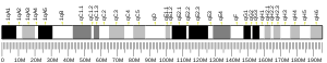
Complement receptor type 1 (CR1) also known as C3b/C4b receptor or CD35 is a protein that in humans is encoded by the CR1 gene.

<span class="mw-page-title-main">CD23</span> Low-affinity" receptor for IgE

CD23, also known as Fc epsilon RII, or FcεRII, is the "low-affinity" receptor for IgE, an antibody isotype involved in allergy and resistance to parasites, and is important in regulation of IgE levels. Unlike many of the antibody receptors, CD23 is a C-type lectin. It is found on mature B cells, activated macrophages, eosinophils, follicular dendritic cells, and platelets.

Co-stimulation is a secondary signal which immune cells rely on to activate an immune response in the presence of an antigen-presenting cell. In the case of T cells, two stimuli are required to fully activate their immune response. During the activation of lymphocytes, co-stimulation is often crucial to the development of an effective immune response. Co-stimulation is required in addition to the antigen-specific signal from their antigen receptors.

<span class="mw-page-title-main">Integrin alpha X</span> Mammalian protein found in Homo sapiens

CD11c, also known as Integrin, alpha X (ITGAX), is a gene that encodes for CD11c.

<span class="mw-page-title-main">Follicular dendritic cells</span> Immune cells found in lymph nodes

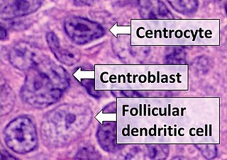
Follicular dendritic cells (FDC) are cells of the immune system found in primary and secondary lymph follicles of the B cell areas of the lymphoid tissue. Unlike dendritic cells (DC), FDCs are not derived from the bone-marrow hematopoietic stem cell, but are of mesenchymal origin. Possible functions of FDC include: organizing lymphoid tissue's cells and microarchitecture, capturing antigen to support B cell, promoting debris removal from germinal centers, and protecting against autoimmunity. Disease processes that FDC may contribute include primary FDC-tumor, chronic inflammatory conditions, HIV-1 infection development, and neuroinvasive scrapie.

<span class="mw-page-title-main">CD19</span> Biomarker for B cell lineage

B-lymphocyte antigen CD19, also known as CD19 molecule, B-Lymphocyte Surface Antigen B4, T-Cell Surface Antigen Leu-12 and CVID3 is a transmembrane protein that in humans is encoded by the gene CD19. In humans, CD19 is expressed in all B lineage cells. Contrary to some early doubts, human plasma cells do express CD19, as confirmed by others. CD19 plays two major roles in human B cells: on the one hand, it acts as an adaptor protein to recruit cytoplasmic signaling proteins to the membrane; on the other, it works within the CD19/CD21 complex to decrease the threshold for B cell receptor signaling pathways. Due to its presence on all B cells, it is a biomarker for B lymphocyte development, lymphoma diagnosis and can be utilized as a target for leukemia immunotherapies.

Macrophage-1 antigen is a complement receptor ("CR3") consisting of CD11b and CD18.

Angioimmunoblastic T-cell lymphoma is a mature T-cell lymphoma of blood or lymph vessel immunoblasts characterized by a polymorphous lymph node infiltrate showing a marked increase in follicular dendritic cells (FDCs) and high endothelial venules (HEVs) and systemic involvement.

<span class="mw-page-title-main">C-C chemokine receptor type 7</span> Protein-coding gene in the species Homo sapiens

C-C chemokine receptor type 7 is a protein that in humans is encoded by the CCR7 gene. Two ligands have been identified for this receptor: the chemokines ligand 19 (CCL19/ELC) and ligand 21 (CCL21). The ligands have similar affinity for the receptor, though CCL19 has been shown to induce internalisation of CCR7 and desensitisation of the cell to CCL19/CCL21 signals. CCR7 is a transmembrane protein with 7 transmembrane domains, which is coupled with heterotrimeric G proteins, which transduce the signal downstream through various signalling cascades. The main function of the receptor is to guide immune cells to immune organs by detecting specific chemokines, which these tissues secrete.

<span class="mw-page-title-main">CD3G</span> Protein-coding gene in the species Homo sapiens

T-cell surface glycoprotein CD3 gamma chain is a protein that in humans is encoded by the CD3G gene.

<span class="mw-page-title-main">CD81</span> Mammalian protein found in Homo sapiens

CD81 molecule, also known as CD81, is a protein which in humans is encoded by the CD81 gene. It is also known as 26 kDa cell surface protein, TAPA-1, and Tetraspanin-28 (Tspan-28).

<span class="mw-page-title-main">SLAMF6</span> Protein-coding gene in humans

SLAM family member 6 is a protein that in humans is encoded by the SLAMF6 gene.

<span class="mw-page-title-main">FCGR2B</span>

Fc fragment of IgG receptor IIb is a low affinity inhibitory receptor for the Fc region of immunoglobulin gamma (IgG). FCGR2B participates in the phagocytosis of immune complexes and in the regulation of antibody production by B lymphocytes.

<span class="mw-page-title-main">CD79B</span> Mammalian protein found in Homo sapiens

CD79b molecule, immunoglobulin-associated beta, also known as CD79B, is a human gene.

VG-1 is a B cell line which was derived from primary effusion lymphoma (PEL). It was first established in 2000 by David T. Scadden’s group at Massachusetts General Hospital. It is infected with Kaposi's sarcoma-associated herpesvirus (KSHV), but negative with Epstein–Barr virus (EBV).

Epstein–Barr virus (EBV) latent membrane protein 2 (LMP2) are two viral proteins of the Epstein–Barr virus. LMP2A/LMP2B are transmembrane proteins that act to block tyrosine kinase signaling. LMP2A is a transmembrane protein that inhibits normal B-cell signal transduction by mimicking an activated B-cell receptor (BCR). The N-terminus domain of LMP2A is tyrosine phosphorylated and associates with Src family protein tyrosine kinases (PTKs) as well as spleen tyrosine kinase (Syk). PTKs and Syk are associated with BCR signal transduction.

The NSG mouse is a brand of immunodeficient laboratory mice, developed and marketed by Jackson Laboratory, which carries the strain NOD.Cg-Prkdcscid Il2rgtm1Wjl/SzJ. NSG branded mice are among the most immunodeficient described to date. NSG branded mice lack mature T cells, B cells, and natural killer (NK) cells. NSG branded mice are also deficient in multiple cytokine signaling pathways, and they have many defects in innate immunity. The compound immunodeficiencies in NSG branded mice permit the engraftment of a wide range of primary human cells, and enable sophisticated modeling of many areas of human biology and disease. NSG branded mice were developed in the laboratory of Dr. Leonard Shultz at Jackson Laboratory, which owns the NSG trade mark.

Sushi domain is an evolutionarily conserved protein domain. It is also known as complement control protein (CCP) modules or short consensus repeats (SCR). The name derives from the visual similarity of the domain to nigiri sushi when the primary structure is drawn showing the loops created by the disulfide bonds.

References

  1. 1 2 3 GRCh38: Ensembl release 89: ENSG00000117322 Ensembl, May 2017
  2. 1 2 3 GRCm38: Ensembl release 89: ENSMUSG00000026616 Ensembl, May 2017
  3. "Human PubMed Reference:". National Center for Biotechnology Information, U.S. National Library of Medicine.
  4. "Mouse PubMed Reference:". National Center for Biotechnology Information, U.S. National Library of Medicine.
  5. 1 2 Frank K, Atkinson JP (2001). "Complement system." In Austen KF, Frank K, Atkinson JP, Cantor H. eds. Samter's Immunologic Diseases, 6th ed. Vol. 1, p. 281–298, Philadelphia: Lippincott Williams & Wilkins, ISBN   0-7817-2120-2.
  6. 1 2 "Entrez Gene: CR2 complement component (3d/Epstein Barr virus) receptor 2".
  7. Bradbury LE, Kansas GS, Levy S, Evans RL, Tedder TF (November 1992). "The CD19/CD21 signal transducing complex of human B lymphocytes includes the target of antiproliferative antibody-1 and Leu-13 molecules". J. Immunol. 149 (9): 2841–50. doi: 10.4049/jimmunol.149.9.2841 . PMID   1383329. S2CID   23655762.
  8. Horváth G, Serru V, Clay D, Billard M, Boucheix C, Rubinstein E (November 1998). "CD19 is linked to the integrin-associated tetraspans CD9, CD81, and CD82". J. Biol. Chem. 273 (46): 30537–43. doi: 10.1074/jbc.273.46.30537 . PMID   9804823.
  9. Abbas AK, Lichtman AH (2003). Cellular and Molecular Immunology, 5th ed. Philadelphia: Saunders, ISBN   0-7216-0008-5
  10. Yefenof E, Klein G, Jondal M, Oldstone MB (June 1976). "Surface markers on human B and T-lymphocytes. IX. Two-color immunofluorescence studies on the association between ebv receptors and complement receptors on the surface of lymphoid cell lines". Int. J. Cancer. 17 (6): 693–700. doi:10.1002/ijc.2910170602. PMID   181330. S2CID   20793968.
  11. Jacobson AC, Weis JH (September 2008). "Comparative functional evolution of human and mouse CR1 and CR2". J. Immunol. 181 (5): 2953–9. doi:10.4049/jimmunol.181.5.2953. PMC   3366432 . PMID   18713965.
  12. Donius LR, Handy JM, Weis JJ, Weis JH (July 2013). "Optimal germinal center B cell activation and T-dependent antibody responses require expression of the mouse complement receptor Cr1". J. Immunol. 191 (1): 434–47. doi:10.4049/jimmunol.1203176. PMC   3707406 . PMID   23733878.
  13. Kurshumliu F, Sadiku-Zehri F, Qerimi A, Vela Z, Jashari F, Bytyci S; et al. (2019). "Divergent immunohistochemical expression of CD21 and CD23 by follicular dendritic cells with increasing grade of follicular lymphoma". World J Surg Oncol. 17 (1): 115. doi: 10.1186/s12957-019-1659-8 . PMC   6610797 . PMID   31269981.{{cite journal}}: CS1 maint: multiple names: authors list (link)
    - This article is distributed under the terms of the Creative Commons Attribution 4.0 International License (http://creativecommons.org/licenses/by/4.0/)
  14. Leong, Anthony S-Y; Cooper, Kumarason; Leong, F Joel W-M (2003). Manual of Diagnostic Cytology (2 ed.). Greenwich Medical Media, Ltd. pp. 93–94. ISBN   978-1-84110-100-2.

Further reading