CD31

Last updated
PECAM1
PECAM1.png
Available structures
PDB Ortholog search: PDBe RCSB
Identifiers
Aliases PECAM1 , CD31, CD31/EndoCAM, GPIIA', PECA1, PECAM-1, endoCAM, platelet and endothelial cell adhesion molecule 1, PCAM-1
External IDs OMIM: 173445 MGI: 97537 HomoloGene: 47925 GeneCards: PECAM1
Orthologs
SpeciesHumanMouse
Entrez
Ensembl
UniProt
RefSeq (mRNA)

NM_000442

NM_001032378
NM_008816
NM_001305157
NM_001305158

RefSeq (protein)

NP_000433

NP_001027550
NP_001292086
NP_001292087
NP_032842

Location (UCSC) Chr 17: 64.32 – 64.41 Mb Chr 11: 106.55 – 106.64 Mb
PubMed search [3] [4]
Wikidata
View/Edit Human View/Edit Mouse

Platelet endothelial cell adhesion molecule (PECAM-1) also known as cluster of differentiation 31 (CD31) is a protein that in humans is encoded by the PECAM1 gene found on chromosome17q23.3. [5] [6] [7] [8] PECAM-1 plays a key role in removing aged neutrophils from the body.

Contents

Structure

PECAM-1 is a highly glycosylated protein with a mass of approximately 130 kDa. [9] The structure of this protein was determined by molecular cloning in 1990, when it was found out that PECAM-1 has an N-terminal domain with 574 amino acids, a transmembrane domain with 19 amino acids and a C-terminal cytoplasmic domain with 118 amino acids. The N-terminal domain consists of six extracellular Ig-like domains. [10]

Interactions

PECAM-1 is a cell-cell adhesion protein [11] which interacts with other PECAM-1 molecules through homophilic interactions or with non-PECAM-1 molecules through heterophilic interactions. [12] Homophilic interactions between PECAM-1 molecules are mediated by antiparallel interactions between extracellular Ig-like domain 1 and Ig-like domain 2. These interactions are regulated by the level of PECAM-1 expression. Homophilic interactions occur, only when the surface expression of PECAM-1 is high. Otherwise, when expression is low, heterophilic interactions occur. [13]

Tissue distribution

CD31 is normally found on endothelial cells, platelets, macrophages and Kupffer cells, granulocytes, lymphocytes (T cells, B cells, and NK cells), megakaryocytes, and osteoclasts.

Immunohistochemistry

Micrograph of an angiosarcoma stained with a CD31 immunostain (dark brown). Epithelioid angiosarcoma - CD31 - intermed mag.jpg
Micrograph of an angiosarcoma stained with a CD31 immunostain (dark brown).

In immunohistochemistry, CD31 is used primarily to demonstrate the presence of endothelial cells in histological tissue sections. This can help to evaluate the degree of tumor angiogenesis, which can imply a rapidly growing tumor. Malignant endothelial cells also commonly retain the antigen, so that CD31 immunohistochemistry can also be used to demonstrate both angiomas and angiosarcomas. It can also be demonstrated in small lymphocytic and lymphoblastic lymphomas, although more specific markers are available for these conditions. [14]

Function

PECAM-1 is found on the surface of platelets, monocytes, neutrophils, and some types of T-cells, and makes up a large portion of endothelial cell intercellular junctions. The encoded protein is a member of the immunoglobulin superfamily and is likely involved in leukocyte transmigration, angiogenesis, and integrin activation. [5] CD31 on endothelial cells binds to the CD38 receptor on natural killer cells for those cells to attach to the endothelium. [15] [16]

Role in signaling

PECAM-1 plays a role in cell signaling. In the cytoplasmic domain of PECAM-1 are serine and tyrosine residues which are suitable for phosphorylation. After the tyrosine is phosphorylated, PECAM-1 recruits Src homology 2 (SH2) domain–containing signaling proteins. These proteins can then initiate signaling pathways. Of all these proteins, the protein most widely reported as interacting with the PECAM-1 cytoplasmic domain is SH2 domain–containing protein-tyrosine phosphatase SHP-2. [17] Signaling through PECAM-1 leads to the activation of neutrophils, monocytes and leukocytes. [18]

Leukocyte transmigration

PECAM-1 is involved in migration of monocytes and neutrophils, [19] natural killer cells, [20] Vδ1+ γδ T lymphocytes [21] and CD34+ hematopoietic progenitor cells [22] through the endothelial cells. Moreover, PECAM-1 is involved in transendothelial migration of recent thymic emigrants to the secondary lymphoid organs. [23] Mechanism of leukocyte transmigration can be explained by creating a homophilic interaction. In this interaction migrating leukocytes express PECAM-1 on the surface and then they react with PECAM-1 on the surface of endothelial cell. [24]

Angiogenesis

PECAM-1 is also important for angiogenesis because it enables the formation of new blood vessels through the cell-cell adhesion. [25]

Role of CD31 in diseases

Cancer

PECAM-1 is expressed by many solid tumor cell lines such as hemangioma, angiosarcoma, Kaposi’s sarcoma, breast carcinoma, glioblastoma, colon carcinoma, skin carcinoma and other tumor cell lines. [26] On the surface of these tumor cells PECAM-1 mediates the adhesion to endothelial cells. [27] PECAM-1 modulates tumor growth by the formation of new endothelial cell tubes. In mice, this process can be inhibited using an anti-PECAM-1 antibody. [28]

Recently, it was found out that elderly patients with gastric cancer have high concentration of PECAM-1 in the serum. That suggests that the use of a serum PECAM-1 level can be a good prognostic marker. [29]

Atherosclerosis

Inhibition of PECAM-1 leads to a reduction of atherosclerotic lesions in mice. [30] That means that PECAM-1 is involved in atherosclerosis. The exact mechanism, how PECAM-1 contributes to atherosclerosis is not known, but there are some theories. PECAM-1 can act as a mechanoresponsive molecule. Or the pathogenesis can be caused by the infiltration of leukocytes mediated by PECAM-1. Finally, polymorphisms in the PECAM-1 gene can lead to the progression of atherosclerosis. [31]

Disseminated intravascular coagulation

Extensive microvascular thrombosis and increased microvascular permeability are main characteristics of disseminated intravascular coagulation, a fatal complication of sepsis. Patients with this devastating condition have high levels of PECAM-1 in the serum indicating PECAM-1 as a good diagnostic marker. Moreover, PECAM-1 can protect from the development of disseminated intravascular coagulation by inhibiting macrophage pyroptosis. [32]

Neuroinflammation

PECAM-1 contributes to at least two of the nervous system diseases, multiple sclerosis and cerebral ischaemia. First signs of multiple sclerosis are defects in the blood brain barrier and leukocyte migration mediated by adhesion molecules such as PECAM-1. Moreover, monocytes in patients with multiple sclerosis express high level of PECAM-1. Cerebral ischaemia is caused by the accumulation of leukocytes, which then infiltrate brain parenchyma and release toxic compounds such as oxygen radicals. Interactions between leukocyte and endothelium are mediated by PECAM-1. High levels of soluble PECAM-1 can be used to diagnose both diseases. Increased PECAM-1 levels indicate damage in the blood brain barrier in patients with multiple sclerosis and high PECAM-1 levels can be used as a short-term prediction of a stroke in patients with cerebral ischaemia. [33]

Related Research Articles

<span class="mw-page-title-main">Cell adhesion</span> Process of cell attachment

Cell adhesion is the process by which cells interact and attach to neighbouring cells through specialised molecules of the cell surface. This process can occur either through direct contact between cell surfaces such as cell junctions or indirect interaction, where cells attach to surrounding extracellular matrix, a gel-like structure containing molecules released by cells into spaces between them. Cells adhesion occurs from the interactions between cell-adhesion molecules (CAMs), transmembrane proteins located on the cell surface. Cell adhesion links cells in different ways and can be involved in signal transduction for cells to detect and respond to changes in the surroundings. Other cellular processes regulated by cell adhesion include cell migration and tissue development in multicellular organisms. Alterations in cell adhesion can disrupt important cellular processes and lead to a variety of diseases, including cancer and arthritis. Cell adhesion is also essential for infectious organisms, such as bacteria or viruses, to cause diseases.

<span class="mw-page-title-main">L1 (protein)</span> Mammalian protein found in Homo sapiens

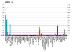
L1, also known as L1CAM, is a transmembrane protein member of the L1 protein family, encoded by the L1CAM gene. This protein, of 200-220 kDa, is a neuronal cell adhesion molecule with a strong implication in cell migration, adhesion, neurite outgrowth, myelination and neuronal differentiation. It also plays a key role in treatment-resistant cancers due to its function. It was first identified in 1984 by M. Schachner who found the protein in post-mitotic mice neurons.

<span class="mw-page-title-main">Selectin</span> Family of cell adhesion molecules

The selectins are a family of cell adhesion molecules. All selectins are single-chain transmembrane glycoproteins that share similar properties to C-type lectins due to a related amino terminus and calcium-dependent binding. Selectins bind to sugar moieties and so are considered to be a type of lectin, cell adhesion proteins that bind sugar polymers.

<span class="mw-page-title-main">P-selectin</span> Type-1 transmembrane protein

P-selectin is a type-1 transmembrane protein that in humans is encoded by the SELP gene.

<span class="mw-page-title-main">ICAM-1</span> Mammalian protein found in Homo sapiens

ICAM-1 also known as CD54 is a protein that in humans is encoded by the ICAM1 gene. This gene encodes a cell surface glycoprotein which is typically expressed on endothelial cells and cells of the immune system. It binds to integrins of type CD11a / CD18, or CD11b / CD18 and is also exploited by rhinovirus as a receptor for entry into respiratory epithelium.

<span class="mw-page-title-main">E-selectin</span> Mammalian protein found in Homo sapiens

E-selectin, also known as CD62 antigen-like family member E (CD62E), endothelial-leukocyte adhesion molecule 1 (ELAM-1), or leukocyte-endothelial cell adhesion molecule 2 (LECAM2), is a selectin cell adhesion molecule expressed only on endothelial cells activated by cytokines. Like other selectins, it plays an important part in inflammation. In humans, E-selectin is encoded by the SELE gene.

<span class="mw-page-title-main">VCAM-1</span> Protein-coding gene in the species Homo sapiens

Vascular cell adhesion protein 1 also known as vascular cell adhesion molecule 1 (VCAM-1) or cluster of differentiation 106 (CD106) is a protein that in humans is encoded by the VCAM1 gene. VCAM-1 functions as a cell adhesion molecule.

<span class="mw-page-title-main">PTPN11</span> Protein-coding gene in the species Homo sapiens

Tyrosine-protein phosphatase non-receptor type 11 (PTPN11) also known as protein-tyrosine phosphatase 1D (PTP-1D), Src homology region 2 domain-containing phosphatase-2 (SHP-2), or protein-tyrosine phosphatase 2C (PTP-2C) is an enzyme that in humans is encoded by the PTPN11 gene. PTPN11 is a protein tyrosine phosphatase (PTP) Shp2.

<span class="mw-page-title-main">CD146</span> Protein-coding gene in the species Homo sapiens

CD146 also known as the melanoma cell adhesion molecule (MCAM) or cell surface glycoprotein MUC18, is a 113kDa cell adhesion molecule currently used as a marker for endothelial cell lineage. In humans, the CD146 protein is encoded by the MCAM gene.

<span class="mw-page-title-main">Leukocyte extravasation</span>

Leukocyte extravasation is the movement of leukocytes out of the circulatory system and towards the site of tissue damage or infection. This process forms part of the innate immune response, involving the recruitment of non-specific leukocytes. Monocytes also use this process in the absence of infection or tissue damage during their development into macrophages.

<span class="mw-page-title-main">PTPN6</span> Protein-coding gene in the species Homo sapiens

Tyrosine-protein phosphatase non-receptor type 6, also known as Src homology region 2 domain-containing phosphatase-1 (SHP-1), is an enzyme that in humans is encoded by the PTPN6 gene.

<span class="mw-page-title-main">F11 receptor</span> Protein-coding gene in the species Homo sapiens

Junctional adhesion molecule A is a protein that in humans is encoded by the F11R gene. It has also been designated as CD321.

<span class="mw-page-title-main">CD93</span>

CD93 is a protein that in humans is encoded by the CD93 gene. CD93 is a C-type lectin transmembrane receptor which plays a role not only in cell–cell adhesion processes but also in host defense.

<span class="mw-page-title-main">JAM3</span> Protein-coding gene in the species Homo sapiens

Junctional adhesion molecule C is a protein that in humans is encoded by the JAM3 gene.

<span class="mw-page-title-main">PTPRB</span> Protein-coding gene in the species Homo sapiens

Receptor-type tyrosine-protein phosphatase beta or VE-PTP is an enzyme specifically expressed in endothelial cells that in humans is encoded by the PTPRB gene.

<span class="mw-page-title-main">PTPRM</span> Protein-coding gene in the species Homo sapiens

Receptor-type tyrosine-protein phosphatase mu is an enzyme that in humans is encoded by the PTPRM gene.

<span class="mw-page-title-main">CD226</span> Protein-coding gene in the species Homo sapiens

CD226, PTA1 or DNAM-1 is a ~65 kDa immunoglobulin-like transmembrane glycoprotein expressed on the surface of natural killer cells, NK T cell, B cells, dendritic cells, hematopoietic precursor cells, platelets, monocytes and T cells.

<span class="mw-page-title-main">TIE1</span>

Tyrosine kinase with immunoglobulin-like and EGF-like domains 1 also known as TIE1 is an angiopoietin receptor which in humans is encoded by the TIE1 gene.

<span class="mw-page-title-main">FERMT3</span> Protein-coding gene in the species Homo sapiens

Fermitin family homolog 3) (FERMT3), also known as kindlin-3 (KIND3), MIG2-like protein (MIG2B), or unc-112-related protein 2 (URP2) is a protein that in humans is encoded by the FERMT3 gene. The kindlin family of proteins, member of the B4.1 superfamily, comprises three conserved protein homologues, kindlin 1, 2, and 3. They each contain a bipartite FERM domain comprising four subdomains F0, F1, F2, and F3 that show homology with the FERM head (H) domain of the cytoskeletal Talin protein. Kindlins have been linked to Kindler syndrome, leukocyte adhesion deficiency, cancer and other acquired human diseases. They are essential in the organisation of focal adhesions that mediate cell-extracellular matrix junctions and are involved in other cellular compartments that control cell-cell contacts and nucleus functioning. Therefore, they are responsible for cell to cell crosstalk via cell-cell contacts and integrin mediated cell adhesion through focal adhesion proteins and as specialised adhesion structures of hematopoietic cells they are also present in podosome's F actin surrounding ring structure. Isoform 2 may act as a repressor of NF-kappa-B and apoptosis

<span class="mw-page-title-main">Endothelial cell anergy</span> Defense mechanism of tumors against immunity

Endothelial cell anergy is a condition during the process of angiogenesis, where endothelial cells, the cells that line the inside of blood vessels, can no longer respond to inflammatory cytokines. These cytokines are necessary to induce the expression of cell adhesion molecules to allow leukocyte infiltration from the blood into the tissue at places of inflammation, such as a tumor. This condition, which protects the tumor from the immune system, is the result of exposure to angiogenic growth factors.

References

  1. 1 2 3 GRCh38: Ensembl release 89: ENSG00000261371 - Ensembl, May 2017
  2. 1 2 3 GRCm38: Ensembl release 89: ENSMUSG00000020717 - Ensembl, May 2017
  3. "Human PubMed Reference:". National Center for Biotechnology Information, U.S. National Library of Medicine.
  4. "Mouse PubMed Reference:". National Center for Biotechnology Information, U.S. National Library of Medicine.
  5. 1 2 "Entrez Gene: platelet/endothelial cell adhesion molecule".
  6. Newman PJ, Berndt MC, Gorski J, White GC, Lyman S, Paddock C, Muller WA (March 1990). "PECAM-1 (CD31) cloning and relation to adhesion molecules of the immunoglobulin gene superfamily". Science. 247 (4947): 1219–1222. Bibcode:1990Sci...247.1219N. doi:10.1126/science.1690453. PMID   1690453.
  7. Gumina RJ, Kirschbaum NE, Rao PN, vanTuinen P, Newman PJ (June 1996). "The human PECAM1 gene maps to 17q23". Genomics. 34 (2): 229–232. doi:10.1006/geno.1996.0272. PMID   8661055.
  8. Xie Y, Muller WA (October 1996). "Fluorescence in situ hybridization mapping of the mouse platelet endothelial cell adhesion molecule-1 (PECAM1) to mouse chromosome 6, region F3-G1". Genomics. 37 (2): 226–228. doi:10.1006/geno.1996.0546. PMID   8921400.
  9. Simmons DL, Walker C, Power C, Pigott R (June 1990). "Molecular cloning of CD31, a putative intercellular adhesion molecule closely related to carcinoembryonic antigen". The Journal of Experimental Medicine. 171 (6): 2147–2152. doi:10.1084/jem.171.6.2147. PMC   2187965 . PMID   2351935.
  10. Newman PJ, Berndt MC, Gorski J, White GC, Lyman S, Paddock C, Muller WA (March 1990). "PECAM-1 (CD31) cloning and relation to adhesion molecules of the immunoglobulin gene superfamily". Science. 247 (4947): 1219–1222. Bibcode:1990Sci...247.1219N. doi:10.1126/science.1690453. PMID   1690453.
  11. Albelda SM, Muller WA, Buck CA, Newman PJ (September 1991). "Molecular and cellular properties of PECAM-1 (endoCAM/CD31): a novel vascular cell-cell adhesion molecule". The Journal of Cell Biology. 114 (5): 1059–1068. doi:10.1083/jcb.114.5.1059. PMC   2289123 . PMID   1874786.
  12. DeLisser HM, Newman PJ, Albelda SM (October 1994). "Molecular and functional aspects of PECAM-1/CD31". Immunology Today. 15 (10): 490–495. doi:10.1016/0167-5699(94)90195-3. PMID   7945775.
  13. Sun J, Williams J, Yan HC, Amin KM, Albelda SM, DeLisser HM (August 1996). "Platelet endothelial cell adhesion molecule-1 (PECAM-1) homophilic adhesion is mediated by immunoglobulin-like domains 1 and 2 and depends on the cytoplasmic domain and the level of surface expression". The Journal of Biological Chemistry. 271 (31): 18561–18570. doi: 10.1074/jbc.271.31.18561 . PMID   8702505.
  14. Leong, Anthony S-Y, Cooper, Kumarason, Leong, F Joel W-M (2003). Manual of Diagnostic Cytology (2 ed.). Greenwich Medical Media, Ltd. p. 103. ISBN   978-1-84110-100-2.
  15. Zambello R, Barilà G, Manni S, Piazza F, Semenzato G (March 2020). "NK cells and CD38: Implication for (Immuno)Therapy in Plasma Cell Dyscrasias". Cells. 9 (3): 768. doi: 10.3390/cells9030768 . PMC   7140687 . PMID   32245149.
  16. Glaría E, Valledor AF (January 2020). "Roles of CD38 in the Immune Response to Infection". Cells. 9 (1): 228. doi: 10.3390/cells9010228 . PMC   7017097 . PMID   31963337.
  17. Newman PJ, Newman DK (June 2003). "Signal transduction pathways mediated by PECAM-1: new roles for an old molecule in platelet and vascular cell biology". Arteriosclerosis, Thrombosis, and Vascular Biology. 23 (6): 953–964. doi: 10.1161/01.ATV.0000071347.69358.D9 . PMID   12689916.
  18. Elias CG, Spellberg JP, Karan-Tamir B, Lin CH, Wang YJ, McKenna PJ, et al. (June 1998). "Ligation of CD31/PECAM-1 modulates the function of lymphocytes, monocytes and neutrophils". European Journal of Immunology. 28 (6): 1948–1958. doi: 10.1002/(SICI)1521-4141(199806)28:06<1948::AID-IMMU1948>3.0.CO;2-C . PMID   9645377. S2CID   32378668.
  19. Muller WA, Weigl SA, Deng X, Phillips DM (August 1993). "PECAM-1 is required for transendothelial migration of leukocytes". The Journal of Experimental Medicine. 178 (2): 449–460. doi:10.1084/jem.178.2.449. PMC   2191108 . PMID   8340753.
  20. Berman ME, Xie Y, Muller WA (February 1996). "Roles of platelet/endothelial cell adhesion molecule-1 (PECAM-1, CD31) in natural killer cell transendothelial migration and beta 2 integrin activation". Journal of Immunology. 156 (4): 1515–1524. doi:10.4049/jimmunol.156.4.1515. PMID   8568255. S2CID   1741600.
  21. Poggi A, Zocchi MR, Carosio R, Ferrero E, Angelini DF, Galgani S, et al. (June 2002). "Transendothelial migratory pathways of V delta 1+TCR gamma delta+ and V delta 2+TCR gamma delta+ T lymphocytes from healthy donors and multiple sclerosis patients: involvement of phosphatidylinositol 3 kinase and calcium calmodulin-dependent kinase II". Journal of Immunology. 168 (12): 6071–6077. doi: 10.4049/jimmunol.168.12.6071 . PMID   12055216. S2CID   82477153.
  22. Voermans C, Rood PM, Hordijk PL, Gerritsen WR, van der Schoot CE (2000). "Adhesion molecules involved in transendothelial migration of human hematopoietic progenitor cells". Stem Cells. 18 (6): 435–443. doi: 10.1634/stemcells.18-6-435 . PMID   11072032. S2CID   37713443.
  23. Kimmig S, Przybylski GK, Schmidt CA, Laurisch K, Möwes B, Radbruch A, Thiel A (March 2002). "Two subsets of naive T helper cells with distinct T cell receptor excision circle content in human adult peripheral blood". The Journal of Experimental Medicine. 195 (6): 789–794. doi:10.1084/jem.20011756. PMC   2193736 . PMID   11901204.
  24. Mamdouh Z, Chen X, Pierini LM, Maxfield FR, Muller WA (February 2003). "Targeted recycling of PECAM from endothelial surface-connected compartments during diapedesis". Nature. 421 (6924): 748–753. Bibcode:2003Natur.421..748M. doi:10.1038/nature01300. PMID   12610627. S2CID   26318819.
  25. DeLisser HM, Christofidou-Solomidou M, Strieter RM, Burdick MD, Robinson CS, Wexler RS, et al. (September 1997). "Involvement of endothelial PECAM-1/CD31 in angiogenesis". The American Journal of Pathology. 151 (3): 671–677. PMC   1857836 . PMID   9284815.
  26. Bergom C, Gao C, Newman PJ (October 2005). "Mechanisms of PECAM-1-mediated cytoprotection and implications for cancer cell survival". Leukemia & Lymphoma. 46 (10): 1409–1421. doi:10.1080/10428190500126091. PMID   16194886. S2CID   25682089.
  27. Tang DG, Chen YQ, Newman PJ, Shi L, Gao X, Diglio CA, Honn KV (October 1993). "Identification of PECAM-1 in solid tumor cells and its potential involvement in tumor cell adhesion to endothelium". The Journal of Biological Chemistry. 268 (30): 22883–22894. doi: 10.1016/S0021-9258(18)41609-2 . PMID   8226797.
  28. Zhou Z, Christofidou-Solomidou M, Garlanda C, DeLisser HM (1999). "Antibody against murine PECAM-1 inhibits tumor angiogenesis in mice". Angiogenesis. 3 (2): 181–188. doi:10.1023/a:1009092107382. PMID   14517436. S2CID   33204411.
  29. Li Y, Guo XB, Wei YH, Kang XL (January 2021). "Serum CXCL13 and PECAM-1 can be used as diagnostic and prognostic markers in elderly patients with gastric cancer". Clinical & Translational Oncology. 23 (1): 130–138. doi:10.1007/s12094-020-02403-w. PMID   32500259. S2CID   219313556.
  30. Stevens HY, Melchior B, Bell KS, Yun S, Yeh JC, Frangos JA (September 2008). "PECAM-1 is a critical mediator of atherosclerosis". Disease Models & Mechanisms. 1 (2–3): 175–81, discussion 179. doi:10.1242/dmm.000547. PMC   2562188 . PMID   19048083.
  31. Woodfin A, Voisin MB, Nourshargh S (December 2007). "PECAM-1: a multi-functional molecule in inflammation and vascular biology". Arteriosclerosis, Thrombosis, and Vascular Biology. 27 (12): 2514–2523. doi: 10.1161/ATVBAHA.107.151456 . PMID   17872453.
  32. Luo L, Xu M, Liao D, Deng J, Mei H, Hu Y (August 2020). "PECAM-1 protects against DIC by dampening inflammatory responses via inhibiting macrophage pyroptosis and restoring vascular barrier integrity". Translational Research. 222: 1–16. doi:10.1016/j.trsl.2020.04.005. PMID   32417429. S2CID   218678428.
  33. Kalinowska A, Losy J (December 2006). "PECAM-1, a key player in neuroinflammation". European Journal of Neurology. 13 (12): 1284–1290. doi:10.1111/j.1468-1331.2006.01640.x. PMID   17116209. S2CID   22437957.

Further reading