CD22

Last updated
CD22
Identifiers
Aliases CD22 , SIGLEC-2, SIGLEC2, CD22 molecule
External IDs OMIM: 107266 MGI: 88322 HomoloGene: 31052 GeneCards: CD22
Orthologs
SpeciesHumanMouse
Entrez
Ensembl
UniProt
RefSeq (mRNA)

NM_001043317
NM_009845

RefSeq (protein)

NP_001172028
NP_001172029
NP_001172030
NP_001265346
NP_001762

Contents

NP_001036782
NP_033975

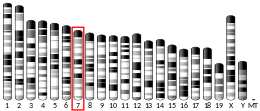
Location (UCSC)n/a Chr 7: 30.56 – 30.58 Mb
PubMed search [2] [3]
Wikidata
View/Edit Human View/Edit Mouse

CD22, or cluster of differentiation-22, is a molecule belonging to the SIGLEC family of lectins. [4] It is found on the surface of mature B cells and to a lesser extent on some immature B cells. Generally speaking, CD22 is a regulatory molecule that prevents the overactivation of the immune system and the development of autoimmune diseases. [5]

CD22 is a sugar binding transmembrane protein, which specifically binds sialic acid with an immunoglobulin (Ig) domain located at its N-terminus. The presence of Ig domains makes CD22 a member of the immunoglobulin superfamily. CD22 functions as an inhibitory receptor for B cell receptor (BCR) signaling. It is also involved in the B cell trafficking to Peyer's patches in mice. [6] In mice, it has been shown that CD22 blockade restores homeostatic microglial phagocytosis in aging brains. [7]

image of microglia Microglial cells.tif
image of microglia

Structure

CD22 is a transmembrane protein with a molecular weight of 140 kDa. The extracellular part of CD22 consists of seven immunoglobulin domains and the intracellular part is formed by 141-amino acid cytoplasmic tail. [8]

Extracellular part

The binding site for ligands is located at the extracellular N-terminus, specifically at the last immunoglobulin domain called the V-like domain. This domain binds to ligands containing sialic acid via α2,6-linkage to the galactose residue. Such ligands are commonly expressed on the surface of erythrocytes, monocytes, cytokine-activated endothelial cells, T cells and B cells. To a lesser extent they are present on soluble IgM and on the soluble plasmatic glycoprotein called haptoglobin. Therefore, CD22 can bind ligands in the cis configuration, when they are on the surface of B cells, or in the trans configuration, when they are on the surface of other cells or on soluble glycoproteins or attached to a cell-associated antigen. However, CD22 is masked on most B-cell surfaces, meaning that it cannot bind exogenous ligands, so cis interaction with glycoprotein ligands on the same cell is preferred. [9]

Trans ligands

Trans interactions between CD22 and its ligands are important for B cell adhesion and migration. Specifically, CD22-deficient mice have been shown to have reduced numbers of recirculating B cells and reduced numbers of IgM-secreting plasma cells in the bone marrow. Together, this implies that CD22 interacting with trans ligands is crucial for the homing of mature, recirculating B cells to the bone marrow. [10]

BCR signaling

The intracellular part of CD22 consists of 6 tyrosine residues which contain both ITIM and ITAM motifs suggesting both inhibitory and activation role in signaling. [11] Because of the tyrosine residues, the cytoplasmic domain of CD22 can be phosphorylated. This happens when the BCR is cross-linked by the antigen. Phosphorylation is mediated by Lyn, a protein tyrosine kinase (PTK) of the Src family found in lipid rafts. [9]

Inhibitory role

After CD22 is phosphorylated, the ITIM motifs provide docking sites for the SH2 domain containing protein tyrosine phosphatase called SHP-1. SHP-1 inhibits mitogen-activated protein kinase (MAPK) and dephosphorylates components of BCR signaling. That means that association of CD22 with SHP-1 leads to the inhibition of BCR signaling. [12] [9]

Activation role

After CD22 is phosphorylated, the ITAM motifs provide docking sites for the SH2 domain of Lyn or other Syk kinase or Src-family tyrosine kinases. Thus, CD22 positively regulates BCR signaling and thereby promotes B cell survival. [9]

Autoimmunity

Single-nucleotide polymorphisms in the CD22 gene lead to a higher likelihood of autoimmune disease. Specifically, some studies show that polymorphisms in the CD22 gene are associated with susceptibility to systemic lupus erythematosus (SLE) and cutaneous systemic sclerosis. In addition, mutations in enzymes involved in the glycosylation of the CD22 ligand may also lead to the susceptibility to autoimmune diseases. Specifically, mutations in the sialic acid esterase were frequently found in patients with rheumatoid arthritis and SLE. This enzyme is essential for deacetylation of the N-glycan sialic acid present in CD22 ligands and is therefore crucial for ligand binding. [13]

BCR signaling & inhibitory role Schematic representation of the CD22 and B-cell receptor signalling process.png
BCR signaling & inhibitory role

As a drug target

Because CD22 is restricted to B cells, it is an excellent target for immunotherapy of B cell malignancies. There are several mechanisms by which this can be achieved, namely monoclonal antibodies, bispecific antibodies, antibody-drug conjugates, radioimmunoconjugates or CAR-T cells. [14]

An immunotoxin, BL22 (CAT-3888), that targets this receptor was developed at the NIH. [15] BL22 was superseded by moxetumomab pasudotox (HA22, CAT-8015). [16] Moxetumomab pasudotox is approved in the EU and USA for treatment of relapsed or refractory hairy cell leukemia. [17] [18]

Inotuzumab

It was shown that antibody-drug conjugates work better than naked antibodies. The reason is that CD22 is rapidly internalized rather than being exposed to the extracellular environment making it more suitable for specific delivery of these conjugates. [19] One of such therapeutics is Inotuzumab, which was approved by the FDA for the treatment of relapsed or refractory B cell acute lymphoblastic leukemia in August 2017. [20] Inotuzumab consists of a CD22-targeting immunoglobulin G4 humanized monoclonal antibody conjugated to calicheamicin. The mechanism by which calicheamicin destroys malignant cells is that it binds to DNA, causing DNA double-strand breaks, and this in turn leads to transcription inhibition. [19]

Interactions

CD22 has been shown to interact with Grb2, [21] [22] PTPN6, [22] [23] [24] [25] [26] LYN, [21] [24] SHC1 [21] and INPP5D. [21]

Related Research Articles

<span class="mw-page-title-main">B cell</span> Type of white blood cell

B cells, also known as B lymphocytes, are a type of white blood cell of the lymphocyte subtype. They function in the humoral immunity component of the adaptive immune system. B cells produce antibody molecules which may be either secreted or inserted into the plasma membrane where they serve as a part of B-cell receptors. When a naïve or memory B cell is activated by an antigen, it proliferates and differentiates into an antibody-secreting effector cell, known as a plasmablast or plasma cell. In addition, B cells present antigens and secrete cytokines. In mammals, including marsupials B cells mature in the bone marrow, which is at the core of most bones. In birds, B cells mature in the bursa of Fabricius, a lymphoid organ where they were first discovered by Chang and Glick, which is why the B stands for bursa and not bone marrow, as commonly believed.

<span class="mw-page-title-main">Fc receptor</span> Surface protein important to the immune system

In immunology, an Fc receptor is a protein found on the surface of certain cells – including, among others, B lymphocytes, follicular dendritic cells, natural killer cells, macrophages, neutrophils, eosinophils, basophils, human platelets, and mast cells – that contribute to the protective functions of the immune system. Its name is derived from its binding specificity for a part of an antibody known as the Fc region. Fc receptors bind to antibodies that are attached to infected cells or invading pathogens. Their activity stimulates phagocytic or cytotoxic cells to destroy microbes, or infected cells by antibody-mediated phagocytosis or antibody-dependent cell-mediated cytotoxicity. Some viruses such as flaviviruses use Fc receptors to help them infect cells, by a mechanism known as antibody-dependent enhancement of infection.

<span class="mw-page-title-main">B-cell receptor</span> Transmembrane protein on the surface of a B cell

The B-cell receptor (BCR) is a transmembrane protein on the surface of a B cell. A B-cell receptor is composed of a membrane-bound immunoglobulin molecule and a signal transduction moiety. The former forms a type 1 transmembrane receptor protein, and is typically located on the outer surface of these lymphocyte cells. Through biochemical signaling and by physically acquiring antigens from the immune synapses, the BCR controls the activation of the B cell. B cells are able to gather and grab antigens by engaging biochemical modules for receptor clustering, cell spreading, generation of pulling forces, and receptor transport, which eventually culminates in endocytosis and antigen presentation. B cells' mechanical activity adheres to a pattern of negative and positive feedbacks that regulate the quantity of removed antigen by manipulating the dynamic of BCR–antigen bonds directly. Particularly, grouping and spreading increase the relation of antigen with BCR, thereby proving sensitivity and amplification. On the other hand, pulling forces delinks the antigen from the BCR, thus testing the quality of antigen binding.

<span class="mw-page-title-main">GRB2</span> Protein-coding gene in the species Homo sapiens

Growth factor receptor-bound protein 2, also known as Grb2, is an adaptor protein involved in signal transduction/cell communication. In humans, the GRB2 protein is encoded by the GRB2 gene.

<span class="mw-page-title-main">CD19</span> Biomarker for B cell lineage

B-lymphocyte antigen CD19, also known as CD19 molecule, B-Lymphocyte Surface Antigen B4, T-Cell Surface Antigen Leu-12 and CVID3 is a transmembrane protein that in humans is encoded by the gene CD19. In humans, CD19 is expressed in all B lineage cells. Contrary to some early doubts, human plasma cells do express CD19, as confirmed by others. CD19 plays two major roles in human B cells: on the one hand, it acts as an adaptor protein to recruit cytoplasmic signaling proteins to the membrane; on the other, it works within the CD19/CD21 complex to decrease the threshold for B cell receptor signaling pathways. Due to its presence on all B cells, it is a biomarker for B lymphocyte development, lymphoma diagnosis and can be utilized as a target for leukemia immunotherapies.

Siglecs(Sialic acid-binding immunoglobulin-type lectins) are cell surface proteins that bind sialic acid. They are found primarily on the surface of immune cells and are a subset of the I-type lectins. There are 14 different mammalian Siglecs, providing an array of different functions based on cell surface receptor-ligand interactions.

<span class="mw-page-title-main">Lymphocyte cytosolic protein 2</span> Protein-coding gene in the species Homo sapiens

Lymphocyte cytosolic protein 2, also known as LCP2 or SLP-76, is a signal-transducing adaptor protein expressed in T cells and myeloid cells and is important in the signaling of T-cell receptors (TCRs). As an adaptor protein, SLP-76 does not have catalytic functions, primarily binding other signaling proteins to form larger signaling complexes. It is a key component of the signaling pathways of receptors with immunoreceptor tyrosine-based activation motifs (ITAMs) such as T-cell receptors, its precursors, and receptors for the Fc regions of certain antibodies. SLP-76 is expressed in T-cells and related lymphocytes like natural killer cells.
<span class="mw-page-title-main">CD33</span> Mammalian protein found in Homo sapiens

CD33 or Siglec-3 is a transmembrane receptor expressed on cells of myeloid lineage. It is usually considered myeloid-specific, but it can also be found on some lymphoid cells.

An immunoreceptor tyrosine-based inhibitory motif (ITIM), is a conserved sequence of amino acids that is found intracellularly in the cytoplasmic domains of many inhibitory receptors of the non-catalytic tyrosine-phosphorylated receptor family found on immune cells. These immune cells include T cells, B cells, NK cells, dendritic cells, macrophages and mast cells. ITIMs have similar structures of S/I/V/LxYxxI/V/L, where x is any amino acid, Y is a tyrosine residue that can be phosphorylated, S is the amino acid serine, I is the amino acid isoleucine, and V is the amino acid valine. ITIMs recruit SH2 domain-containing phosphatases, which inhibit cellular activation. ITIM-containing receptors often serve to target immunoreceptor tyrosine-based activation motif (ITAM)-containing receptors, resulting in an innate inhibition mechanism within cells. ITIM bearing receptors have important role in regulation of immune system allowing negative regulation at different levels of the immune response.
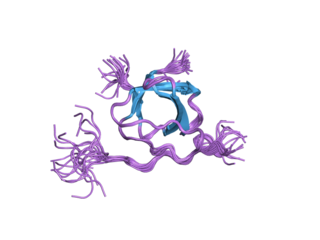
<span class="mw-page-title-main">LYN</span> Mammalian protein found in Homo sapiens

Tyrosine-protein kinase Lyn is a protein that in humans is encoded by the LYN gene.
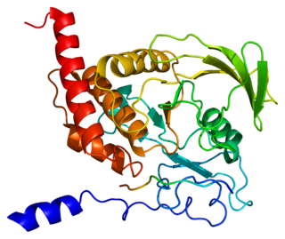
<span class="mw-page-title-main">PTPN6</span> Protein-coding gene in humans

Tyrosine-protein phosphatase non-receptor type 6, also known as Src homology region 2 domain-containing phosphatase-1 (SHP-1), is an enzyme that in humans is encoded by the PTPN6 gene.

<span class="mw-page-title-main">INPP5D</span> Protein-coding gene in the species Homo sapiens

Src homology 2 (SH2) domain containing inositol polyphosphate 5-phosphatase 1(SHIP1) is an enzyme with phosphatase activity. SHIP1 is structured by multiple domain and is encoded by the INPP5D gene in humans. SHIP1 is expressed predominantly by hematopoietic cells but also, for example, by osteoblasts and endothelial cells. This phosphatase is important for the regulation of cellular activation. Not only catalytic but also adaptor activities of this protein are involved in this process. Its movement from the cytosol to the cytoplasmic membrane, where predominantly performs its function, is mediated by tyrosine phosphorylation of the intracellular chains of cell surface receptors that SHIP1 binds. Insufficient regulation of SHIP1 leads to different pathologies.

<span class="mw-page-title-main">B-cell linker</span> Mammalian protein found in Homo sapiens

B-cell linker (BLNK) protein is expressed in B cells and macrophages and plays a large role in B cell receptor signaling. Like all adaptor proteins, BLNK has no known intrinsic enzymatic activity. Its function is to temporally and spatially coordinate and regulate downstream signaling effectors in B cell receptor (BCR) signaling, which is important in B cell development. Binding of these downstream effectors is dependent on BLNK phosphorylation. BLNK is encoded by the BLNK gene and is also known as SLP-65, BASH, and BCA.

<span class="mw-page-title-main">LILRB4</span> Protein-coding gene in the species Homo sapiens

Leukocyte immunoglobulin-like receptor subfamily B member 4 is a protein that in humans is encoded by the LILRB4 gene.
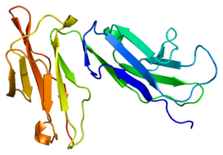
<span class="mw-page-title-main">FCGR2B</span>

Fc fragment of IgG receptor IIb is a low affinity inhibitory receptor for the Fc region of immunoglobulin gamma (IgG). FCGR2B participates in the phagocytosis of immune complexes and in the regulation of antibody production by B lymphocytes.

<span class="mw-page-title-main">SIGLEC8</span> Protein-coding gene in the species Homo sapiens

Sialic acid-binding Ig-like lectin 8 is a protein that in humans is encoded by the SIGLEC8 gene. This gene is located on chromosome 19q13.4, about 330 kb downstream of the SIGLEC9 gene. Within the siglec family of transmembrane proteins, Siglec-8 belongs to the CD33-related siglec subfamily, a subfamily that has undergone rapid evolution.

<span class="mw-page-title-main">PILRB</span> Protein-coding gene in the species Homo sapiens

Paired immunoglobulin-like type 2 receptor beta is a protein that in humans is encoded by the PILRB gene.

<span class="mw-page-title-main">SIGLEC10</span> Protein-coding gene in the species Homo sapiens

Sialic acid-binding Ig-like lectin 10 is a protein that in humans is encoded by the SIGLEC10 gene. Siglec-G is often referred to as the murine paralog of human Siglec-10

<span class="mw-page-title-main">CD79A</span> Mammalian protein found in Homo sapiens

Cluster of differentiation CD79A also known as B-cell antigen receptor complex-associated protein alpha chain and MB-1 membrane glycoprotein, is a protein that in humans is encoded by the CD79A gene.

<span class="mw-page-title-main">SIGLEC6</span> Protein-coding gene in the species Homo sapiens

Sialic acid-binding Ig-like lectin 6 is a protein that in humans is encoded by the SIGLEC6 gene. The gene was originally named CD33L (CD33-like) due to similarities between these genes but later became known as OB-BP1 due to its ability to bind to this factor and, finally, SIGLEC6 as the sixth member of the SIGLEC family of receptors to be identified. The protein has also been given the CD designation CD327.

References

  1. 1 2 3 GRCm38: Ensembl release 89: ENSMUSG00000030577 - Ensembl, May 2017
  2. "Human PubMed Reference:". National Center for Biotechnology Information, U.S. National Library of Medicine.
  3. "Mouse PubMed Reference:". National Center for Biotechnology Information, U.S. National Library of Medicine.
  4. Crocker PR, Clark EA, Filbin M, Gordon S, Jones Y, Kehrl JH, et al. (February 1998). "Siglecs: a family of sialic-acid binding lectins". Glycobiology. 8 (2): v. doi:10.1093/oxfordjournals.glycob.a018832. PMID   9498912.
  5. Hatta Y, Tsuchiya N, Matsushita M, Shiota M, Hagiwara K, Tokunaga K (April 1999). "Identification of the gene variations in human CD22". Immunogenetics. 49 (4): 280–6. doi:10.1007/s002510050494. PMID   10079291. S2CID   22947237.
  6. Lee M, Kiefel H, LaJevic MD, Macauley MS, Kawashima H, O'Hara E, et al. (October 2014). "Transcriptional programs of lymphoid tissue capillary and high endothelium reveal control mechanisms for lymphocyte homing". Nature Immunology. 15 (10): 982–95. doi:10.1038/ni.2983. PMC   4222088 . PMID   25173345.
  7. Pluvinage JV, Wyss-Coray T, et al. (April 11, 2019). "CD22 blockade restores homeostatic microglial phagocytosis in aging brains". Nature. 568 (7751): 187–192. Bibcode:2019Natur.568..187P. doi:10.1038/s41586-019-1088-4. PMC   6574119 . PMID   30944478.
  8. Tedder TF, Tuscano J, Sato S, Kehrl JH (1997). "CD22, a B lymphocyte-specific adhesion molecule that regulates antigen receptor signaling". Annual Review of Immunology. 15: 481–504. doi:10.1146/annurev.immunol.15.1.481. ISSN   0732-0582. PMID   9143697.
  9. 1 2 3 4 Walker JA, Smith KG (March 2008). "CD22: an inhibitory enigma". Immunology. 123 (3): 314–325. doi:10.1111/j.1365-2567.2007.02752.x. ISSN   1365-2567. PMC   2433339 . PMID   18067554.
  10. Nitschke L (July 2009). "CD22 and Siglec-G: B-cell inhibitory receptors with distinct functions". Immunological Reviews. 230 (1): 128–143. doi:10.1111/j.1600-065X.2009.00801.x. ISSN   1600-065X. PMID   19594633. S2CID   205825220.
  11. Poe JC, Fujimoto M, Jansen PJ, Miller AS, Tedder TF (2000-06-09). "CD22 forms a quaternary complex with SHIP, Grb2, and Shc. A pathway for regulation of B lymphocyte antigen receptor-induced calcium flux". The Journal of Biological Chemistry. 275 (23): 17420–17427. doi: 10.1074/jbc.M001892200 . ISSN   0021-9258. PMID   10748054.
  12. Sato S, Tuscano JM, Inaoki M, Tedder TF (August 1998). "CD22 negatively and positively regulates signal transduction through the B lymphocyte antigen receptor". Seminars in Immunology. 10 (4): 287–297. doi:10.1006/smim.1998.0121. ISSN   1044-5323. PMID   9695185.
  13. Clark EA, Giltiay NV (2018). "CD22: A Regulator of Innate and Adaptive B Cell Responses and Autoimmunity". Frontiers in Immunology. 9: 2235. doi: 10.3389/fimmu.2018.02235 . ISSN   1664-3224. PMC   6173129 . PMID   30323814.
  14. Shah NN, Sokol L (2021). "Targeting CD22 for the Treatment of B-Cell Malignancies". ImmunoTargets and Therapy. 10: 225–236. doi: 10.2147/ITT.S288546 . ISSN   2253-1556. PMC   8275043 . PMID   34262884.
  15. Clinical trial number NCT00074048 for "BL22 Immunotoxin in Treating Patients Previously Treated With Cladribine for Hairy Cell Leukemia" at ClinicalTrials.gov
  16. http://www.cambridgeantibody.com/__data/assets/pdf_file/10857/CAT-3888,_CAT-8015_and_CAT-5001_Nov06.pdf Archived 2007-02-27 at the Wayback Machine CAT URL Redirects to Medimmune home page
  17. "Lumoxiti EPAR". European Medicines Agency (EMA). 9 December 2020. Retrieved 16 July 2021..
  18. "Moxetumomab pasudotox-tdfk FDA Approval". U.S. Food and Drug Administration (FDA). Retrieved 20 April 2020.
  19. 1 2 Wynne J, Wright D, Stock W (2019-01-08). "Inotuzumab: from preclinical development to success in B-cell acute lymphoblastic leukemia". Blood Advances. 3 (1): 96–104. doi:10.1182/bloodadvances.2018026211. ISSN   2473-9537. PMC   6325303 . PMID   30622147.
  20. Research Cf (2019-02-09). "FDA approves inotuzumab ozogamicin for relapsed or refractory B-cell precursor ALL". FDA.
  21. 1 2 3 4 Poe JC, Fujimoto M, Jansen PJ, Miller AS, Tedder TF (June 2000). "CD22 forms a quaternary complex with SHIP, Grb2, and Shc. A pathway for regulation of B lymphocyte antigen receptor-induced calcium flux". The Journal of Biological Chemistry. 275 (23): 17420–7. doi: 10.1074/jbc.M001892200 . PMID   10748054.
  22. 1 2 Otipoby KL, Draves KE, Clark EA (November 2001). "CD22 regulates B cell receptor-mediated signals via two domains that independently recruit Grb2 and SHP-1". The Journal of Biological Chemistry. 276 (47): 44315–22. doi: 10.1074/jbc.M105446200 . PMID   11551923.
  23. Blasioli J, Paust S, Thomas ML (January 1999). "Definition of the sites of interaction between the protein tyrosine phosphatase SHP-1 and CD22". The Journal of Biological Chemistry. 274 (4): 2303–7. doi: 10.1074/jbc.274.4.2303 . PMID   9890995.
  24. 1 2 Greer SF, Justement LB (May 1999). "CD45 regulates tyrosine phosphorylation of CD22 and its association with the protein tyrosine phosphatase SHP-1". Journal of Immunology. 162 (9): 5278–86. doi: 10.4049/jimmunol.162.9.5278 . PMID   10228003. S2CID   2223820.
  25. Law CL, Sidorenko SP, Chandran KA, Zhao Z, Shen SH, Fischer EH, et al. (February 1996). "CD22 associates with protein tyrosine phosphatase 1C, Syk, and phospholipase C-gamma(1) upon B cell activation". The Journal of Experimental Medicine. 183 (2): 547–60. doi:10.1084/jem.183.2.547. PMC   2192439 . PMID   8627166.
  26. Adachi T, Wienands J, Wakabayashi C, Yakura H, Reth M, Tsubata T (July 2001). "SHP-1 requires inhibitory co-receptors to down-modulate B cell antigen receptor-mediated phosphorylation of cellular substrates". The Journal of Biological Chemistry. 276 (28): 26648–55. doi: 10.1074/jbc.M100997200 . PMID   11356834.