Cell surface proteins, part of the acquired immune system
Major histocompatibility complex molecule
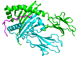
Major histocompatibility complex protein (class I) in orange and pink, with a presented peptide in red. Membrane in grey. The transmembrane and cytoplasmic domains are shown in cartoon form. (PDB: 1hsa)
The major histocompatibility complex (MHC) is a large locus on vertebrate DNA containing a set of closely linked polymorphic genes that code for cell surface proteins essential for the adaptive immune system. These cell surface proteins are called MHC molecules.
The name of this locus comes from its discovery through the study of transplanted tissue compatibility.[1] Later studies revealed that tissue rejection due to incompatibility is only a facet of the full function of MHC molecules: binding an antigen derived from self-proteins, or from pathogens, and bringing the antigen presentation to the cell surface for recognition by the appropriate T-cells.[2] MHC molecules mediate the interactions of leukocytes, also called white blood cells (WBCs), with other leukocytes or with body cells. The MHC determines donor compatibility for organ transplant, as well as one's susceptibility to autoimmune diseases.
In a cell, protein molecules of the host's own phenotype or of other biologic entities are continually synthesized and degraded. Each MHC molecule on the cell surface displays a small peptide (a molecular fraction of a protein) called an epitope.[3] The presented self-antigens prevent an organism's immune system from targeting its own cells. The presentation of pathogen-derived proteins results in the elimination of the infected cell by the immune system.
Diversity of an individual's self-antigen presentation, mediated by MHC self-antigens, is attained in at least three ways: (1) an organism's MHC repertoire is polygenic (via multiple, interacting genes); (2) MHC expression is codominant (from both sets of inherited alleles); (3) MHC gene variants are highly polymorphic (diversely varying from organism to organism within a species).[4]Sexual selection has been observed in male mice choosing to mate with females with different MHCs.[5] Also, at least for MHC I presentation, there has been evidence of antigenic peptide splicing, which can combine peptides from different proteins, vastly increasing antigen diversity.[6]
Discovery
The first descriptions of the MHC were made by British immunologistPeter Gorer in 1936.[7] MHC genes were first identified in inbred mice strains. Clarence Little transplanted tumors across different strains and found rejection of transplanted tumors according to strains of host versus donor.[8]George Snell selectively bred two mouse strains, attained a new strain nearly identical to one of the progenitor strains, but differing crucially in histocompatibility—that is, tissue compatibility upon transplantation—and thereupon identified an MHC locus.[9] Later Jean Dausset demonstrated the existence of MHC genes in humans and described the first human leucocyte antigen, the protein which we call now HLA-A2. Some years laterBaruj Benacerraf showed that polymorphic MHC genes not only determine an individual’s unique constitution of antigens but also regulate the interaction among the various cells of the immunological system. These three scientists have been awarded the 1980 Nobel Prize in Physiology or Medicine[10] for their discoveries concerning “genetically determined structures on the cell surface that regulate immunological reactions”.
The first fully sequenced and annotated MHC was published for humans in 1999 by a consortium of sequencing centers from the UK, USA and Japan in Nature.[11] It was a "virtual MHC" since it was a mosaic from different individuals. A much shorter MHC locus from chickens was published in the same issue of Nature.[12] Many other species have been sequenced and the evolution of the MHC was studied, e.g. in the gray short-tailed opossum (Monodelphis domestica), a marsupial, MHC spans 3.95 Mb, yielding 114 genes, 87 shared with humans.[13] Marsupial MHC genotypic variation lies between eutherian mammals and birds, taken as the minimal MHC encoding, but is closer in organization to that of nonmammals. The IPD-MHC Database[14] was created which provides a centralised repository for sequences of the Major Histocompatibility Complex (MHC) from a number of different species. The database contains 77 species for the release from 2019-12-19.
Genes
The MHC locus is present in all jawed vertebrates; it is assumed to have arisen about 450 million years ago.[15] Despite the difference in the number of genes included in the MHC of different species, the overall organization of the locus is rather similar. Usual MHC contains about a hundred genes and pseudogenes, not all of them are involved in immunity. In humans, the MHC region occurs on chromosome 6, between the flanking genetic markersMOG and COL11A2 (from 6p22.1 to 6p21.3 about 29Mb to 33Mb on the hg38 assembly), and contains 224 genes spanning 3.6 megabase pairs (3 600 000 bases).[11] About half have known immune functions. The human MHC is also called the HLA (human leukocyte antigen) complex (often just the HLA). Similarly, there is SLA (Swine leukocyte antigens), BoLA (Bovine leukocyte antigens), DLA for dogs, etc. However, historically, the MHC in mice is called the Histocompatibility system 2 or just the H-2, in rats – RT1, and in chicken – B-locus.[citation needed]
The MHC gene family is divided into three subgroups: MHC class I, MHC class II, and MHC class III. Among all those genes present in MHC, there are two types of genes coding for the proteins MHC class I molecules and MHC class II molecules that are directly involved in the antigen presentation. These genes are highly polymorphic, 19031 alleles of class I HLA, and 7183 of class II HLA are deposited for human in the IMGT database.[16]
(1) peptide-binding proteins and (2) proteins assisting antigen loading onto MHC class II's peptide-binding proteins (such as MHC II DM, MHC II DQ, MHC II DR, and MHC II DP).
Two chains, called α & β, whose ligands are the CD4 receptors borne by helper T cells.
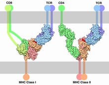
The first crystal structure of Class I MHC molecule, human HLA-A2, was published in 1989.[17] The structure revealed that MHC-I molecules are heterodimers, they have polymorphic heavy α-subunit whose gene occurs inside the MHC locus and small invariant β2 microglobulin subunit whose gene is located usually outside of it. Polymorphic heavy chain of MHC-I molecule contains N-terminal extra-cellular region composed by three domains, α1, α2, and α3, transmembrane helix to hold MHC-I molecule on the cell surface and short cytoplasmic tail. Two domains, α1 and α2 form deep peptide-binding groove between two long α-helices and the floor of the groove formed by eight β-strands. Immunoglobulin-like domain α3 involved in the interaction with CD8 co-receptor. β2 microglobulin provides stability of the complex and participates in the recognition of peptide-MHC class I complex by CD8 co-receptor.[18] The peptide is non-covalently bound to MHC-I, it is held by the several pockets on the floor of the peptide-binding groove. Amino acid side-chains that are most polymorphic in human alleles fill up the central and widest portion of the binding groove, while conserved side-chains are clustered at the narrower ends of the groove.
Schematic view of MHC class I and MHC class II molecules
Classical MHC molecules present epitopes to the TCRs of CD8+ T lymphocytes. Nonclassical molecules (MHC class IB) exhibit limited polymorphism, expression patterns, and presented antigens; this group is subdivided into a group encoded within MHC loci (e.g., HLA-E, -F, -G), as well as those not (e.g., stress ligands such as ULBPs, Rae1, and H60); the antigen/ligand for many of these molecules remain unknown, but they can interact with each of CD8+ T cells, NKT cells, and NK cells. The evolutionary oldest nonclassical MHC class I lineage in human was deduced to be the lineage that includes the CD1 and PROCR (alias EPCR) molecules and this lineage may have been established before the origin of tetrapod species.[19] However, the only nonclassical MHC class I lineage for which evidence exists that it was established before the evolutionary separation of Actinopterygii (ray-finned fish) and Sarcopterygii (lobe-finned fish plus tetrapods) is lineage Z of which members are found, together in each species with classical MHC class I, in lungfish and throughout ray-finned fishes;[20] why the Z lineage was well conserved in ray-finned fish but lost in tetrapods is not understood.
MHC class II can be conditionally expressed by all cell types, but normally occurs only on "professional" antigen-presenting cells (APCs): macrophages, B cells, and especially dendritic cells (DCs). An APC takes up an antigenic protein, performs antigen processing, and returns a molecular fraction of it—a fraction termed the epitope—and displays it on the APC's surface coupled within an MHC class II molecule (antigen presentation). On the cell's surface, the epitope can be recognized by immunologic structures like T-cell receptors (TCRs). The molecular region which binds to the epitope is the paratope.
On surfaces of helper T cells are CD4 receptors, as well as TCRs. When a naive helper T cell's CD4 molecule docks to an APC's MHC class II molecule, its TCR can meet and bind the epitope coupled within the MHC class II. This event primes the naive T cell. According to the local milieu, that is, the balance of cytokines secreted by APCs in the microenvironment, the naive helper T cell (Th0) polarizes into either a memory Th cell or an effector Th cell of phenotype either type 1 (Th1), type 2 (Th2), type 17 (Th17), or regulatory/suppressor (Treg), as so far identified, the Th cell's terminal differentiation.
MHC class II thus mediates immunization to—or, if APCs polarize Th0 cells principally to Treg cells, immune tolerance of—an antigen. The polarization during primary exposure to an antigen is key in determining a number of chronic diseases, such as inflammatory bowel diseases and asthma, by skewing the immune response that memory Th cells coordinate when their memory recall is triggered upon secondary exposure to similar antigens. B cells express MHC class II to present antigens to Th0, but when their B cell receptors bind matching epitopes, interactions which are not mediated by MHC, these activated B cells secrete soluble immunoglobulins: antibody molecules mediating humoral immunity.
Class II MHC molecules are also heterodimers, genes for both α and β subunits are polymorphic and located within MHC class II subregion. Peptide-binding groove of MHC-II molecules is forms by N-terminal domains of both subunits of the heterodimer, α1 and β1, unlike MHC-I molecules, where two domains of the same chain are involved. In addition, both subunits of MHC-II contain transmembrane helix and immunoglobulin domains α2 or β2 that can be recognized by CD4 co-receptors.[21] In this way MHC molecules chaperone which type of lymphocytes may bind to the given antigen with high affinity, since different lymphocytes express different T-Cell Receptor (TCR) co-receptors.
MHC class II molecules in humans have five to six isotypes. Classical molecules present peptides to CD4+ lymphocytes. Nonclassical molecules, accessories, with intracellular functions, are not exposed on cell membranes, but in internal membranes, assisting with the loading of antigenic peptides onto classic MHC class II molecules. The important nonclassical MHC class II molecule DM is only found from the evolutionary level of lungfish,[22] although also in more primitive fishes both classical and nonclassical MHC class II are found.[23][24]
Class III molecules have physiologic roles unlike classes I and II, but are encoded between them in the short arm of human chromosome 6. Class III molecules include several secreted proteins with immune functions: components of the complement system (such as C2, C4, and B factor), cytokines (such as TNF-α, LTA, and LTB), and heat shock proteins.
Function
MHC is the tissue-antigen that allows the immune system (more specifically T cells) to bind to, recognize, and tolerate itself (autorecognition). MHC is also the chaperone for intracellular peptides that are complexed with MHCs and presented to T cell receptors (TCRs) as potential foreign antigens. MHC interacts with TCR and its co-receptors to optimize binding conditions for the TCR-antigen interaction, in terms of antigen binding affinity and specificity, and signal transduction effectiveness.
Essentially, the MHC-peptide complex is a complex of auto-antigen/allo-antigen. Upon binding, T cells should in principle tolerate the auto-antigen, but activate when exposed to the allo-antigen. Disease states occur when this principle is disrupted.
Autoimmune reaction: Having some MHC molecules increases the risk of autoimmune diseases more than having others. HLA-B27 is an example. It is unclear how exactly having the HLA-B27 tissue type increases the risk of ankylosing spondylitis and other associated inflammatory diseases, but mechanisms involving aberrant antigen presentation or T cell activation have been hypothesized.
Tissue allorecognition: MHC molecules in complex with peptide epitopes are essentially ligands for TCRs. T cells become activated by binding to the peptide-binding grooves of any MHC molecule that they were not trained to recognize during positive selection in the thymus.
Antigen processing and presentation
MHC class I pathway: Proteins in the cytosol are degraded by the proteasome, liberating peptides internalized by TAP channel in the endoplasmic reticulum, there associating with MHC-I molecules freshly synthesized. MHC-I/peptide complexes enter Golgi apparatus, are glycosylated, enter secretory vesicles, fuse with the cell membrane, and externalize on the cell membrane interacting with T lymphocytes.
Peptides are processed and presented by two classical pathways:
In MHC class II, phagocytes such as macrophages and immature dendritic cells take up entities by phagocytosis into phagosomes—though B cells exhibit the more general endocytosis into endosomes—which fuse with lysosomes whose acidic enzymes cleave the uptaken protein into many different peptides. Via physicochemical dynamics in molecular interaction with the particular MHC class II variants borne by the host, encoded in the host's genome, a particular peptide exhibits immunodominance and loads onto MHC class II molecules. These are trafficked to and externalized on the cell surface.[27]
In MHC class I, any nucleated cell normally presents cytosolic peptides, mostly self peptides derived from protein turnover and defective ribosomal products. During viral infection, intracellular microorganism infection, or cancerous transformation, such proteins degraded in the proteosome are as well loaded onto MHC class I molecules and displayed on the cell surface. T lymphocytes can detect a peptide displayed at 0.1–1% of the MHC molecules.
Peptide binding for Class I and Class II MHC molecules, showing the binding of peptides between the alpha-helix walls, upon a beta-sheet base. The difference in binding positions is shown. Class I primarily makes contact with backbone residues at the Carboxy and amino terminal regions, while Class II primarily makes contacts along the length of the residue backbone. The precise location of binding residues is determined by the MHC allele.
Table 2. Characteristics of the antigen processing pathways
Characteristic
MHC-I pathway
MHC-II pathway
Composition of the stable peptide-MHC complex
Polymorphic chain α and β2 microglobulin, peptide bound to α chain
In their development in the thymus, T lymphocytes are selected to recognize MHC molecules of the host, but not recognize other self antigens. Following selection, each T lymphocyte shows dual specificity: The TCR recognizes self MHC, but only non-self antigens.
MHC restriction occurs during lymphocyte development in the thymus through a process known as positive selection. T cells that do not receive a positive survival signal — mediated mainly by thymic epithelial cells presenting self peptides bound to MHC molecules — to their TCR undergo apoptosis. Positive selection ensures that mature T cells can functionally recognize MHC molecules in the periphery (i.e. elsewhere in the body).
MHC molecules enable immune system surveillance of the population of protein molecules in a host cell, and greater MHC diversity permits greater diversity of antigen presentation. In 1976, Yamazaki et al demonstrated a sexual selectionmate choice by male mice for females of a different MHC. Similar results have been obtained with fish.[29] Some data find lower rates of early pregnancy loss in human couples of dissimilar MHC genes.[30]
MHC may be related to mate choice in some human populations, a theory that found support by studies by Ober and colleagues in 1997,[31] as well as by Chaix and colleagues in 2008.[32] However, the latter findings have been controversial.[33] If it exists, the phenomenon might be mediated by olfaction, as MHC phenotype appears strongly involved in the strength and pleasantness of perceived odour of compounds from sweat. Fatty acid esters—such as methyl undecanoate, methyl decanoate, methyl nonanoate, methyl octanoate, and methyl hexanoate—show strong connection to MHC.[34]
In 1995, Claus Wedekind found that in a group of female college students who smelled T-shirts worn by male students for two nights (without deodorant, cologne, or scented soaps), by far most women chose shirts worn by men of dissimilar MHCs, a preference reversed if the women were on oral contraceptives.[35] In 2005 in a group of 58 subjects, women were more indecisive when presented with MHCs like their own,[36] although with oral contraceptives, the women showed no particular preference.[37] No studies show the extent to which odor preference determines mate selection (or vice versa).
Evolutionary diversity
Most mammals have MHC variants similar to those of humans, who bear great allelic diversity, especially among the nine classical genes—seemingly due largely to gene duplication—though human MHC regions have many pseudogenes.[38] The most diverse loci, namely HLA-A, HLA-B, and HLA-C, have roughly 6000, 7200, and 5800 known alleles, respectively.[39] Many HLA alleles are ancient, sometimes of closer homology to a chimpanzee MHC alleles than to some other human alleles of the same gene.
MHC allelic diversity has challenged evolutionary biologists for explanation. Most posit balancing selection (see polymorphism (biology)), which is any natural selection process whereby no single allele is absolutely most fit, such as frequency-dependent selection[40] and heterozygote advantage. Pathogenic coevolution, as a type of balancing selection, posits that common alleles are under greatest pathogenic pressure, driving positive selection of uncommon alleles—moving targets, so to say, for pathogens. As pathogenic pressure on the previously common alleles decreases, their frequency in the population stabilizes, and remain circulating in a large population.[41]Genetic drift is also a major driving force in some species.[42][43] It is possible that the combined effects of some or all of these factors cause the genetic diversity.[44]
MHC diversity has also been suggested as a possible indicator for conservation, because large, stable populations tend to display greater MHC diversity, than smaller, isolated populations.[45][46] Small, fragmented populations that have experienced a population bottleneck typically have lower MHC diversity. For example, relatively low MHC diversity has been observed in the cheetah (Acinonyx jubatus),[47]Eurasian beaver (Castor fiber),[48] and giant panda (Ailuropoda melanoleuca).[49] In 2007 low MHC diversity was attributed a role in disease susceptibility in the Tasmanian devil (Sarcophilus harrisii), native to the isolated island of Tasmania, such that an antigen of a transmissible tumor, involved in devil facial tumour disease, appears to be recognized as a self antigen.[50] To offset inbreeding, efforts to sustain genetic diversity in populations of endangered species and of captive animals have been suggested.
In ray-finned fish like rainbow trout, allelic polymorphism in MHC class II is reminiscent of that in mammals and predominantly maps to the peptide binding groove.[51] However, in MHC class I of many teleost fishes, the allelic polymorphism is much more extreme than in mammals in the sense that the sequence identity levels between alleles can be very low and the variation extends far beyond the peptide binding groove.[51][52][20] It has been speculated that this type of MHC class I allelic variation contributes to allograft rejection, which may be especially important in fish to avoid grafting of cancer cells through their mucosal skin.[53]
The MHC locus (6p21.3) has 3 other paralogous loci in the human genome, namely 19pl3.1, 9q33–q34, and 1q21–q25. It is believed that the loci arouse from the two-round duplications in vertebrates of a single ProtoMHC locus, and the new domain organizations of the MHC genes were a result of later cis-duplication and exon shuffling in a process termed "the MHC Big Bang."[54] Genes in this locus are apparently linked to intracellular intrinsic immunity in the basal MetazoanTrichoplax adhaerens.[55]
In transplant rejection
In a transplant procedure, as of an organ or stem cells, MHC molecules themselves act as antigens and can provoke immune response in the recipient, thus causing transplant rejection. MHC molecules were identified and named after their role in transplant rejection between mice of different strains, though it took over 20 years to clarify MHC's role in presenting peptide antigens to cytotoxic T lymphocytes (CTLs).[56]
Each human cell expresses six MHC class I alleles (one HLA-A, -B, and -C allele from each parent) and six to eight MHC class II alleles (one HLA-DP and -DQ, and one or two HLA-DR from each parent, and combinations of these). The MHC variation in the human population is high, at least 350 alleles for HLA-A genes, 620 alleles for HLA-B, 400 alleles for DR, and 90 alleles for DQ. Any two individuals who are not identical twins, triplets, or higher order multiple births, will express differing MHC molecules. All MHC molecules can mediate transplant rejection, but HLA-C and HLA-DP, showing low polymorphism, seem least important.[clarification needed]
When maturing in the thymus, T lymphocytes are selected for their TCR incapacity to recognize self antigens, yet T lymphocytes can react against the donor MHC's peptide-binding groove, the variable region of MHC holding the presented antigen's epitope for recognition by TCR, the matching paratope. T lymphocytes of the recipient take the incompatible peptide-binding groove as nonself antigen. [clarification needed]
Transplant rejection has various types known to be mediated by MHC (HLA):
Hyperacute rejection occurs when, before the transplantation, the recipient has preformed anti-HLA antibodies, perhaps by previous blood transfusions (donor tissue that includes lymphocytes expressing HLA molecules), by anti-HLA generated during pregnancy (directed at the father's HLA displayed by the fetus), or by previous transplantation;
Acute cellular rejection occurs when the recipient's T lymphocytes are activated by the donor tissue, causing damage via mechanisms such as direct cytotoxicity from CD8 cells.
Acute humoral rejection and chronic disfunction occurs when the recipient's anti-HLA antibodies form directed at HLA molecules present on endothelial cells of the transplanted tissue.
In all of the above situations, immunity is directed at the transplanted organ, sustaining lesions. A cross-reaction test between potential donor cells and recipient serum seeks to detect presence of preformed anti-HLA antibodies in the potential recipient that recognize donor HLA molecules, so as to prevent hyperacute rejection. In normal circumstances, compatibility between HLA-A, -B, and -DR molecules is assessed. The higher the number of incompatibilities, the lower the five-year survival rate. Global databases of donor information enhance the search for compatible donors.
The involvement in allogeneic transplant rejection appears to be an ancient feature of MHC molecules, because also in fish associations between transplant rejections and (mis-)matching of MHC class I[57][58] and MHC class II[59] were observed.
Human MHC class I and II are also called human leukocyte antigen (HLA). To clarify the usage, some of the biomedical literature uses HLA to refer specifically to the HLA protein molecules and reserves MHC for the region of the genome that encodes for this molecule, but this is not a consistent convention.
The most studied HLA genes are the nine classical MHC genes: HLA-A, HLA-B, HLA-C, HLA-DPA1, HLA-DPB1, HLA-DQA1, HLA-DQB1, HLA-DRA, and HLA-DRB1. In humans, the MHC gene cluster is divided into three regions: classes I, II, and III. The A, B and C genes belong to MHC class I, whereas the six D genes belong to class II.
MHC alleles are expressed in codominant fashion.[60] This means the alleles (variants) inherited from both parents are expressed equally:
Each person carries 2 alleles of each of the 3 class-I genes, (HLA-A, HLA-B and HLA-C), and so can express six different types of MHC-I (see figure).
In the class-II locus, each person inherits a pair of HLA-DP genes (DPA1 and DPB1, which encode α and β chains), a couple of genes HLA-DQ (DQA1 and DQB1, for α and β chains), one gene HLA-DRα (DRA1), and one or more genes HLA-DRβ (DRB1 and DRB3, -4 or -5). That means that one heterozygous individual can inherit six or eight functioning class-II alleles, three or more from each parent. The role of DQA2 or DQB2 is not verified. The DRB2, DRB6, DRB7, DRB8 and DRB9 are pseudogenes.
The set of alleles that is present in each chromosome is called the MHC haplotype. In humans, each HLA allele is named with a number. For instance, for a given individual, his haplotype might be HLA-A2, HLA-B5, HLA-DR3, etc... Each heterozygous individual will have two MHC haplotypes, one each from the paternal and maternal chromosomes.
The MHC genes are highly polymorphic; many different alleles exist in the different individuals inside a population. The polymorphism is so high, in a mixed population (nonendogamic), no two individuals have exactly the same set of MHC molecules, with the exception of identical twins.
The polymorphic regions in each allele are located in the region for peptide contact. Of all the peptides that could be displayed by MHC, only a subset will bind strongly enough to any given HLA allele, so by carrying two alleles for each gene, each encoding specificity for unique antigens, a much larger set of peptides can be presented.
On the other hand, inside a population, the presence of many different alleles ensures there will always be an individual with a specific MHC molecule able to load the correct peptide to recognize a specific microbe. The evolution of the MHC polymorphism ensures that a population will not succumb to a new pathogen or a mutated one, because at least some individuals will be able to develop an adequate immune response to win over the pathogen. The variations in the MHC molecules (responsible for the polymorphism) are the result of the inheritance of different MHC molecules, and they are not induced by recombination, as it is the case for the antigen receptors.
Because of the high levels of allelic diversity found within its genes, MHC has also attracted the attention of many evolutionary biologists.[61]
↑ "The Nobel Prize in Physiology or Medicine 1980". 10 October 1980. The Nobel Assembly of Karolinska Institutet has decided today to award the Nobel Prize in Physiology or Medicine for 1980 jointly to Baruj Benacerraf, Jean Dausset and George Snell
↑ Kulski JK, Shiina T, Anzai T, Kohara S, Inoko H (December 2002). "Comparative genomic analysis of the MHC: the evolution of class I duplication blocks, diversity and complexity from shark to man". Immunological Reviews. 190: 95–122. doi:10.1034/j.1600-065x.2002.19008.x. PMID12493009. S2CID41765680.
↑ Saper MA, Bjorkman PJ, Wiley DC (May 1991). "Refined structure of the human histocompatibility antigen HLA-A2 at 2.6 A resolution". Journal of Molecular Biology. 219 (2): 277–319. doi:10.1016/0022-2836(91)90567-p. PMID2038058.
↑ Dijkstra JM, Yamaguchi T, Grimholt U (July 2018). "Conservation of sequence motifs suggests that the nonclassical MHC class I lineages CD1/PROCR and UT were established before the emergence of tetrapod species". Immunogenetics. 70 (7): 459–476. doi:10.1007/s00251-017-1050-2. PMID29270774. S2CID24591879.
↑ Dijkstra JM, Yamaguchi T (March 2019). "Ancient features of the MHC class II presentation pathway, and a model for the possible origin of MHC molecules". Immunogenetics. 71 (3): 233–249. doi:10.1007/s00251-018-1090-2. PMID30377750. S2CID53110357.
↑ Santos PS, Schinemann JA, Gabardo J, Bicalho MD (April 2005). "New evidence that the MHC influences odor perception in humans: a study with 58 Southern Brazilian students". Hormones and Behavior. 47 (4): 384–8. doi:10.1016/j.yhbeh.2004.11.005. PMID15777804. S2CID8568275.
↑ Apanius V, Penn D, Slev PR, Ruff LR, Potts WK (2017). "The Nature of Selection on the Major Histocompatibility Complex". Critical Reviews in Immunology. 37 (2–6): 75–120. doi:10.1615/CritRevImmunol.v37.i2-6.10. PMID29773018.
↑ Abbas AB, Lichtman AH (2009). "Ch.10 Immune responses against tumors and transplant". Basic Immunology. Functions and disorders of the immune system (3rded.). Saunders (Elsevier). ISBN978-1-4160-4688-2.
↑ Sarder MR, Fischer U, Dijkstra JM, Kiryu I, Yoshiura Y, Azuma T, etal. (August 2003). "The MHC class I linkage group is a major determinant in the in vivo rejection of allogeneic erythrocytes in rainbow trout (Oncorhynchus mykiss)". Immunogenetics. 55 (5): 315–24. doi:10.1007/s00251-003-0587-4. PMID12879308. S2CID21437633.
↑ Quiniou SM, Wilson M, Bengtén E, Waldbieser GC, Clem LW, Miller NW (2005). "MHC RFLP analyses in channel catfish full-sibling families: identification of the role of MHC molecules in spontaneous allogeneic cytotoxic responses". Developmental and Comparative Immunology. 29 (5): 457–67. doi:10.1016/j.dci.2004.08.008. PMID15707666.
↑ Cardwell TN, Sheffer RJ, Hedrick PW (August 2001). "MHC variation and tissue transplantation in fish". The Journal of Heredity. 92 (4): 305–8. doi:10.1093/jhered/92.4.305. PMID11535641.
↑ Abbas AB, Lichtman AH (2009). "Ch.3 Antigen capture and presentation to lymphocytes". Basic Immunology. Functions and disorders of the immune system (3rded.). Saunders (Elsevier). ISBN978-1-4160-4688-2.
In immunology, an antigen (Ag) is a molecule, moiety, foreign particulate matter, or an allergen, such as pollen, that can bind to a specific antibody or T-cell receptor. The presence of antigens in the body may trigger an immune response.
Histocompatibility, or tissue compatibility, is the property of having the same, or sufficiently similar, alleles of a set of genes called human leukocyte antigens (HLA), or major histocompatibility complex (MHC). Each individual expresses many unique HLA proteins on the surface of their cells, which signal to the immune system whether a cell is part of the self or an invading organism. T cells recognize foreign HLA molecules and trigger an immune response to destroy the foreign cells. Histocompatibility testing is most relevant for topics related to whole organ, tissue, or stem cell transplants, where the similarity or difference between the donor's HLA alleles and the recipient's triggers the immune system to reject the transplant. The wide variety of potential HLA alleles lead to unique combinations in individuals and make matching difficult.
The human leukocyte antigen (HLA) system or complex of genes on chromosome 6 in humans which encode cell-surface proteins responsible for regulation of the immune system. The HLA system is also known as the human version of the major histocompatibility complex (MHC) found in many animals.
Antigen processing, or the cytosolic pathway, is an immunological process that prepares antigens for presentation to special cells of the immune system called T lymphocytes. It is considered to be a stage of antigen presentation pathways. This process involves two distinct pathways for processing of antigens from an organism's own (self) proteins or intracellular pathogens, or from phagocytosed pathogens ; subsequent presentation of these antigens on class I or class II major histocompatibility complex (MHC) molecules is dependent on which pathway is used. Both MHC class I and II are required to bind antigens before they are stably expressed on a cell surface. MHC I antigen presentation typically involves the endogenous pathway of antigen processing, and MHC II antigen presentation involves the exogenous pathway of antigen processing. Cross-presentation involves parts of the exogenous and the endogenous pathways but ultimately involves the latter portion of the endogenous pathway.
MHC class I molecules are one of two primary classes of major histocompatibility complex (MHC) molecules and are found on the cell surface of all nucleated cells in the bodies of vertebrates. They also occur on platelets, but not on red blood cells. Their function is to display peptide fragments of proteins from within the cell to cytotoxic T cells; this will trigger an immediate response from the immune system against a particular non-self antigen displayed with the help of an MHC class I protein. Because MHC class I molecules present peptides derived from cytosolic proteins, the pathway of MHC class I presentation is often called cytosolic or endogenous pathway.
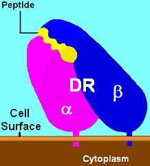
HLA-DR is an MHC class II cell surface receptor encoded by the human leukocyte antigen complex on chromosome 6 region 6p21.31. The complex of HLA-DR and peptide, generally between 9 and 30 amino acids in length, constitutes a ligand for the T-cell receptor (TCR). HLA were originally defined as cell surface antigens that mediate graft-versus-host disease. Identification of these antigens has led to greater success and longevity in organ transplant.
HLA class II histocompatibility antigen, DRB1 beta chain is a protein that in humans is encoded by the HLA-DRB1 gene. DRB1 encodes the most prevalent beta subunit of HLA-DR. DRB1 alleles, especially those encoding amino acid sequence changes at positions 11 and 13, are associated risk of rheumatoid arthritis.
HLA class II histocompatibility antigen, DR alpha chain is a protein that in humans is encoded by the HLA-DRA gene. HLA-DRA encodes the alpha subunit of HLA-DR. Unlike the alpha chains of other Human MHC class II molecules, the alpha subunit is practically invariable. However it can pair with, in any individual, the beta chain from 3 different DR beta loci, DRB1, and two of any DRB3, DRB4, or DRB5 alleles. Thus there is the potential that any given individual can form 4 different HLA-DR isoforms.
Antigen presentation is a vital immune process that is essential for T cell immune response triggering. Because T cells recognize only fragmented antigens displayed on cell surfaces, antigen processing must occur before the antigen fragment can be recognized by a T-cell receptor. Specifically, the fragment, bound to the major histocompatibility complex (MHC), is transported to the surface of the cell, a process known as presentation. If there has been an infection with viruses or bacteria, the cell will present an endogenous or exogenous peptide fragment derived from the antigen by MHC molecules. There are two types of MHC molecules which differ in the behaviour of the antigens: MHC class I molecules (MHC-I) bind peptides from the cell cytosol, while peptides generated in the endocytic vesicles after internalisation are bound to MHC class II (MHC-II). Cellular membranes separate these two cellular environments - intracellular and extracellular. Each T cell can only recognize tens to hundreds of copies of a unique sequence of a single peptide among thousands of other peptides presented on the same cell, because an MHC molecule in one cell can bind to quite a large range of peptides. Predicting which antigens will be presented to the immune system by a certain MHC/HLA type is difficult, but the technology involved is improving.
MHC Class II molecules are a class of major histocompatibility complex (MHC) molecules normally found only on professional antigen-presenting cells such as dendritic cells, mononuclear phagocytes, some endothelial cells, thymic epithelial cells, and B cells. These cells are important in initiating immune responses.
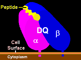
HLA-DQ (DQ) is a cell surface receptor protein found on antigen-presenting cells. It is an αβ heterodimer of type MHC class II. The α and β chains are encoded by two loci, HLA-DQA1 and HLA-DQB1, that are adjacent to each other on chromosome band 6p21.3. Both α-chain and β-chain vary greatly. A person often produces two α-chain and two β-chain variants and thus 4 isoforms of DQ. The DQ loci are in close genetic linkage to HLA-DR, and less closely linked to HLA-DP, HLA-A, HLA-B and HLA-C.
HLA-DP is a protein/peptide-antigen receptor and graft-versus-host disease antigen that is composed of 2 subunits, DPα and DPβ. DPα and DPβ are encoded by two loci, HLA-DPA1 and HLA-DPB1, that are found in the MHC Class II region in the Human Leukocyte Antigen complex on human chromosome 6 . Less is known about HLA-DP relative to HLA-DQ and HLA-DR but the sequencing of DP types and determination of more frequent haplotypes has progressed greatly within the last few years.
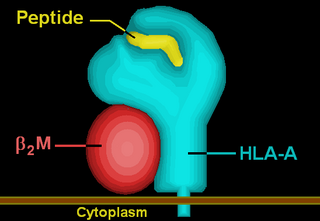
HLA-A is a group of human leukocyte antigens (HLA) that are encoded by the HLA-A locus, which is located at human chromosome 6p21.3. HLA is a major histocompatibility complex (MHC) antigen specific to humans. HLA-A is one of three major types of human MHC class I transmembrane proteins. The others are HLA-B and HLA-C. The protein is a heterodimer, and is composed of a heavy α chain and smaller β chain. The α chain is encoded by a variant HLA-A gene, and the β chain (β2-microglobulin) is an invariant β2 microglobulin molecule. The β2 microglobulin protein is encoded by the B2M gene, which is located at chromosome 15q21.1 in humans.
Minor histocompatibility antigen are peptides presented on the cellular surface of donated organs that are known to give an immunological response in some organ transplants. They cause problems of rejection less frequently than those of the major histocompatibility complex (MHC). Minor histocompatibility antigens (MiHAs) are diverse, short segments of proteins and are referred to as peptides. These peptides are normally around 9-12 amino acids in length and are bound to both the major histocompatibility complex (MHC) class I and class II proteins. Peptide sequences can differ among individuals and these differences arise from SNPs in the coding region of genes, gene deletions, frameshift mutations, or insertions. About a third of the characterized MiHAs come from the Y chromosome. Prior to becoming a short peptide sequence, the proteins expressed by these polymorphic or diverse genes need to be digested in the proteasome into shorter peptides. These endogenous or self peptides are then transported into the endoplasmic reticulum with a peptide transporter pump called TAP where they encounter and bind to the MHC class I molecule. This contrasts with MHC class II molecules's antigens which are peptides derived from phagocytosis/endocytosis and molecular degradation of non-self entities' proteins, usually by antigen-presenting cells. MiHA antigens are either ubiquitously expressed in most tissue like skin and intestines or restrictively expressed in the immune cells.
Major histocompatibility complex, class II, DR beta 4, also known as HLA-DRB4, is a human gene.
HLA class II histocompatibility antigen, DRB5 beta chain is a protein that in humans is encoded by the HLA-DRB5 gene.
Major histocompatibility complex, class II, DQ beta 1, also known as HLA-DQB1, is a human gene and also denotes the genetic locus that contains this gene. The protein encoded by this gene is one of two proteins that are required to form the DQ heterodimer, a cell surface receptor essential to the function of the immune system.
Major histocompatibility complex, class II, DQ alpha 1, also known as HLA-DQA1, is a human gene present on short arm of chromosome 6 (6p21.3) and also denotes the genetic locus which contains this gene. The protein encoded by this gene is one of two proteins that are required to form the DQ heterodimer, a cell surface receptor essential to the function of the immune system.
HLA class II histocompatibility antigen, DRB3-1 beta chain is a protein that in humans is encoded by the HLA-DRB3 gene.
HLA class I histocompatibility antigen, alpha chain F is a protein that in humans is encoded by the HLA-F gene. It is an empty intracellular molecule that encodes a non-classical heavy chain anchored to the membrane and forming a heterodimer with a β-2 microglobulin light chain. It belongs to the HLA class I heavy chain paralogues that separate from most of the HLA heavy chains. HLA-F is localized in the endoplasmic reticulum and Golgi apparatus, and is also unique in the sense that it exhibits few polymorphisms in the human population relative to the other HLA genes; however, there have been found different isoforms from numerous transcript variants found for the HLA-F gene. Its pathways include IFN-gamma signaling and CDK-mediated phosphorylation and removal of the Saccharomycescerevisiae Cdc6 protein, which is crucial for functional DNA replication.
This page is based on this Wikipedia article Text is available under the CC BY-SA 4.0 license; additional terms may apply. Images, videos and audio are available under their respective licenses.