MHC class II

Last updated
MHC Class II
MHC Class 2.svg
Schematic representation of MHC class II
Identifiers
SymbolMHC Class II
Membranome 63

MHC Class II molecules are a class of major histocompatibility complex (MHC) molecules normally found only on professional antigen-presenting cells such as dendritic cells, macrophages, some endothelial cells, thymic epithelial cells, and B cells. These cells are important in initiating immune responses.

Contents

The antigens presented by class II peptides are derived from extracellular proteins (not cytosolic as in MHC class I).

Loading of a MHC class II molecule occurs by phagocytosis; extracellular proteins are endocytosed, digested in lysosomes, and the resulting epitopic peptide fragments are loaded onto MHC class II molecules prior to their migration to the cell surface.

In humans, the MHC class II protein complex is encoded by the human leukocyte antigen gene complex (HLA). HLAs corresponding to MHC class II are HLA-DP, HLA-DM, HLA-DOA, HLA-DOB, HLA-DQ, and HLA-DR.

Mutations in the HLA gene complex can lead to bare lymphocyte syndrome (BLS), which is a type of MHC class II deficiency.

Structure

Like MHC class I molecules, class II molecules are also heterodimers, but in this case consist of two homogenous peptides, an α and β chain, both of which are encoded in the MHC. [1] The subdesignation α1, α2, etc. refers to separate domains within the HLA gene; each domain is usually encoded by a different exon within the gene, and some genes have further domains that encode leader sequences, transmembrane sequences, etc. These molecules have both extracellular regions as well as a transmembrane sequence and a cytoplasmic tail. The α1 and β1 regions of the chains come together to make a membrane-distal peptide-binding domain, while the α2 and β2 regions, the remaining extracellular parts of the chains, form a membrane-proximal immunoglobulin-like domain. The antigen binding groove, where the antigen or peptide binds, is made up of two α-helixes walls and β-sheet. [2]

Because the antigen-binding groove of MHC class II molecules is open at both ends while the corresponding groove on class I molecules is closed at each end, the antigens presented by MHC class II molecules are longer, generally between 15 and 24 amino acid residues long.

Expression

These molecules are constitutively expressed in professional, immune antigen-presenting cells, but may also be induced on other cells by interferon γ. [3] They are expressed on the epithelial cells in the thymus and on APCs in the periphery. MHC class II expression is closely regulated in APCs by CIITA, which is the MHC class II transactivator. CIITA is solely expressed on professional APCs; however, non-professional APCs can also regulate CIITA activity and MHC II expression. As mentioned interferon γ (IFN γ ) triggers the expression of CIITA and is also responsible for converting monocytes which are MHC class II negative cells into functional APCs that express MHC class II on their surfaces. [4]

MHC class II is also expressed on group 3 innate lymphoid cells.

Importance

Having MHC class II molecules present proper peptides that are bound stably is essential for overall immune function.

[5] Because class II MHC is loaded with extracellular proteins, it is mainly concerned with presentation of extracellular pathogens (for example, bacteria that might be infecting a wound or the blood). Class II molecules interact mainly with immune cells, like the T helper cell (CD4+). The peptide presented regulates how T cells respond to an infection. [5] Stable peptide binding is essential to prevent detachment and degradation of a peptide, which could occur without secure attachment to the MHC molecule. [5] This would prevent T cell recognition of the antigen, T cell recruitment, and a proper immune response. [5] The triggered appropriate immune response may include localized inflammation and swelling due to recruitment of phagocytes or may lead to a full-force antibody immune response due to activation of B cells.

Synthesis

During synthesis of class II MHC in the endoplasmic reticulum, the α and β chains are produced and complexed with a special polypeptide known as the invariant chain. [6] The nascent MHC class II protein in the rough ER has its peptide-binding cleft blocked by the invariant chain (Ii; a trimer) to prevent it from binding cellular peptides or peptides from the endogenous pathway (such as those that would be loaded onto class I MHC).

The invariant chain also facilitates the export of class II MHC from the ER to the Golgi apparatus, followed by fusion with a late endosome containing endocytosed, degraded proteins. The invariant chain is then broken down in stages by proteases called cathepsins, leaving only a small fragment known as CLIP which maintains blockage of the peptide binding cleft on the MHC molecule. A MHC class II-like structure, HLA-DM, facilitates CLIP removal and allows the binding of peptides with higher affinities. The stable class II MHC is then presented on the cell surface.

Recycling of MHC class II complexes

After MHC class II complexes are synthesized and presented on APCs they are unable to be expressed on the cell surface indefinitely, due to the internalization of the plasma membrane by the APCs(antigen presenting cells). In some cells, antigens bind to recycled MHC class II molecules while they are in the early endosomes, while other cells such as dendritic cells internalize antigens via receptor-mediated endocytosis and create MHC class II molecules plus peptide in the endosomal-lysosomal antigen processing compartment which is independent of the synthesis of new MHC class II complexes. These suggest that after the antigen is internalized, already existent MHC class II complexes on mature dendritic cells can be recycled and developed into new MHC class II molecules plus peptide. [4]

Antigen processing and presentation

Unlike MHC I, MHC II is meant to present extracellular pathogens rather than intracellular. Furthermore, the first step is to acquire the pathogen through phagocytosis. The pathogen is then broken down in a lysosome and a desired component is then acquired and loaded onto a MHC II molecule. The MHC II molecule then travels to the surface to present the antigen to a helper T cell. MHC II activate helper T cells which help release cytokines and other things which will help induce other cells which help to combat the pathogens outside the cells.

Genes

AlphaBeta
HLA-DM HLA-DMA HLA-DMB
HLA-DO HLA-DOA HLA-DOB
HLA-DP HLA-DPA1 HLA-DPB1
HLA-DQ HLA-DQA1, HLA-DQA2 HLA-DQB1, HLA-DQB2
HLA-DR HLA-DRA HLA-DRB1, HLA-DRB3, HLA-DRB4, HLA-DRB5

Pathways controlling MHC class II antigen presentation

Pathway: PSD4–ARL14/ARF7–MYO1E

Molecules involved

Several molecules are involved in this pathway. [7]

  • PIK3R2 [8] and PIP5K1A [9] are two kinases that create substrates for PSD4.
  • PSD4 [10] [11] (Pleckstrin and Sec7 Domain containing 4) is a GEF (Guanine nucleotide Exchange Factor) that loads ARL14/ARF7 with GTP.
  • ARL14/ARF7 [12] is a Small GTPase protein that is selectively expressed in immune cells. This protein is localized within MHC-II compartments in immature dendritic cells.
  • ARF7EP [13] is an effector of ARL14/ARF7 that interacts with MYO1E.
  • MYO1E [14] is a protein that controls MHC-II compartments with an actin-based mechanism.

Pathway

PIK3R2 and PIP5K1A are two kinases that phosphorylate Phosphatidylinositol (PIP) providing PSD4 with substrates for its GTP loading ability. PSD4 as a guanine exchange factor, loads ARL14/ARF7 with GTP. Subsequently, ARF7EP interacts with MYO1E which binds itself to actin myofibers. Altogether, this complex contributes to maintain MHC-II loaded vesicles within the immature dendritic cell, impeding its translocation to the cell membrane.

Pathway showing how MHC-II distribution is controlled within Immature Dendritic Cells. PSD4.ARL14.MYO1E PATHWAY.jpg
Pathway showing how MHC-II distribution is controlled within Immature Dendritic Cells.

Bare lymphocyte syndrome

One type of MHC class II deficiency, also called bare lymphocyte syndrome, is due to mutations in the genes that code for transcription factors that regulate the expression of the MHC class II genes. [15] It results in the depletion of CD4 T cells and some immunoglobulin isotypes even though there are normal levels of both CD8 Cells and B cells present. Deficient MHC class II molecules are unable to present antigens to T cells and properly activate T cells. T cells are then unable to proliferate, and secrete cytokines which normally participate in the immune response. Not only do the deficient MHC class II molecules affect the activation and proliferation of T cells but also the rest of the immune response cascade which includes B cells. Therefore, with this decrease in the number of T cells, the T cells cannot interact and activate the B cells. Normally when B cells are activated they divide, proliferate and differentiate, which includes the differentiation of these cells into plasma cells which are responsible for producing antibodies. [16] However, when there is a deficiency in MHC class II molecules B cells are not activated and cannot differentiate into plasma cells which causes them to be deficient in antibodies which are unable to perform as they are expected. The only current form of treatment is a bone-marrow transplant however even this does not cure the disease and most patients do not live past age ten. [17]

MHC class II and Type I diabetes

MHC class II genes and molecules are related to a multitude of different diseases, one of which being Type I diabetes. HLA class II genes are the most important genes associated with the risk of inheriting Type I diabetes, accounting for about 40-50% of heritability. Alleles of these genes that affect peptide binding to the MHC class II molecules seem to impact Type I diabetes risk the most. Specific allele polymorphisms have been identified to increase the risk (such as DRB1 and DQB1). Others have been associated with a resistance to the disease. [18]

See also

Related Research Articles

<span class="mw-page-title-main">Major histocompatibility complex</span> Cell surface proteins, part of the acquired immune system

The major histocompatibility complex (MHC) is a large locus on vertebrate DNA containing a set of closely linked polymorphic genes that code for cell surface proteins essential for the adaptive immune system. These cell surface proteins are called MHC molecules.

A class II gene is a type of gene that codes for a protein. Class II genes are transcribed by RNAP II.

Antigen processing, or the cytosolic pathway, is an immunological process that prepares antigens for presentation to special cells of the immune system called T lymphocytes. It is considered to be a stage of antigen presentation pathways. This process involves two distinct pathways for processing of antigens from an organism's own (self) proteins or intracellular pathogens, or from phagocytosed pathogens ; subsequent presentation of these antigens on class I or class II major histocompatibility complex (MHC) molecules is dependent on which pathway is used. Both MHC class I and II are required to bind antigens before they are stably expressed on a cell surface. MHC I antigen presentation typically involves the endogenous pathway of antigen processing, and MHC II antigen presentation involves the exogenous pathway of antigen processing. Cross-presentation involves parts of the exogenous and the endogenous pathways but ultimately involves the latter portion of the endogenous pathway.

<span class="mw-page-title-main">Antigen-presenting cell</span> Cell that displays antigen bound by MHC proteins on its surface

An antigen-presenting cell (APC) or accessory cell is a cell that displays an antigen bound by major histocompatibility complex (MHC) proteins on its surface; this process is known as antigen presentation. T cells may recognize these complexes using their T cell receptors (TCRs). APCs process antigens and present them to T cells.

<span class="mw-page-title-main">MHC class I</span> Protein of the immune system

MHC class I molecules are one of two primary classes of major histocompatibility complex (MHC) molecules and are found on the cell surface of all nucleated cells in the bodies of vertebrates. They also occur on platelets, but not on red blood cells. Their function is to display peptide fragments of proteins from within the cell to cytotoxic T cells; this will trigger an immediate response from the immune system against a particular non-self antigen displayed with the help of an MHC class I protein. Because MHC class I molecules present peptides derived from cytosolic proteins, the pathway of MHC class I presentation is often called cytosolic or endogenous pathway.

<span class="mw-page-title-main">HLA-DRB1</span> Protein-coding gene in the species Homo sapiens

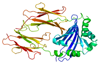
HLA class II histocompatibility antigen, DRB1 beta chain is a protein that in humans is encoded by the HLA-DRB1 gene. DRB1 encodes the most prevalent beta subunit of HLA-DR. DRB1 alleles, especially those encoding amino acid sequence changes at positions 11 and 13, are associated risk of rheumatoid arthritis.

<span class="mw-page-title-main">HLA-DRA</span> Protein-coding gene in the species Homo sapiens

HLA class II histocompatibility antigen, DR alpha chain is a protein that in humans is encoded by the HLA-DRA gene. HLA-DRA encodes the alpha subunit of HLA-DR. Unlike the alpha chains of other Human MHC class II molecules, the alpha subunit is practically invariable. However it can pair with, in any individual, the beta chain from 3 different DR beta loci, DRB1, and two of any DRB3, DRB4, or DRB5 alleles. Thus there is the potential that any given individual can form 4 different HLA-DR isoforms.

<span class="mw-page-title-main">Antigen presentation</span> Vital immune process that is essential for T cell immune response triggering

Antigen presentation is a vital immune process that is essential for T cell immune response triggering. Because T cells recognize only fragmented antigens displayed on cell surfaces, antigen processing must occur before the antigen fragment can be recognized by a T-cell receptor. Specifically, the fragment, bound to the major histocompatibility complex (MHC), is transported to the surface of the cell, a process known as presentation. If there has been an infection with viruses or bacteria, the cell will present an endogenous or exogenous peptide fragment derived from the antigen by MHC molecules. There are two types of MHC molecules which differ in the behaviour of the antigens: MHC class I molecules (MHC-I) bind peptides from the cell cytosol, while peptides generated in the endocytic vesicles after internalisation are bound to MHC class II (MHC-II). Cellular membranes separate these two cellular environments - intracellular and extracellular. Each T cell can only recognize tens to hundreds of copies of a unique sequence of a single peptide among thousands of other peptides presented on the same cell, because an MHC molecule in one cell can bind to quite a large range of peptides. Predicting which antigens will be presented to the immune system by a certain MHC/HLA type is difficult, but the technology involved is improving.

<span class="mw-page-title-main">CLIP (protein)</span>

CLIP or Class II-associated invariant chain peptide is the part of the invariant chain (Ii) that binds to the peptide binding groove of MHC class II and remains there until the MHC receptor is fully assembled. CLIP is one of the most prevalent self peptides found in the thymic cortex of most antigen-presenting cells. The purpose of CLIP is to prevent the degradation of MHC II dimers before antigenic peptides bind, and to prevent autoimmunity.

<span class="mw-page-title-main">Minor histocompatibility antigen</span>

Minor histocompatibility antigen are peptides presented on the cellular surface of donated organs that are known to give an immunological response in some organ transplants. They cause problems of rejection less frequently than those of the major histocompatibility complex (MHC). Minor histocompatibility antigens (MiHAs) are diverse, short segments of proteins and are referred to as peptides. These peptides are normally around 9-12 amino acids in length and are bound to both the major histocompatibility complex (MHC) class I and class II proteins. Peptide sequences can differ among individuals and these differences arise from SNPs in the coding region of genes, gene deletions, frameshift mutations, or insertions. About a third of the characterized MiHAs come from the Y chromosome. Prior to becoming a short peptide sequence, the proteins expressed by these polymorphic or diverse genes need to be digested in the proteasome into shorter peptides. These endogenous or self peptides are then transported into the endoplasmic reticulum with a peptide transporter pump called TAP where they encounter and bind to the MHC class I molecule. This contrasts with MHC class II molecules's antigens which are peptides derived from phagocytosis/endocytosis and molecular degradation of non-self entities' proteins, usually by antigen-presenting cells. MiHA antigens are either ubiquitously expressed in most tissue like skin and intestines or restrictively expressed in the immune cells.

<span class="mw-page-title-main">HLA-DM</span>

HLA-DM is an intracellular protein involved in the mechanism of antigen presentation on antigen presenting cells (APCs) of the immune system. It does this by assisting in peptide loading of major histocompatibility complex (MHC) class II membrane-bound proteins. HLA-DM is encoded by the genes HLA-DMA and HLA-DMB.

<span class="mw-page-title-main">HLA-DRB4</span> Protein-coding gene in the species Homo sapiens

Major histocompatibility complex, class II, DR beta 4, also known as HLA-DRB4, is a human gene.

<span class="mw-page-title-main">HLA-DPB1</span> Protein-coding gene in the species Homo sapiens

HLA class II histocompatibility antigen, DP(W2) beta chain is a protein that in humans is encoded by the HLA-DPB1 gene.

<span class="mw-page-title-main">HLA-DRB3</span> Protein-coding gene in the species Homo sapiens

HLA class II histocompatibility antigen, DRB3-1 beta chain is a protein that in humans is encoded by the HLA-DRB3 gene.

<span class="mw-page-title-main">HLA-F</span> Protein-coding gene in the species Homo sapiens

HLA class I histocompatibility antigen, alpha chain F is a protein that in humans is encoded by the HLA-F gene. It is an empty intracellular molecule that encodes a non-classical heavy chain anchored to the membrane and forming a heterodimer with a β-2 microglobulin light chain. It belongs to the HLA class I heavy chain paralogues that separate from most of the HLA heavy chains. HLA-F is localized in the endoplasmic reticulum and Golgi apparatus, and is also unique in the sense that it exhibits few polymorphisms in the human population relative to the other HLA genes; however, there have been found different isoforms from numerous transcript variants found for the HLA-F gene. Its pathways include IFN-gamma signaling and CDK-mediated phosphorylation and removal of the Saccharomycescerevisiae Cdc6 protein, which is crucial for functional DNA replication.

<span class="mw-page-title-main">HLA-DMB</span> Protein-coding gene in the species Homo sapiens

HLA class II histocompatibility antigen, DM beta chain is a protein that in humans is encoded by the HLA-DMB gene.

<span class="mw-page-title-main">HLA-DMA</span> Protein-coding gene in the species Homo sapiens

HLA class II histocompatibility antigen, DM alpha chain is a protein that in humans is encoded by the HLA-DMA gene.

<span class="mw-page-title-main">HLA-DOB</span> Protein-coding gene in the species Homo sapiens

HLA class II histocompatibility antigen, DO beta chain is a protein that in humans is encoded by the HLA-DOB gene.

Major histocompatibility complex, class II, DP alpha 1, also known as HLA-DPA1, is a human gene.

<span class="mw-page-title-main">CD1b</span> Mammalian protein found in Homo sapiens

T-cell surface glycoprotein CD1b is a protein that in humans is encoded by the CD1B gene.

References

  1. "Histocompatibility". Archived from the original on 2008-12-26. Retrieved 2009-01-21.
  2. Jones EY, Fugger L, Strominger JL, Siebold C (April 2006). "MHC class II proteins and disease: a structural perspective". Nature Reviews. Immunology. 6 (4): 271–82. doi:10.1038/nri1805. PMID   16557259. S2CID   131777.
  3. Ting JP, Trowsdale J (April 2002). "Genetic control of MHC class II expression". Cell. 109 Suppl (2): S21-33. doi: 10.1016/s0092-8674(02)00696-7 . PMID   11983150.
  4. 1 2 Roche PA, Furuta K (April 2015). "The ins and outs of MHC class II-mediated antigen processing and presentation". Nature Reviews. Immunology. 15 (4): 203–16. doi:10.1038/nri3818. PMC   6314495 . PMID   25720354.
  5. 1 2 3 4 Owen JA, Punt J, Stranford SA, Jones PP, Kuby J (2013). Kuby Immunology (7th ed.). New York: W H Freeman & Co. ISBN   978-1-4641-1991-0. OCLC   820117219.
  6. Cresswell, Peter (1996-02-23). "Invariant Chain Structure and MHC Class II Function". Cell. 84 (4): 505–507. doi: 10.1016/S0092-8674(00)81025-9 . ISSN   0092-8674. PMID   8598037. S2CID   8199773.
  7. Paul P, van den Hoorn T, Jongsma ML, Bakker MJ, Hengeveld R, Janssen L, Cresswell P, Egan DA, van Ham M, Ten Brinke A, Ovaa H, Beijersbergen RL, Kuijl C, Neefjes J (April 2011). "A Genome-wide multidimensional RNAi screen reveals pathways controlling MHC class II antigen presentation". Cell. 145 (2): 268–83. doi: 10.1016/j.cell.2011.03.023 . PMID   21458045.
  8. "PIK3R2 phosphoinositide-3-kinase, regulatory subunit 2 (beta) [Homo sapiens (human)&#93". Entrez Gene.
  9. "PIP5K1A phosphatidylinositol-4-phosphate 5-kinase, type I, alpha [Homo sapiens (human)". Entrez Gene.
  10. PSD4 pleckstrin and Sec7 domain containing 4 [Homo sapiens (human)] - Gene - NCBI
  11. Prigent M, Dubois T, Raposo G, Derrien V, Tenza D, Rossé C, Camonis J, Chavrier P (December 2003). "ARF6 controls post-endocytic recycling through its downstream exocyst complex effector". The Journal of Cell Biology. 163 (5): 1111–21. doi:10.1083/jcb.200305029. PMC   2173613 . PMID   14662749.
  12. "ARL14 ADP-ribosylation factor-like 14 [Homo sapiens (human)". Entrez Gene.
  13. "ARL14EP ADP-ribosylation factor-like 14 effector protein [Homo sapiens (human)". Entrez Gene.
  14. "MYO1E myosin IE [Homo sapiens (human)". Entrez Gene.
  15. Steimle V, Otten LA, Zufferey M, Mach B (June 2007). "Complementation cloning of an MHC class II transactivator mutated in hereditary MHC class II deficiency (or bare lymphocyte syndrome). 1993". Journal of Immunology. 178 (11): 6677–88. PMID   17513710.
  16. Mak TW, Saunders ME (2006). The immune response basic and clinical principles. Amsterdam: Elsevier/Academic. ISBN   978-0-12-088451-3. OCLC   986987876.
  17. Serrano-Martín MM, Moreno-Pérez D, García-Martín FJ, Jurado-Ortiz A (March 2007). "[Major histocompatibility complex class II deficiency]". Anales de Pediatria (in Spanish). 66 (3): 305–8. doi:10.1157/13099694. PMID   17349258.
  18. Xie Z, Chang C, Zhou Z (October 2014). "Molecular mechanisms in autoimmune type 1 diabetes: a critical review". Clinical Reviews in Allergy & Immunology. 47 (2): 174–92. doi:10.1007/s12016-014-8422-2. PMID   24752371. S2CID   26085603.