Arrestin

Last updated
S-antigen; retina and pineal gland (arrestin)
1CF1.png
Crystallographic structure of the bovine arrestin-S. [1]
Identifiers
Symbol SAG
Alt. symbolsarrestin-1
NCBI gene 6295
HGNC 10521
OMIM 181031
RefSeq NM_000541
UniProt P10523
Other data
Locus Chr. 2 q37.1
Search for
Structures Swiss-model
Domains InterPro
arrestin beta 1
Identifiers
Symbol ARRB1
Alt. symbolsARR1, arrestin-2
NCBI gene 408
HGNC 711
OMIM 107940
RefSeq NM_004041
UniProt P49407
Other data
Locus Chr. 11 q13
Search for
Structures Swiss-model
Domains InterPro
arrestin beta 2
Identifiers
Symbol ARRB2
Alt. symbolsARR2, arrestin-3
NCBI gene 409
HGNC 712
OMIM 107941
RefSeq NM_004313
UniProt P32121
Other data
Locus Chr. 17 p13
Search for
Structures Swiss-model
Domains InterPro
arrestin 3, retinal (X-arrestin)
Identifiers
Symbol ARR3
Alt. symbolsARRX, arrestin-4
NCBI gene 407
HGNC 710
OMIM 301770
RefSeq NM_004312
UniProt P36575
Other data
Locus Chr. X q
Search for
Structures Swiss-model
Domains InterPro

Arrestins (abbreviated Arr) are a small family of proteins important for regulating signal transduction at G protein-coupled receptors. [2] [3] Arrestins were first discovered as a part of a conserved two-step mechanism for regulating the activity of G protein-coupled receptors (GPCRs) in the visual rhodopsin system by Hermann Kühn, Scott Hall, and Ursula Wilden [4] and in the β-adrenergic system by Martin J. Lohse and co-workers. [5] [6]

Contents

Function

In response to a stimulus, GPCRs activate heterotrimeric G proteins. In order to turn off this response, or adapt to a persistent stimulus, active receptors need to be desensitized. The first step in desensitization is phosphorylation of the receptor by a class of serine/threonine kinases called G protein coupled receptor kinases (GRKs). GRK phosphorylation specifically prepares the activated receptor for arrestin binding. Arrestin binding to the receptor blocks further G protein-mediated signaling and targets receptors for internalization, and redirects signaling to alternative G protein-independent pathways, such as β-arrestin signaling. [7] [8] [9] [10] [6] In addition to GPCRs, arrestins bind to other classes of cell surface receptors and a variety of other signaling proteins. [11]

Subtypes

Mammals express four arrestin subtypes and each arrestin subtype is known by multiple aliases. The systematic arrestin name (1-4) plus the most widely used aliases for each arrestin subtype are listed in bold below:

Fish and other vertebrates appear to have only three arrestins: no equivalent of arrestin-2, which is the most abundant non-visual subtype in mammals, was cloned so far. The proto-chordate Ciona intestinalis (sea squirt) has only one arrestin, which serves as visual in its mobile larva with highly developed eyes, and becomes generic non-visual in the blind sessile adult. Conserved positions of multiple introns in its gene and those of our arrestin subtypes suggest that they all evolved from this ancestral arrestin. [12] Lower invertebrates, such as roundworm Caenorhabditis elegans , also have only one arrestin. Insects have arr1 and arr2, originally termed “visual arrestins” because they are expressed in photoreceptors, and one non-visual subtype (kurtz in Drosophila ). Later arr1 and arr2 were found to play an important role in olfactory neurons and renamed “sensory”. Fungi have distant arrestin relatives involved in pH sensing.

Tissue distribution

One or more arrestin is expressed in virtually every eukaryotic cell. In mammals, arrestin-1 and arrestin-4 are largely confined to photoreceptors, whereas arrestin-2 and arrestin-3 are ubiquitous. Neurons have the highest expression level of both non-visual subtypes. In neuronal precursors both are expressed at comparable levels, whereas in mature neurons arrestin-2 is present at 10-20 fold higher levels than arrestin-3.

Mechanism

Arrestins block GPCR coupling to G proteins in two ways. First, arrestin binding to the cytoplasmic face of the receptor occludes the binding site for heterotrimeric G-protein, preventing its activation (desensitization). [13] Second, arrestin links the receptor to elements of the internalization machinery, clathrin and clathrin adaptor AP2, which promotes receptor internalization via coated pits and subsequent transport to internal compartments, called endosomes. Subsequently, the receptor could be either directed to degradation compartments (lysosomes) or recycled back to the plasma membrane where it can again signal. The strength of arrestin-receptor interaction plays a role in this choice: tighter complexes tend to increase the probability of receptor degradation (Class B), whereas more transient complexes favor recycling (Class A), although this “rule” is far from absolute. [2] More recently direct interactions between Gi/o family G proteins and Arrestin were discovered downstream of multiple receptors, regardless of canonical G protein coupling. [14] These recent findings introduce a GPCR signaling mechanism distinct from canonical G protein activation and β-arrestin desensitization in which GPCRs cause the formation of Gαi:β-arrestin signaling complexes.

Structure

Arrestins are elongated molecules, in which several intra-molecular interactions hold the relative orientation of the two domains. Unstimulated cell arrestins are localized in the cytoplasm in a basal “inactive” conformation. Active phosphorylated GPCRs recruit arrestin to the plasma membrane. Receptor binding induces a global conformational change that involves the movement of the two arrestin domains and the release of its C-terminal tail that contains clathrin and AP2 binding sites. Increased accessibility of these sites in receptor-bound arrestin targets the arrestin-receptor complex to the coated pit. Arrestins also bind microtubules (part of the cellular “skeleton”), where they assume yet another conformation, different from both free and receptor-bound form. Microtubule-bound arrestins recruit certain proteins to the cytoskeleton, which affects their activity and/or redirects it to microtubule-associated proteins.

Arrestins shuttle between cell nucleus and cytoplasm. Their nuclear functions are not fully understood, but it was shown that all four mammalian arrestin subtypes remove some of their partners, such as protein kinase JNK3 or the ubiquitin ligase Mdm2, from the nucleus. Arrestins also modify gene expression by enhancing transcription of certain genes.

Arrestin (or S-antigen), N-terminal domain
PDB 1cf1 EBI.jpg
Structure of arrestin from bovine rod outer segments. [1]
Identifiers
SymbolArrestin_N
Pfam PF00339
Pfam clan CL0135
InterPro IPR011021
PROSITE PDOC00267
SCOP2 1cf1 / SCOPe / SUPFAM
Available protein structures:
Pfam   structures / ECOD  
PDB RCSB PDB; PDBe; PDBj
PDBsum structure summary
PDB 1ayr , 1cf1 , 1g4m , 1g4r , 1jsy , 1zsh
Arrestin (or S-antigen), C-terminal domain
PDB 1g4m EBI.jpg
Structure of bovine beta-arrestin. [15]
Identifiers
SymbolArrestin_C
Pfam PF02752
Pfam clan CL0135
InterPro IPR011022
SCOP2 1cf1 / SCOPe / SUPFAM
Available protein structures:
Pfam   structures / ECOD  
PDB RCSB PDB; PDBe; PDBj
PDBsum structure summary
PDB 1ayr , 1cf1 , 1g4m , 1g4r , 1jsy , 1suj , 1zsh

Related Research Articles

<span class="mw-page-title-main">G protein-coupled receptor</span> Class of cell surface receptors coupled to G-protein-associated intracellular signaling

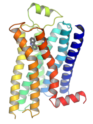
G protein-coupled receptors (GPCRs), also known as seven-(pass)-transmembrane domain receptors, 7TM receptors, heptahelical receptors, serpentine receptors, and G protein-linked receptors (GPLR), form a large group of evolutionarily related proteins that are cell surface receptors that detect molecules outside the cell and activate cellular responses. They are coupled with G proteins. They pass through the cell membrane seven times in form of six loops of amino acid residues, which is why they are sometimes referred to as seven-transmembrane receptors. Ligands can bind either to the extracellular N-terminus and loops or to the binding site within transmembrane helices. They are all activated by agonists, although a spontaneous auto-activation of an empty receptor has also been observed.

<span class="mw-page-title-main">Rhodopsin</span> Light-sensitive receptor protein

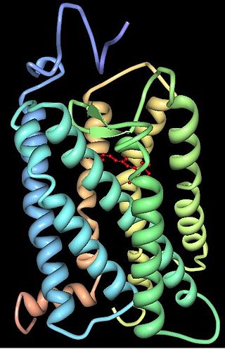
Rhodopsin, also known as visual purple, is a protein encoded by the RHO gene and a G-protein-coupled receptor (GPCR). It is the opsin of the rod cells in the retina and a light-sensitive receptor protein that triggers visual phototransduction in rods. Rhodopsin mediates dim light vision and thus is extremely sensitive to light. When rhodopsin is exposed to light, it immediately photobleaches. In humans, it is regenerated fully in about 30 minutes, after which the rods are more sensitive. Defects in the rhodopsin gene cause eye diseases such as retinitis pigmentosa and congenital stationary night blindness.

<span class="mw-page-title-main">Rod cell</span> Photoreceptor cells that can function in lower light better than cone cells

Rod cells are photoreceptor cells in the retina of the eye that can function in lower light better than the other type of visual photoreceptor, cone cells. Rods are usually found concentrated at the outer edges of the retina and are used in peripheral vision. On average, there are approximately 92 million rod cells in the human retina. Rod cells are more sensitive than cone cells and are almost entirely responsible for night vision. However, rods have little role in color vision, which is the main reason why colors are much less apparent in dim light.

<span class="mw-page-title-main">Beta-1 adrenergic receptor</span> Protein-coding gene in the species Homo sapiens

The beta-1 adrenergic receptor, also known as ADRB1, can either refer to the protein-encoding gene or one of the four adrenergic receptors. It is a G-protein coupled receptor associated with the Gs heterotrimeric G-protein that is expressed predominantly in cardiac tissue. In addition to cardiac tissue, beta-1 adrenergic receptors are also expressed in the cerebral cortex.

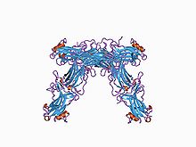
The actions of vasopressin are mediated by stimulation of tissue-specific G protein-coupled receptors (GPCRs) called vasopressin receptors that are classified into the V1 (V1A), V2, and V3 (V1B) receptor subtypes. These three subtypes differ in localization, function and signal transduction mechanisms.

<span class="mw-page-title-main">G protein-coupled receptor kinase</span>

G protein-coupled receptor kinases are a family of protein kinases within the AGC group of kinases. Like all AGC kinases, GRKs use ATP to add phosphate to Serine and Threonine residues in specific locations of target proteins. In particular, GRKs phosphorylate intracellular domains of G protein-coupled receptors (GPCRs). GRKs function in tandem with arrestin proteins to regulate the sensitivity of GPCRs for stimulating downstream heterotrimeric G protein and G protein-independent signaling pathways.

<span class="mw-page-title-main">G protein-coupled receptor kinase 2</span> Enzyme

G-protein-coupled receptor kinase 2 (GRK2) is an enzyme that in humans is encoded by the ADRBK1 gene. GRK2 was initially called Beta-adrenergic receptor kinase, and is a member of the G protein-coupled receptor kinase subfamily of the Ser/Thr protein kinases that is most highly similar to GRK3(βARK2).

Rhodopsin kinase is a serine/threonine-specific protein kinase involved in phototransduction. This enzyme catalyses the following chemical reaction:

<span class="mw-page-title-main">Arrestin beta 2</span> Protein-coding gene in the species Homo sapiens

Beta-arrestin-2, also known as arrestin beta-2, is an intracellular protein that in humans is encoded by the ARRB2 gene.

<span class="mw-page-title-main">Arrestin beta 1</span> Human protein and coding gene

Arrestin, beta 1, also known as ARRB1, is a protein which in humans is encoded by the ARRB1 gene.

<span class="mw-page-title-main">GRK6</span> Protein-coding gene in the species Homo sapiens

This gene encodes a member of the G protein-coupled receptor kinase subfamily of the Ser/Thr protein kinase family, and is most highly similar to GRK4 and GRK5. The protein phosphorylates the activated forms of G protein-coupled receptors to regulate their signaling.

<span class="mw-page-title-main">GRK5</span> Protein-coding gene in the species Homo sapiens

G protein-coupled receptor kinase 5 is a member of the G protein-coupled receptor kinase subfamily of the Ser/Thr protein kinases, and is most highly similar to GRK4 and GRK6. The protein phosphorylates the activated forms of G protein-coupled receptors to regulate their signaling.

<span class="mw-page-title-main">SAG (gene)</span>

S-arrestin is a protein that in humans is encoded by the SAG gene.

<span class="mw-page-title-main">GRK4</span> Protein-coding gene in the species Homo sapiens

G protein-coupled receptor kinase 4 (GRK4) is an enzyme that in humans is encoded by the GRK4 gene.

<span class="mw-page-title-main">ARR3</span> Protein-coding gene in the species Homo sapiens

Arrestin-C, also known as retinal cone arrestin-3, is a protein that in humans is encoded by the ARR3 gene.

<span class="mw-page-title-main">Homologous desensitization</span> When a receptor decreases its response to an agonist at high concentration

Homologous desensitization occurs when a receptor decreases its response to an agonist at high concentration. It is a process through which, after prolonged agonist exposure, the receptor is uncoupled from its signaling cascade and thus the cellular effect of receptor activation is attenuated.

<span class="mw-page-title-main">G beta-gamma complex</span>

The G beta-gamma complex (Gβγ) is a tightly bound dimeric protein complex, composed of one Gβ and one Gγ subunit, and is a component of heterotrimeric G proteins. Heterotrimeric G proteins, also called guanosine nucleotide-binding proteins, consist of three subunits, called alpha, beta, and gamma subunits, or Gα, Gβ, and Gγ. When a G protein-coupled receptor (GPCR) is activated, Gα dissociates from Gβγ, allowing both subunits to perform their respective downstream signaling effects. One of the major functions of Gβγ is the inhibition of the Gα subunit.

G-protein-coupled receptor kinase 7 is a serine/threonine-specific protein kinase involved in phototransduction. This enzyme catalyses the phosphorylation of cone (color) photopsins in retinal cones during high acuity color vision primarily in the fovea.

Heterologous desensitization is the term for the unresponsiveness of cells to one or more agonists to which they are normally responsive. Typically, desensitization is a receptor-based phenomenon in which one receptor type, when bound to its ligand, becomes unable to further influence the signalling pathways by which it regulates cells and, in the case of cell surface membrane receptors, may thereafter be internalized. The desensitized receptor is degraded or freed of its activating ligand and re-cycled to a state where it is again able to respond to cognate ligands by activating its signalling pathways.

<span class="mw-page-title-main">G protein-coupled receptor kinase 3</span> Protein-coding gene in the species Homo sapiens

G-protein-coupled receptor kinase 3 (GRK3) is an enzyme that in humans is encoded by the ADRBK2 gene. GRK3 was initially called Beta-adrenergic receptor kinase 2 (βARK-2), and is a member of the G protein-coupled receptor kinase subfamily of the Ser/Thr protein kinases that is most highly similar to GRK2.

References

  1. 1 2 PDB: 1CF1 ; Hirsch JA, Schubert C, Gurevich VV, Sigler PB (April 1999). "The 2.8 A crystal structure of visual arrestin: a model for arrestin's regulation". Cell. 97 (2): 257–69. doi: 10.1016/S0092-8674(00)80735-7 . PMID   10219246. S2CID   17124300.
  2. 1 2 Moore CA, Milano SK, Benovic JL (2007). "Regulation of receptor trafficking by GRKs and arrestins". Annual Review of Physiology. 69: 451–82. doi:10.1146/annurev.physiol.69.022405.154712. PMID   17037978.
  3. Lefkowitz RJ, Shenoy SK (April 2005). "Transduction of receptor signals by beta-arrestins". Science. 308 (5721): 512–7. Bibcode:2005Sci...308..512L. doi:10.1126/science.1109237. PMID   15845844. S2CID   26931077.
  4. Wilden U, Hall SW, Kühn H (March 1986). "Phosphodiesterase activation by photoexcited rhodopsin is quenched when rhodopsin is phosphorylated and binds the intrinsic 48-kDa protein of rod outer segments". Proceedings of the National Academy of Sciences of the United States of America. 83 (5): 1174–8. Bibcode:1986PNAS...83.1174W. doi: 10.1073/pnas.83.5.1174 . PMC   323037 . PMID   3006038.
  5. Lohse MJ, Benovic JL, Codina J, Caron MG, Lefkowitz RJ (June 1990). "beta-Arrestin: a protein that regulates beta-adrenergic receptor function". Science. 248 (4962): 1547–50. Bibcode:1990Sci...248.1547L. doi:10.1126/science.2163110. PMID   2163110.
  6. 1 2 Gurevich VV, Gurevich EV (June 2006). "The structural basis of arrestin-mediated regulation of G-protein-coupled receptors". Pharmacology & Therapeutics. 110 (3): 465–502. doi:10.1016/j.pharmthera.2005.09.008. PMC   2562282 . PMID   16460808.
  7. Smith JS, Lefkowitz RJ, Rajagopal S (January 2018). "Biased signalling: from simple switches to allosteric microprocessors". Nature Reviews. Drug Discovery. 17 (4): 243–260. doi:10.1038/nrd.2017.229. PMC   5936084 . PMID   29302067.
  8. Cahill TJ, Thomsen AR, Tarrasch JT, Plouffe B, Nguyen AH, Yang F, et al. (February 2017). "Distinct conformations of GPCR-β-arrestin complexes mediate desensitization, signaling, and endocytosis". Proceedings of the National Academy of Sciences of the United States of America. 114 (10): 2562–2567. Bibcode:2017PNAS..114.2562C. doi: 10.1073/pnas.1701529114 . PMC   5347553 . PMID   28223524.
  9. Kumari P, Srivastava A, Banerjee R, Ghosh E, Gupta P, Ranjan R, Chen X, Gupta B, Gupta C, Jaiman D, Shukla AK (November 2016). "Functional competence of a partially engaged GPCR-β-arrestin complex". Nature Communications. 7: 13416. Bibcode:2016NatCo...713416K. doi:10.1038/ncomms13416. PMC   5105198 . PMID   27827372.
  10. Kumari P, Srivastava A, Ghosh E, Ranjan R, Dogra S, Yadav PN, Shukla AK (April 2017). "Core engagement with β-arrestin is dispensable for agonist-induced vasopressin receptor endocytosis and ERK activation". Molecular Biology of the Cell. 28 (8): 1003–10. doi:10.1091/mbc.E16-12-0818. PMC   5391177 . PMID   28228552.
  11. Gurevich VV, Gurevich EV (February 2004). "The molecular acrobatics of arrestin activation". Trends in Pharmacological Sciences. 25 (2): 105–11. doi:10.1016/j.tips.2003.12.008. PMID   15102497.
  12. Gurevich EV, Gurevich VV (2006). "Arrestins: ubiquitous regulators of cellular signaling pathways". Genome Biology. 7 (9): 236. doi:10.1186/gb-2006-7-9-236. PMC   1794542 . PMID   17020596.
  13. Kang Y, Zhou XE, Gao X, He Y, Liu W, Ishchenko A, et al. (July 2015). "Crystal structure of rhodopsin bound to arrestin by femtosecond X-ray laser". Nature. 523 (7562): 561–7. Bibcode:2015Natur.523..561K. doi:10.1038/nature14656. PMC   4521999 . PMID   26200343.
  14. Smith JS, Pack TF, et al. (2021). "Noncanonical scaffolding of Gαi and β-arrestin by G protein–coupled receptors". Science. 371 (Ahead of print): eaay1833. doi:10.1126/science.aay1833. PMC   8005335 . PMID   33479120.
  15. Han M, Gurevich VV, Vishnivetskiy SA, Sigler PB, Schubert C (September 2001). "Crystal structure of beta-arrestin at 1.9 A: possible mechanism of receptor binding and membrane Translocation". Structure. 9 (9): 869–80. doi: 10.1016/S0969-2126(01)00644-X . PMID   11566136.