See also
Wikimedia Commons has media related to Cell adhesion molecules .
Cell adhesion molecules (CAMs) are a subset of cell surface proteins [1] that are involved in the binding of cells with other cells or with the extracellular matrix (ECM), in a process called cell adhesion. [2] In essence, CAMs help cells stick to each other and to their surroundings. CAMs are crucial components in maintaining tissue structure and function. In fully developed animals, these molecules play an integral role in generating force and movement and consequently ensuring that organs are able to execute their functions normally. [3] In addition to serving as "molecular glue", CAMs play important roles in the cellular mechanisms of growth, contact inhibition, and apoptosis. Aberrant expression of CAMs may result in a wide range of pathologies, ranging from frostbite to cancer. [4]
CAMs are typically single-pass transmembrane receptors [5] and are composed of three conserved domains: an intracellular domain that interacts with the cytoskeleton, a transmembrane domain, and an extracellular domain. These proteins can interact in several different ways. [6] The first method is through homophilic binding, where CAMs bind with the same CAMs. They are also capable of heterophilic binding, meaning a CAM on one cell will bind with different CAMs on another cell.
There are four major superfamilies or groups of CAMs: the immunoglobulin super family of cell adhesion molecules (IgCAMs), Cadherins, Integrins, and the Superfamily of C-type of lectin-like domains proteins (CTLDs). Proteoglycans are also considered to be a class of CAMs.
One classification system involves the distinction between calcium-independent CAMs and calcium-dependent CAMs. [7] The Ig-superfamily CAMs do not depend on Ca2+ while integrins, cadherins and selectins depend on Ca2+. In addition, integrins participate in cell–matrix interactions, while other CAM families participate in cell–cell interactions. [8]
Immunoglobulin superfamily CAMs (IgSF CAMs) is regarded as the most diverse superfamily of CAMs. This family is characterized by their extracellular domains containing Ig-like domains. The Ig domains are then followed by Fibronectin type III domain repeats and IgSFs are anchored to the membrane by a GPI moiety. This family is involved in both homophilic or heterophilic binding and has the ability to bind integrins or different IgSF CAMs.[ citation needed ]
Integrins, one of the major classes of receptors within the ECM, [9] mediate cell–ECM interactions with collagen, fibrinogen, fibronectin, and vitronectin. [10] Integrins provide essential links between the extracellular environment and the intracellular signalling pathways, which can play roles in cell behaviours such as apoptosis, differentiation, survival, and transcription. [11]
Integrins are heterodimeric, as they consist of an alpha and beta subunit. [12] There are currently 18 alpha subunits and 8 beta subunits, which combine to make up 24 different integrin combinations. [10] Within each of the alpha and beta subunits there is a large extracellular domain, a transmembrane domain and a short cytoplasmic domain. [13] The extracellular domain is where the ligand binds through the use of divalent cations. The integrins contain multiple divalent cation binding sites in the extracellular domain [14] ). The integrin cation binding sites can be occupied by Ca2+ or by Mn2+ ions. Cations are necessary but not sufficient for integrins to convert from the inactive bent conformation into the active extended conformation. Both the presence of cations bound to the multiple cation binding sites is required, along with the direct physical association with ECM ligands for integrins to attain the extended structure and concomitant activation. [15] Thus, rise in extracellular Ca2+ ions may serve to prime the integrin heterodimer. The release of intracellular Ca2+ have been shown to be important for integrin inside-out activation. [16] However, extracellular Ca2+ binding may exert different effects depending on the type of integrin and the cation concentration. [17] Integrins regulate their activity within the body by changing conformation. Most exist at rest in a low affinity state, which can be altered to high affinity through an external agonist which causes a conformational change within the integrin, increasing their affinity. [11]
An example of this is the aggregation of platelets; [11] Agonists such as thrombin or collagen trigger the integrin into its high affinity state, which causes increased fibrinogen binding, causing platelet aggregation.
The cadherins are homophilic Ca2+
-dependent glycoproteins. [18] The classic cadherins (E-, N- and P-) are concentrated at the intermediate cell junctions, which link to the actin filament network through specific linking proteins called catenins. [18]
Cadherins are notable in embryonic development. For example, cadherins are crucial in gastrulation for the formation of the mesoderm, endoderm, and ectoderm. Cadherins also contribute significantly to the development of the nervous system. The distinct temporal and spatial localization of cadherins implicates these molecules as major players in the process of synaptic stabilization. Each cadherin exhibits a unique pattern of tissue distribution that is carefully controlled by calcium. The diverse family of cadherins include epithelial (E-cadherins), placental (P-cadherins), neural (N-cadherins), retinal (R-cadherins), brain (B-cadherins and T-cadherins), and muscle (M-cadherins). [18] Many cell types express combinations of cadherin types.
The extracellular domain has major repeats called extracellular cadherin domains (ECD). Sequences involved in Ca2+
binding between the ECDs are necessary for cell adhesion. The cytoplasmic domain has specific regions where catenin proteins bind. [19]
The selectins are a family of heterophilic CAMs that are dependent on fucosylated carbohydrates, e.g., mucins for binding. The three family members are E-selectin (endothelial), L-selectin (leukocyte), and P-selectin (platelet). The best-characterized ligand for the three selectins is P-selectin glycoprotein ligand-1 (PSGL-1), which is a mucin-type glycoprotein expressed on all white blood cells. Selectins have been implicated in several roles but they are especially important in the immune system by helping white blood cell homing and trafficking. [20]
The variety in CAMs leads to diverse functionality of these proteins in the biological setting. One of the CAMS that are particularly important in the lymphocyte homing is addressin. [21] Lymphocyte homing is a key process occurring in a strong immune system. It controls the process of circulating lymphocytes adhering to particular regions and organs of the body. [22] The process is highly regulated by cell adhesion molecules, particularly, the addressin also known as MADCAM1. This antigen is known for its role in tissue-specific adhesion of lymphocytes to high endothelium venules. [23] Through these interactions they play a crucial role in orchestrating circulating lymphocytes.
CAM function in cancer metastasis, inflammation, and thrombosis makes it a viable therapeutic target that is currently being considered. For example, they block the metastatic cancer cells' ability to extravasate and home to secondary sites. This has been successfully demonstrated in metastatic melanoma that hones to the lungs. In mice, when antibodies directed against CAMs in the lung endothelium were used as treatment there was a significant reduction in the number of metastatic sites. [24]

Integrins are transmembrane receptors that help cell-cell and cell-extracellular matrix (ECM) adhesion. Upon ligand binding, integrins activate signal transduction pathways that mediate cellular signals such as regulation of the cell cycle, organization of the intracellular cytoskeleton, and movement of new receptors to the cell membrane. The presence of integrins allows rapid and flexible responses to events at the cell surface.

Cell adhesion is the process by which cells interact and attach to neighbouring cells through specialised molecules of the cell surface. This process can occur either through direct contact between cell surfaces such as cell junctions or indirect interaction, where cells attach to surrounding extracellular matrix, a gel-like structure containing molecules released by cells into spaces between them. Cells adhesion occurs from the interactions between cell-adhesion molecules (CAMs), transmembrane proteins located on the cell surface. Cell adhesion links cells in different ways and can be involved in signal transduction for cells to detect and respond to changes in the surroundings. Other cellular processes regulated by cell adhesion include cell migration and tissue development in multicellular organisms. Alterations in cell adhesion can disrupt important cellular processes and lead to a variety of diseases, including cancer and arthritis. Cell adhesion is also essential for infectious organisms, such as bacteria or viruses, to cause diseases.

Cadherins (named for "calcium-dependent adhesion") are cell adhesion molecules important in forming adherens junctions that let cells adhere to each other. Cadherins are a class of type-1 transmembrane proteins, and they depend on calcium (Ca2+) ions to function, hence their name. Cell-cell adhesion is mediated by extracellular cadherin domains, whereas the intracellular cytoplasmic tail associates with numerous adaptors and signaling proteins, collectively referred to as the cadherin adhesome.
The L1 family is a family of cell adhesion molecules that includes four different L1-like proteins. They are members of the immunoglobulin superfamily. The members of the L1-family in humans are called L1 or L1cam, CHL1, Neurofascin and NRCAM. L1 family members are found on neurons, especially on their axons. Sometimes they are found on glia, such as Schwann cells, radial glia and Bergmann glia cells and, as such, are important for neural cell migration during development. L1 family members are expressed throughout the vertebrate and invertebrate kingdoms.

Cell junctions or junctional complexes, are a class of cellular structures consisting of multiprotein complexes that provide contact or adhesion between neighboring cells or between a cell and the extracellular matrix in animals. They also maintain the paracellular barrier of epithelia and control paracellular transport. Cell junctions are especially abundant in epithelial tissues. Combined with cell adhesion molecules and extracellular matrix, cell junctions help hold animal cells together.

Neural cell adhesion molecule (NCAM), also called CD56, is a homophilic binding glycoprotein expressed on the surface of neurons, glia and skeletal muscle. Although CD56 is often considered a marker of neural lineage commitment due to its discovery site, CD56 expression is also found in, among others, the hematopoietic system. Here, the expression of CD56 is mostly associated with, but not limited to, natural killer cells. CD56 has been detected on other lymphoid cells, including gamma delta (γδ) Τ cells and activated CD8+ T cells, as well as on dendritic cells. NCAM has been implicated as having a role in cell–cell adhesion, neurite outgrowth, synaptic plasticity, and learning and memory.

The selectins are a family of cell adhesion molecules. All selectins are single-chain transmembrane glycoproteins that share similar properties to C-type lectins due to a related amino terminus and calcium-dependent binding. Selectins bind to sugar moieties and so are considered to be a type of lectin, cell adhesion proteins that bind sugar polymers.
L-selectin, also known as CD62L, is a cell adhesion molecule found on the cell surface of leukocytes, and the blastocyst. It is coded for in the human by the SELL gene. L-selectin belongs to the selectin family of proteins, which recognize sialylated carbohydrate groups containing a Sialyl LewisX (sLeX) determinant. L-selectin plays an important role in both the innate and adaptive immune responses by facilitating leukocyte-endothelial cell adhesion events. These tethering interactions are essential for the trafficking of monocytes and neutrophils into inflamed tissue as well as the homing of lymphocytes to secondary lymphoid organs. L-selectin is also expressed by lymphoid primed hematopoietic stem cells and may participate in the migration of these stem cells to the primary lymphoid organs. In addition to its function in the immune response, L-selectin is expressed on embryonic cells and facilitates the attachment of the blastocyst to the endometrial endothelium during human embryo implantation.

Mucosal vascular addressin cell adhesion molecule 1 (MAdCAM-1) is a protein that in humans is encoded by the MADCAM1 gene. The protein encoded by this gene is an endothelial cell adhesion molecule that interacts preferentially with the leukocyte beta7 integrin LPAM-1, L-selectin, and VLA-4 on myeloid cells to direct leukocytes into mucosal and inflamed tissues. It is a member of the immunoglobulin superfamily and is similar to ICAM-1 and VCAM-1.
CD22, or cluster of differentiation-22, is a molecule belonging to the SIGLEC family of lectins. It is found on the surface of mature B cells and to a lesser extent on some immature B cells. Generally speaking, CD22 is a regulatory molecule that prevents the overactivation of the immune system and the development of autoimmune diseases.
Lymphocyte homing receptors are cell adhesion molecules expressed on lymphocyte cell membranes that recognize addressins on target tissues. Lymphocyte homing refers to adhesion of the circulating lymphocytes in blood to specialized endothelial cells within lymphoid organs. These diverse tissue-specific adhesion molecules on lymphocytes and on endothelial cells contribute to the development of specialized immune responses.

Leukocyte extravasation is the movement of leukocytes out of the circulatory system and towards the site of tissue damage or infection. This process forms part of the innate immune response, involving the recruitment of non-specific leukocytes. Monocytes also use this process in the absence of infection or tissue damage during their development into macrophages.

Receptor-type tyrosine-protein phosphatase mu is an enzyme that in humans is encoded by the PTPRM gene.
Nectins and Nectin-like molecules (Necl) are families of cellular adhesion molecules involved in Ca2+-independent cellular adhesion.
IgSF CAMs are cell adhesion molecules that belong to Immunoglobulin superfamily. It is regarded as the most diverse superfamily of CAMs. This family is characterized by their extracellular domains containing Ig-like domains. The Ig domains are then followed by Fibronectin type III domain repeats and IgSFs are anchored to the membrane by a GPI moiety. This family is involved in both homophilic or heterophilic binding and has the ability to bind integrins or different IgSF CAMs.

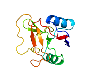
In molecular biology, the adhesin molecule (immunoglobulin-like) is a protein domain. This domain is found in mucosal vascular addressin cell adhesion molecule 1 proteins (MAdCAM-1). These are cell adhesion molecules expressed on the endothelium in mucosa that guide the specific homing of lymphocytes into mucosal tissues. MAdCAM-1 belongs to a subclass of the immunoglobulin superfamily (IgSF), the members of which are ligands for integrins. The crystal structure of this domain has been reported; it adopts an immunoglobulin-like beta-sandwich structure, with seven strands arranged in two beta-sheets in a Greek-key topology.
Gut-specific homing is the mechanism by which activated T cells and antibody-secreting cells (ASCs) are targeted to both inflamed and non-inflamed regions of the gut in order to provide an effective immune response. This process relies on the key interaction between the integrin α4β7 and the addressin MadCAM-1 on the surfaces of the appropriate cells. Additionally, this interaction is strengthened by the presence of CCR9, a chemokine receptor, which interacts with TECK. Vitamin A-derived retinoic acid regulates the expression of these cell surface proteins.

Synaptic stabilization is crucial in the developing and adult nervous systems and is considered a result of the late phase of long-term potentiation (LTP). The mechanism involves strengthening and maintaining active synapses through increased expression of cytoskeletal and extracellular matrix elements and postsynaptic scaffold proteins, while pruning less active ones. For example, cell adhesion molecules (CAMs) play a large role in synaptic maintenance and stabilization. Gerald Edelman discovered CAMs and studied their function during development, which showed CAMs are required for cell migration and the formation of the entire nervous system. In the adult nervous system, CAMs play an integral role in synaptic plasticity relating to learning and memory.

A junctional adhesion molecule (JAM) is a protein that is a member of the immunoglobulin superfamily, and is expressed in a variety of different tissues, such as leukocytes, platelets, and epithelial and endothelial cells. They have been shown to regulate signal complex assembly on both their cytoplasmic and extracellular domains through interaction with scaffolding that contains a PDZ domain and adjacent cell's receptors, respectively. JAMs adhere to adjacent cells through interactions with integrins LFA-1 and Mac-1, which are contained in leukocyte β2 and α4β1, which is contained in β1. JAMs have many influences on leukocyte-endothelial cell interactions, which are primarily moderated by the integrins discussed above. They interact in their cytoplasmic domain with scaffold proteins that contain a PDZ domain, which are common protein interaction modules that target short amino acid sequences at the C-terminus of proteins, to form tight junctions in both epithelial and endothelial cells as polarity is gained in the cell.
Integrin α4β7 is an integrin heterodimer composed of CD49d (alpha-4) subunit and beta-7 subunit noncovalently linked. LPAM-1 is expressed on the cell surface of leukocytes. This receptor is involved in lymphocyte trafficking pathway to site of inflammation in intestinal tissues.
{{cite journal}}: Cite journal requires |journal= (help)