Selectin

Last updated
Selectin
Pselectin.PNG
Crystallographic structure of P-selectin lectin bound to sugar, shown in sticks. [1]
Identifiers
SymbolSelectin
InterPro IPR002396
Membranome 12

The selectins ( cluster of differentiation 62 or CD62) are a family of cell adhesion molecules (or CAMs). All selectins are single-chain transmembrane glycoproteins that share similar properties to C-type lectins due to a related amino terminus and calcium-dependent binding. [2] Selectins bind to sugar moieties and so are considered to be a type of lectin, cell adhesion proteins that bind sugar polymers. [3]

Contents

Structure

All three known members of the selectin family (L-, E-, and P-selectin) share a similar cassette structure: an N-terminal, calcium-dependent lectin domain, an epidermal growth factor (EGF)-like domain, a variable number of consensus repeat units (2, 6, and 9 for L-, E-, and P-selectin, respectively), a transmembrane domain (TM) and an intracellular cytoplasmic tail (cyto). The transmembrane and cytoplasmic parts are not conserved across the selectins being responsible for their targeting to different compartments. [4] Though they share common elements, their tissue distribution and binding kinetics are quite different, reflecting their divergent roles in various pathophysiological processes. [5]

Types

There are three subsets of selectins:

L-selectin is the smallest of the vascular selectins, expressed on all granulocytes and monocytes and on most lymphocytes, can be found in most leukocytes. P-selectin, the largest selectin, is stored in α-granules of platelets and in Weibel–Palade bodies of endothelial cells, and is translocated to the cell surface of activated endothelial cells and platelets. E-selectin is not expressed under baseline conditions, except in skin microvessels, but is rapidly induced by inflammatory cytokines.

These three types share a significant degree of sequence homology among themselves (except in the transmembrane and cytoplasmic domains) and between species. Analysis of this homology has revealed that the lectin domain, which binds sugars, is most conserved, suggesting that the three selectins bind similar sugar structures. The cytoplasmic and transmembrane domains are highly conserved between species, but not conserved across the selectins. These parts of the selectin molecules are responsible for their targeting to different compartments: P-selectin to secretory granules, E-selectin to the plasma membrane, and L-selectin to the tips of microfolds on leukocytes. [4]

Etymology

The name selectin comes from the words "selected" and "lectins," which are a type of carbohydrate-recognizing protein. [6]

Function

Selectins are involved in constitutive lymphocyte homing, and in chronic and acute inflammation processes, including post-ischemic inflammation in muscle, kidney and heart, skin inflammation, atherosclerosis, glomerulonephritis and lupus erythematosus [4] and cancer metastasis.

During an inflammatory response, P-selectin is expressed on endothelial cells first, followed by E-selectin later. Stimuli such as histamine and thrombin cause endothelial cells to mobilize immediate release of preformed P-selectin from Weible-Palade bodies inside the cell. Cytokines such as TNF-alpha stimulate transcription and translation of E-selectin and additional P-selectin, which account for the delay of several hours. [7]

As the leukocyte rolls along the blood vessel wall, the distal lectin-like domain of the selectin binds to certain carbohydrate groups presented on proteins (such as PSGL-1) on the leukocyte, which slows the cell and allows it to leave the blood vessel and enter the site of infection. The low-affinity nature of selectins is what allows the characteristic "rolling" action attributed to leukocytes during the leukocyte adhesion cascade. [2]

Each selectin has a carbohydrate recognition domain that mediates binding to specific glycans on apposing cells. They have remarkably similar protein folds and carbohydrate binding residues, [1] leading to overlap in the glycans to which they bind.

Selectins bind to the sialyl Lewis X (SLex) determinant “NeuAcα2-3Galβ1-4(Fucα1-3)GlcNAc.” However, SLex, per se, does not constitute an effective selectin receptor. Instead, SLex and related sialylated, fucosylated glycans are components of more extensive binding determinants. [8]

The best-characterized ligand for the three selectins is P-selectin glycoprotein ligand-1 (PSGL-1), which is a mucin-type glycoprotein expressed on all white blood cells.

Neutrophils and eosinophils bind to E-selectin. One of the reported ligands for E-selectin is the sialylated Lewis X antigen (SLex). Eosinophils, like neutrophils, use sialylated, protease-resistant structures to bind to E-selectin, although the eosinophil expresses much lower levels of these structures on its surface. [9]

Ligands for P-selectin on eosinophils and neutrophils are similar sialylated, protease-sensitive, endo-beta-galactosidase-resistant structures, clearly different from those reported for E-selectin, and suggest disparate roles for P-selectin and E-selectin during recruitment during inflammatory responses. [10]

Bonding mechanisms

Selectins have hinge domains, allowing them to undergo rapid conformational changes in the nanosecond range between ‘open’ and ‘closed’ conformations. Shear stress on the selectin molecule causes it to favor the ‘open’ conformation. [11]

In leukocyte rolling, the ‘open’ conformation of the selectin allows it to bind to inward sialyl Lewis molecules farther up along the PSGL-1 chain, increasing overall binding affinity—if the selectin-sialyl Lewis bond breaks, it can slide and form new bonds with the other sialyl Lewis molecules down the chain. In the ‘closed’ conformation, however, the selectin is only able to bind to one sialyl Lewis molecule, and thus has greatly reduced binding affinity.

The result of such is that selectins exhibit catch and slip bond behavior—under low shear stresses, their bonding affinities are actually increased by an increase in tensile force applied to the bond because of more selectins preferring the ‘open’ conformation. At high stresses, the binding affinities are still reduced because the selectin-ligand bond is still a normal slip bond. It is thought that this shear stress threshold helps select for the right diameter of blood vessels to initiate leukocyte extravasation, and may also help prevent inappropriate leukocyte aggregation during vascular stasis. [12]

Role in cancer

It is becoming evident that selectin may play a role in inflammation and progression of cancer. [4] Tumor cells exploit the selectin-dependent mechanisms mediating cell tethering and rolling interactions through recognition of carbohydrate ligands on tumor cell to enhance distant organ metastasis, [13] [14] showing ‘leukocyte mimicry’. [15]

A number of studies have shown increased expression of carbohydrate ligands on metastatic tumor, [16] enhanced E-selectin expression on the surface of endothelial vessels at the site at tumor metastasis, [17] and the capacity of metastatic tumor cells to roll and adhere to endothelial cells, indicating the role of selectins in metastasis. [18] In addition to E-selectin, the role of P-selectin (expressed on platelets) and L-selectin (on leukocytes) in cancer dissemination has been suggested in the way that they interact with circulating cancer cells at an early stage of metastasis. [19] [20]

Organ selectivity

The selectins and selectin ligands determine the organ selectivity of metastasis. Several factors may explain the seed and soil theory or homing of metastasis. In particular, genetic regulation and activation of specific chemokines, cytokines and proteases may direct metastasis to a preferred organ. In fact, the extravasation of circulating tumor cells in the host organ requires successive adhesive interactions between endothelial cells and their ligands or counter-receptors present on the cancer cells. Metastatic cells that show a high propensity to metastasize to certain organs adhere at higher rates to venular endothelial cells isolated from these target sites. Moreover, they invade the target tissue at higher rates and respond better to paracrine growth factors released from the target site.

Typically, the cancer cell/endothelial cell interactions imply first a selectin-mediated initial attachment and rolling of the circulating cancer cells on the endothelium. The rolling cancer cells then become activated by locally released chemokines present at the surface of endothelial cells. This triggers the activation of integrins from the cancer cells allowing their firmer adhesion to members of the Ig-CAM family such as ICAM, initiating the transendothelial migration and extravasation processes.[72]

The appropriate set of endothelial receptors is sometimes not expressed constitutively and the cancer cells have to trigger their expression. In this context, the culture supernatants of cancer cells can trigger the expression of E- selectin by endothelial cells suggesting that cancer cells may release by themselves cytokines such as TNF-α, IL-1β or INF-γ that will directly activate endothelial cells to express E-selectin, P-selectin, ICAM-2 or VCAM. On the other hand, several studies further show that cancer cells may initiate the expression of endothelial adhesion molecules in a more indirect ways.

Since the adhesion of several cancer cells to endothelium requires the presence of endothelial selectins as well as sialyl Lewis carbohydrates on cancer cells, the degree of expression of selectins on the vascular wall and the presence of the appropriate ligand on cancer cells are determinant for their adhesion and extravasation into a specific organ. The differential selectin expression profile on endothelium and the specific interactions of selectins expressed by endothelial cells of potential target organs and their ligands expressed on cancer cells are major determinants that underlie the organ-specific distribution of metastases.

Research

Selectins are involved in projects to treat osteoporosis, a disease that occurs when bone-creating cells called osteoblasts become too scarce. Osteoblasts develop from stem cells, and scientists hope to eventually be able to treat osteoporosis by adding stem cells to a patient’s bone marrow. Researchers have developed a way to use selectins to direct stem cells introduced into the vascular system to the bone marrow. [21] E-selectins are constitutively expressed in the bone marrow, and researchers have shown that tagging stem cells with a certain glycoprotein causes these cells to migrate to the bone marrow. Thus, selectins may someday be essential to a regenerative therapy for osteoporosis. [22]

See also

Related Research Articles

<span class="mw-page-title-main">Cell adhesion</span> Process of cell attachment

Cell adhesion is the process by which cells interact and attach to neighbouring cells through specialised molecules of the cell surface. This process can occur either through direct contact between cell surfaces such as cell junctions or indirect interaction, where cells attach to surrounding extracellular matrix, a gel-like structure containing molecules released by cells into spaces between them. Cells adhesion occurs from the interactions between cell-adhesion molecules (CAMs), transmembrane proteins located on the cell surface. Cell adhesion links cells in different ways and can be involved in signal transduction for cells to detect and respond to changes in the surroundings. Other cellular processes regulated by cell adhesion include cell migration and tissue development in multicellular organisms. Alterations in cell adhesion can disrupt important cellular processes and lead to a variety of diseases, including cancer and arthritis. Cell adhesion is also essential for infectious organisms, such as bacteria or viruses, to cause diseases.

Cell adhesion molecules (CAMs) are a subset of cell surface proteins that are involved in the binding of cells with other cells or with the extracellular matrix (ECM), in a process called cell adhesion. In essence, CAMs help cells stick to each other and to their surroundings. CAMs are crucial components in maintaining tissue structure and function. In fully developed animals, these molecules play an integral role in generating force and movement and consequently ensuring that organs are able to execute their functions normally. In addition to serving as "molecular glue", CAMs play important roles in the cellular mechanisms of growth, contact inhibition, and apoptosis. Aberrant expression of CAMs may result in a wide range of pathologies, ranging from frostbite to cancer.

<span class="mw-page-title-main">P-selectin glycoprotein ligand-1</span> Protein-coding gene in the species Homo sapiens

Selectin P ligand, also known as SELPLG or CD162, is a human gene.

<span class="mw-page-title-main">CD44</span> Cell-surface glycoprotein

The CD44 antigen is a cell-surface glycoprotein involved in cell–cell interactions, cell adhesion and migration. In humans, the CD44 antigen is encoded by the CD44 gene on chromosome 11. CD44 has been referred to as HCAM, Pgp-1, Hermes antigen, lymphocyte homing receptor, ECM-III, and HUTCH-1.

<span class="mw-page-title-main">P-selectin</span> Type-1 transmembrane protein

P-selectin is a type-1 transmembrane protein that in humans is encoded by the SELP gene.

<span class="mw-page-title-main">Versican</span> Protein-coding gene in the species Homo sapiens

Versican is a large extracellular matrix proteoglycan that is present in a variety of human tissues. It is encoded by the VCAN gene.

<span class="mw-page-title-main">ICAM-1</span> Mammalian protein found in Homo sapiens

ICAM-1 also known as CD54 is a protein that in humans is encoded by the ICAM1 gene. This gene encodes a cell surface glycoprotein which is typically expressed on endothelial cells and cells of the immune system. It binds to integrins of type CD11a / CD18, or CD11b / CD18 and is also exploited by rhinovirus as a receptor for entry into respiratory epithelium.

<span class="mw-page-title-main">L-selectin</span> Mammalian protein found in Homo sapiens

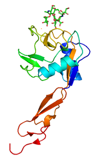
L-selectin, also known as CD62L, is a cell adhesion molecule found on the cell surface of leukocytes, and the blastocyst. It is coded for in the human by the SELL gene. L-selectin belongs to the selectin family of proteins, which recognize sialylated carbohydrate groups containing a Sialyl LewisX (sLeX) determinant. L-selectin plays an important role in both the innate and adaptive immune responses by facilitating leukocyte-endothelial cell adhesion events. These tethering interactions are essential for the trafficking of monocytes and neutrophils into inflamed tissue as well as the homing of lymphocytes to secondary lymphoid organs. L-selectin is also expressed by lymphoid primed hematopoietic stem cells and may participate in the migration of these stem cells to the primary lymphoid organs. In addition to its function in the immune response, L-selectin is expressed on embryonic cells and facilitates the attachment of the blastocyst to the endometrial endothelium during human embryo implantation.

<span class="mw-page-title-main">E-selectin</span>

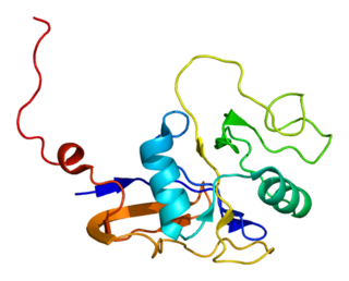
E-selectin, also known as CD62 antigen-like family member E (CD62E), endothelial-leukocyte adhesion molecule 1 (ELAM-1), or leukocyte-endothelial cell adhesion molecule 2 (LECAM2), is a selectin cell adhesion molecule expressed only on endothelial cells activated by cytokines. Like other selectins, it plays an important part in inflammation. In humans, E-selectin is encoded by the SELE gene.

High endothelial venules (HEV) are specialized post-capillary venules characterized by plump endothelial cells as opposed to the usual flatter endothelial cells found in regular venules. HEVs enable lymphocytes circulating in the blood to directly enter a lymph node.

Sialyl-Lewis <sup>X</sup> Chemical compound

Sialyl LewisX (sLeX), also known as cluster of differentiation 15s (CD15s) or stage-specific embryonic antigen 1 (SSEA-1), is a tetrasaccharide carbohydrate which is usually attached to O-glycans on the surface of cells. It is known to play a vital role in cell-to-cell recognition processes. It is also the means by which an egg attracts sperm; first, to stick to it, then bond with it and eventually form a zygote.

<span class="mw-page-title-main">Galectin</span> Protein family binding to β-galactoside sugars

Galectins are a class of proteins that bind specifically to β-galactoside sugars, such as N-acetyllactosamine, which can be bound to proteins by either N-linked or O-linked glycosylation. They are also termed S-type lectins due to their dependency on disulphide bonds for stability and carbohydrate binding. There have been about 15 galectins discovered in mammals, encoded by the LGALS genes, which are numbered in a consecutive manner. Only galectin-1, -2, -3, -4, -7, -7B, -8, -9, -9B, 9C, -10, -12, -13, -14, and -16 have been identified in humans. Galectin-5 and -6 are found in rodents, whereas galectin-11 and -15 are uniquely found in sheep and goats. Members of the galectin family have also been discovered in other mammals, birds, amphibians, fish, nematodes, sponges, and some fungi. Unlike the majority of lectins they are not membrane bound, but soluble proteins with both intra- and extracellular functions. They have distinct but overlapping distributions but found primarily in the cytosol, nucleus, extracellular matrix or in circulation. Although many galectins must be secreted, they do not have a typical signal peptide required for classical secretion. The mechanism and reason for this non-classical secretion pathway is unknown.

The mannose receptor is a C-type lectin primarily present on the surface of macrophages, immature dendritic cells and liver sinusoidal endothelial cells, but is also expressed on the surface of skin cells such as human dermal fibroblasts and keratinocytes. It is the first member of a family of endocytic receptors that includes Endo180 (CD280), M-type PLA2R, and DEC-205 (CD205).

Glycosylation-dependent cell adhesion molecule-1 (GLYCAM1) is a proteoglycan ligand expressed on cells of the high endothelial venules in lymphoid tissues. It is the ligand for the receptor L-selectin allowing for naive lymphocytes to exit the bloodstream into lymphoid tissues. GLYCAM1 binds to L-selectin by presenting one or more O-linked carbohydrates to the lectin domain of the leukocyte cell surface selectin. Data suggests that GLYCAM1 is a hormone-regulated milk protein that is part of the milk mucin complex.

<span class="mw-page-title-main">Integrin beta 2</span> Mammalian protein found in Homo sapiens

In molecular biology, CD18 is an integrin beta chain protein that is encoded by the ITGB2 gene in humans. Upon binding with one of a number of alpha chains, CD18 is capable of forming multiple heterodimers, which play significant roles in cellular adhesion and cell surface signaling, as well as important roles in immune responses. CD18 also exists in soluble, ligand binding forms. Deficiencies in CD18 expression can lead to adhesion defects in circulating white blood cells in humans, reducing the immune system's ability to fight off foreign invaders.

<span class="mw-page-title-main">Leukocyte extravasation</span>

Leukocyte extravasation is the movement of leukocytes out of the circulatory system and towards the site of tissue damage or infection. This process forms part of the innate immune response, involving the recruitment of non-specific leukocytes. Monocytes also use this process in the absence of infection or tissue damage during their development into macrophages.

<span class="mw-page-title-main">FUT7</span> Protein-coding gene in the species Homo sapiens

Alpha-(1,3)-fucosyltransferase is an enzyme that in humans is encoded by the FUT7 gene.

<span class="mw-page-title-main">SIGLEC8</span> Protein-coding gene in the species Homo sapiens

Sialic acid-binding Ig-like lectin 8 is a protein that in humans is encoded by the SIGLEC8 gene. This gene is located on chromosome 19q13.4, about 330 kb downstream of the SIGLEC9 gene. Within the siglec family of transmembrane proteins, Siglec-8 belongs to the CD33-related siglec subfamily, a subfamily that has undergone rapid evolution.

A catch bond is a type of noncovalent bond whose dissociation lifetime increases with tensile force applied to the bond. Normally, bond lifetimes are expected to diminish with force. In the case of catch bonds, the lifetime of the bond actually increases up to a maximum before it decreases like in a normal bond. Catch bonds work in a way that is conceptually similar to that of a Chinese finger trap. While catch bonds are strengthened by an increase in force, the force increase is not necessary for the bond to work. Catch bonds were suspected for many years to play a role in the rolling of leukocytes, being strong enough to roll in presence of high forces caused by high shear stresses, while avoiding getting stuck in capillaries where the fluid flow, and therefore shear stress, is low. The existence of catch bonds was debated for many years until strong evidence of their existence was found in bacteria. Definite proof of their existence came shortly thereafter in leukocytes.

<span class="mw-page-title-main">Endothelial cell anergy</span> Defense mechanism of tumors against immunity

Endothelial cell anergy is a condition during the process of angiogenesis, where endothelial cells, the cells that line the inside of blood vessels, can no longer respond to inflammatory cytokines. These cytokines are necessary to induce the expression of cell adhesion molecules to allow leukocyte infiltration from the blood into the tissue at places of inflammation, such as a tumor. This condition, which protects the tumor from the immune system, is the result of exposure to angiogenic growth factors.

References

  1. 1 2 PDB: 1G1R ; Somers WS, Tang J, Shaw GD, Camphausen RT (October 2000). "Insights into the molecular basis of leukocyte tethering and rolling revealed by structures of P- and E-selectin bound to SLe(X) and PSGL-1". Cell. 103 (3): 467–79. doi: 10.1016/S0092-8674(00)00138-0 . PMID   11081633. S2CID   12719907.
  2. 1 2 Cotran; Kumar, Collins (1998). Robbins Pathologic Basis of Disease. Philadelphia: W.B Saunders Company. ISBN   978-0-7216-7335-6.
  3. Parham, Peter (2005). The immune system (2nd ed.). New York: Garland Science. pp.  244–245. ISBN   978-0-8153-4093-5.
  4. 1 2 3 4 Ley K (June 2003). "The role of selectins in inflammation and disease". Trends in Molecular Medicine. 9 (6): 263–8. CiteSeerX   10.1.1.407.6232 . doi:10.1016/S1471-4914(03)00071-6. PMID   12829015.
  5. Cheung LS, Raman PS, Balzer EM, Wirtz D, Konstantopoulos K (February 2011). "Biophysics of selectin-ligand interactions in inflammation and cancer". Physical Biology. 8 (1): 015013. Bibcode:2011PhBio...8a5013S. doi:10.1088/1478-3975/8/1/015013. PMID   21301059. S2CID   3909905.
  6. Kappelmayer J, Nagy B (2017). "The Interaction of Selectins and PSGL-1 as a Key Component in Thrombus Formation and Cancer Progression". BioMed Research International. 2017: 6138145. doi: 10.1155/2017/6138145 . PMC   5478826 . PMID   28680883.
  7. Jennette, J. Charles; Falk, Ronald J. (2008). "Immunologic Mechanisms of Vasculitis". Seldin and Giebisch's the Kidney. pp. 2315–2338. doi:10.1016/B978-012088488-9.50085-1. ISBN   9780120884889.
  8. Nimrichter L, Burdick MM, Aoki K, Laroy W, Fierro MA, Hudson SA, Von Seggern CE, Cotter RJ, Bochner BS, Tiemeyer M, Konstantopoulos K, Schnaar RL (November 2008). "E-selectin receptors on human leukocytes". Blood. 112 (9): 3744–52. doi:10.1182/blood-2008-04-149641. PMC   2572800 . PMID   18579791.
  9. Bochner BS, Sterbinsky SA, Bickel CA, Werfel S, Wein M, Newman W (January 1994). "Differences between human eosinophils and neutrophils in the function and expression of sialic acid-containing counterligands for E-selectin". Journal of Immunology. 152 (2): 774–82. doi: 10.4049/jimmunol.152.2.774 . PMID   7506734. S2CID   45677380.
  10. Wein M, Sterbinsky SA, Bickel CA, Schleimer RP, Bochner BS (March 1995). "Comparison of human eosinophil and neutrophil ligands for P-selectin: ligands for P-selectin differ from those for E-selectin". American Journal of Respiratory Cell and Molecular Biology. 12 (3): 315–9. doi:10.1165/ajrcmb.12.3.7532979. PMID   7532979.
  11. Thomas W (September 2006). "For catch bonds, it all hinges on the interdomain region". The Journal of Cell Biology. 174 (7): 911–3. doi:10.1083/jcb.200609029. PMC   2064382 . PMID   17000873.
  12. Yago T, Wu J, Wey CD, Klopocki AG, Zhu C, McEver RP (September 2004). "Catch bonds govern adhesion through L-selectin at threshold shear". The Journal of Cell Biology. 166 (6): 913–23. doi:10.1083/jcb.200403144. PMC   2172126 . PMID   15364963.
  13. Barthel SR, Gavino JD, Descheny L, Dimitroff CJ (November 2007). "Targeting selectins and selectin ligands in inflammation and cancer". Expert Opinion on Therapeutic Targets. 11 (11): 1473–91. doi:10.1517/14728222.11.11.1473. PMC   2559865 . PMID   18028011.
  14. St Hill CA (June 2011). "Interactions between endothelial selectins and cancer cells regulate metastasis". Frontiers in Bioscience. 16 (9): 3233–51. doi: 10.2741/3909 . PMID   21622232.
  15. Witz IP (2006). "Tumor-Microenvironment Interactions". Tumor-microenvironment interactions: the selectin-selectin ligand axis in tumor-endothelium cross talk. Cancer Treatment and Research. Vol. 130. pp. 125–40. doi:10.1007/0-387-26283-0_6. ISBN   978-0-387-26282-6. PMID   16610706.
  16. Nakamori S, Kameyama M, Imaoka S, Furukawa H, Ishikawa O, Sasaki Y, Izumi Y, Irimura T (April 1997). "Involvement of carbohydrate antigen sialyl Lewis(x) in colorectal cancer metastasis". Diseases of the Colon and Rectum. 40 (4): 420–31. doi:10.1007/BF02258386. PMID   9106690. S2CID   24770173.
  17. Matsuura N, Narita T, Mitsuoka C, Kimura N, Kannagi R, Imai T, Funahashi H, Takagi H (1997). "Increased concentration of soluble E-selectin in the sera of breast cancer patients". Anticancer Research. 17 (2B): 1367–72. PMID   9137500.
  18. Gout S, Morin C, Houle F, Huot J (September 2006). "Death receptor-3, a new E-Selectin counter-receptor that confers migration and survival advantages to colon carcinoma cells by triggering p38 and ERK MAPK activation". Cancer Research. 66 (18): 9117–24. doi: 10.1158/0008-5472.CAN-05-4605 . PMID   16982754.
  19. Borsig L, Wong R, Hynes RO, Varki NM, Varki A (February 2002). "Synergistic effects of L- and P-selectin in facilitating tumor metastasis can involve non-mucin ligands and implicate leukocytes as enhancers of metastasis". Proceedings of the National Academy of Sciences of the United States of America. 99 (4): 2193–8. Bibcode:2002PNAS...99.2193B. doi: 10.1073/pnas.261704098 . PMC   122341 . PMID   11854515.
  20. Peeters CF, Ruers TJ, Westphal JR, de Waal RM (February 2005). "Progressive loss of endothelial P-selectin expression with increasing malignancy in colorectal cancer". Laboratory Investigation; A Journal of Technical Methods and Pathology. 85 (2): 248–56. doi: 10.1038/labinvest.3700217 . PMID   15640834.
  21. In the lab of Robert Sackstein Harvard University
  22. Sackstein Lab