Dysferlin

Last updated
DYSF
4ihb pdb.png
Available structures
PDB Ortholog search: PDBe RCSB
Identifiers
Aliases DYSF , FER1L1, LGMD2B, MMD1, dysferlin, LGMDR2
External IDs OMIM: 603009 MGI: 1349385 HomoloGene: 20748 GeneCards: DYSF
Orthologs
SpeciesHumanMouse
Entrez
Ensembl
UniProt
RefSeq (mRNA)

NM_001077694
NM_021469
NM_001310152

RefSeq (protein)
Location (UCSC) Chr 2: 71.45 – 71.69 Mb Chr 6: 83.99 – 84.19 Mb
PubMed search [3] [4]
Wikidata
View/Edit Human View/Edit Mouse

Dysferlin also known as dystrophy-associated fer-1-like protein is a protein that in humans is encoded by the DYSF gene. [5] Dysferlin is linked with plasma membrane repair., [6] stabilization of calcium signaling [7] [8] [9] and the development of the T-tubule system of the muscle [10] A defect in the DYSF gene, located on chromosome 2p12-14, results in several types of muscular dystrophy; including Miyoshi myopathy (MM), Limb-girdle muscular dystrophy type 2B (LGMD2B) and Distal Myopathy (DM). A reduction or absence of dysferlin, termed dysferlinopathy, usually becomes apparent in the third or fourth decade of life and is characterised by weakness and wasting of various voluntary skeletal muscles. [11] Pathogenic mutations leading to dysferlinopathy can occur throughout the DYSF gene.

Structure

Ferlin family
Identifiers
SymbolDysferlin
OPM superfamily 452
OPM protein 4cah
Membranome 205

The human dysferlin protein is a 237 kilodalton type-II transmembrane protein. [12] [13] [14] [15] [16] It contains a large intracellular cytoplasmic N-terminal domain, an extreme C-terminal transmembrane domain, and a short C-terminal extracellular domain. The cytosolic domain of dysferlin is composed of seven highly conserved C2 domains (C2A-G) which are conserved across several proteins within the ferlin family, including dysferlin homolog myoferlin. [17] [18] [13] In fact, the C2 domain at any given position is more similar to the C2 domain at the corresponding position within other ferlin family members than the adjacent C2 domain within the same protein. This suggests that each individual C2 domain may in fact play a specific role in dysferlin function and each has in fact been shown to be required for two of dysferlin's roles stabilization of calcium signaling and membrane repair. [19] Mutations in each of these domains can cause dysferlinopathy. A crystal structure of the C2A domain of human dysferlin has been solved, and reveals that the C2A domain changes conformation when interacting with calcium ions, [13] which is consistent with a growing body of evidence suggesting that the C2A domain plays a role in calcium-dependent lipid binding. [20] Its ability to stabilize calcium signaling in the intact dysferlin protein depends on its calcium binding activity. [21] In addition to the C2 domains, dysferlin also contains "FerA" and "DysF" domains. Mutations in both FerA [22] and DysF [23] can cause muscular dystrophies. DysF domain has an interesting structure as in contains one DysF domain within another DysF domain, a result of gene duplication; however, the function of this domain is currently unknown. [23] FerA domain is conserved among all members of ferlin protein family. FerA domain is a four helix bundle and it can interact with membrane, usually in a calcium-dependent manner. [22]

Function

The most intensively studied role for dysferlin is in a cellular process called membrane repair. Membrane repair is a critical mechanism by which cells are able to seal dramatic wounds to the plasma membrane. Muscle is thought to be particularly prone to membrane wounds given that muscle cells transmit high force and undergo cycles of contraction. Dysferlin is highly expressed in muscle, and is homologous to the ferlin family of proteins, which are thought to regulate membrane fusion across a wide variety of species and cell types. [24] Several lines of evidence suggest that dysferlin may be involved in membrane repair in muscle. First, dysferlin-deficient muscle fibers show accumulation of vesicles (which are critical for membrane repair in non-muscle cell types) near membrane lesions, indicating that dysferlin may be required for fusion of repair vesicles with the plasma membrane. Further, dysferlin-deficient muscle fibers take up extracellular dyes to a greater extent than wild-type muscle fibers following laser-induced wounding in-vitro. [25] Dysferlin is also markedly enriched at membrane lesions with several additional proteins thought to be involved in membrane resealing, including annexin and MG53. [26] Exactly how dysferlin contributes to membrane resealing is not clear, but biochemical evidence indicates that dysferlin may bind lipids in a calcium-dependent manner, consistent with a role for dysferlin in regulating fusion of repair vesicles with the sarcolemma during membrane repair. [27] Furthermore, live-cell imaging of dysferlin-eGFP expressing myotubes indicates that dysferlin localizes to a cellular compartment that responds to injury by forming large dysferlin-containing vesicles, and formation of these vesicles may contribute to wound repair. [28] Dysferlin may also be involved in Alzheimer's disease pathogenesis. [29]

Another well studied role for dysferlin is in stabilization of calcium signaling, especially following a mild injury. This approach was based on two observations: that muscle lacking dysferlin that is injured by eccentric contractions can repair its plasma membrane, or sarcolemma, as efficiently as healthy muscle can, [30] and that most of the dysferlin in healthy muscle is concentrated in the transverse tubules at triad junctions, [31] [32] where calcium release is regulated. Destabilization of signaling in dysferlinopathic muscle can result in the generation of calcium waves, [33] which can contribute to the disease pathology. Nearly every change in dysferlin that affects membrane repair also destabilizes calcium signaling, [34] suggesting that these two activities are closely linked. Remarkably, however, membrane repair requires calcium ions, whereas calcium ions contribute to the destabilization of signaling when dysferlin is absent or mutated. [35] These paradoxical results have yet to be reconciled.

Interactions

Dysferlin has been shown to bind to itself, to form dimers and perhaps larger oligomers. [36] It can also has been shown to interact with Caveolin 3 in skeletal muscle, [37] and this interaction is thought to retain dysferlin within the plasma membrane. [38] Dysferlin also interacts with MG53, and a functional interaction between dysferlin, caveolin-3 and MG53 is thought to be critical for membrane repair in skeletal muscle. [39]

Related Research Articles

<span class="mw-page-title-main">Titin</span> Largest-known protein in human muscles

Titin is a protein that in humans is encoded by the TTN gene. Titin is a giant protein, greater than 1 µm in length, that functions as a molecular spring that is responsible for the passive elasticity of muscle. It comprises 244 individually folded protein domains connected by unstructured peptide sequences. These domains unfold when the protein is stretched and refold when the tension is removed.

In molecular biology, caveolins are a family of integral membrane proteins that are the principal components of caveolae membranes and involved in receptor-independent endocytosis. Caveolins may act as scaffolding proteins within caveolar membranes by compartmentalizing and concentrating signaling molecules. They also induce positive (inward) membrane curvature by way of oligomerization, and hairpin insertion. Various classes of signaling molecules, including G-protein subunits, receptor and non-receptor tyrosine kinases, endothelial nitric oxide synthase (eNOS), and small GTPases, bind Cav-1 through its 'caveolin-scaffolding domain'.

<span class="mw-page-title-main">Caveolin 3</span> Protein-coding gene in the species Homo sapiens

Caveolin-3 is a protein that in humans is encoded by the CAV3 gene. Alternative splicing has been identified for this locus, with inclusion or exclusion of a differentially spliced intron. In addition, transcripts utilize multiple polyA sites and contain two potential translation initiation sites.

<span class="mw-page-title-main">Distal myopathy</span> Medical condition

Distal myopathy is a group of rare genetic disorders that cause muscle damage and weakness, predominantly in the hands and/or feet. Mutation of many different genes can be causative. Many types involve dysferlin.

<span class="mw-page-title-main">Emerin</span> Protein-coding gene in humans

Emerin is a protein that in humans is encoded by the EMD gene, also known as the STA gene. Emerin, together with LEMD3, is a LEM domain-containing integral protein of the inner nuclear membrane in vertebrates. Emerin is highly expressed in cardiac and skeletal muscle. In cardiac muscle, emerin localizes to adherens junctions within intercalated discs where it appears to function in mechanotransduction of cellular strain and in beta-catenin signaling. Mutations in emerin cause X-linked recessive Emery–Dreifuss muscular dystrophy, cardiac conduction abnormalities and dilated cardiomyopathy.

<span class="mw-page-title-main">Laminopathy</span> Medical condition

Laminopathies are a group of rare genetic disorders caused by mutations in genes encoding proteins of the nuclear lamina. They are included in the more generic term nuclear envelopathies that was coined in 2000 for diseases associated with defects of the nuclear envelope. Since the first reports of laminopathies in the late 1990s, increased research efforts have started to uncover the vital role of nuclear envelope proteins in cell and tissue integrity in animals.

<span class="mw-page-title-main">SK3</span> Protein-coding gene

SK3 also known as KCa2.3 is a protein that in humans is encoded by the KCNN3 gene.

<span class="mw-page-title-main">Fukutin</span> Mammalian protein found in Homo sapiens

Fukutin is a eukaryotic protein necessary for the maintenance of muscle integrity, cortical histogenesis, and normal ocular development. Mutations in the fukutin gene have been shown to result in Fukuyama congenital muscular dystrophy (FCMD) characterised by brain malformation - one of the most common autosomal-recessive disorders in Japan. In humans this protein is encoded by the FCMD gene, located on chromosome 9q31. Human fukutin exhibits a length of 461 amino acids and a predicted molecular mass of 53.7 kDa.

<span class="mw-page-title-main">FHL1</span> Mammalian protein found in humans

Four and a half LIM domains protein 1 is a protein that in humans is encoded by the FHL1 gene.

Calpain-3 is a protein that in humans is encoded by the CAPN3 gene.

<span class="mw-page-title-main">FLNC (gene)</span> Protein-coding gene in the species Homo sapiens

Filamin-C (FLN-C) also known as actin-binding-like protein (ABPL) or filamin-2 (FLN2) is a protein that in humans is encoded by the FLNC gene. Filamin-C is mainly expressed in cardiac and skeletal muscles, and functions at Z-discs and in subsarcolemmal regions.

<span class="mw-page-title-main">ATP2A1</span> Protein-coding gene in the species Homo sapiens

Sarcoplasmic/endoplasmic reticulum calcium ATPase 1 (SERCA1) also known as Calcium pump 1, is an enzyme that in humans is encoded by the ATP2A1 gene.

<span class="mw-page-title-main">Integrin alpha 7</span>

Alpha-7 integrin is a protein that in humans is encoded by the ITGA7 gene. Alpha-7 integrin is critical for modulating cell-matrix interactions. Alpha-7 integrin is highly expressed in cardiac muscle, skeletal muscle and smooth muscle cells, and localizes to Z-disc and costamere structures. Mutations in ITGA7 have been associated with congenital myopathies and noncompaction cardiomyopathy, and altered expression levels of alpha-7 integrin have been identified in various forms of muscular dystrophy.

<span class="mw-page-title-main">Delta-sarcoglycan</span> Mammalian protein found in Homo sapiens

Delta-sarcoglycan is a protein that in humans is encoded by the SGCD gene.

<span class="mw-page-title-main">SGCA</span> Protein-coding gene in the species Homo sapiens

Alpha-sarcoglycan is a protein that in humans is encoded by the SGCA gene.

<span class="mw-page-title-main">Ryanodine receptor 1</span> Protein and coding gene in humans

Ryanodine receptor 1 (RYR-1) also known as skeletal muscle calcium release channel or skeletal muscle-type ryanodine receptor is one of a class of ryanodine receptors and a protein found primarily in skeletal muscle. In humans, it is encoded by the RYR1 gene.

Collagen VI (ColVI) is a type of collagen primarily associated with the extracellular matrix of skeletal muscle. ColVI maintains regularity in muscle function and stabilizes the cell membrane. It is synthesized by a complex, multistep pathway that leads to the formation of a unique network of linked microfilaments located in the extracellular matrix (ECM). ColVI plays a vital role in numerous cell types, including chondrocytes, neurons, myocytes, fibroblasts, and cardiomyocytes. ColVI molecules are made up of three alpha chains: α1(VI), α2(VI), and α3(VI). It is encoded by 6 genes: COL6A1, COL6A2, COL6A3, COL6A4, COL6A5, and COL6A6. The chain lengths of α1(VI) and α2(VI) are about 1,000 amino acids. The chain length of α3(VI) is roughly a third larger than those of α1(VI) and α2(VI), and it consists of several spliced variants within the range of 2,500 to 3,100 amino acids.

<span class="mw-page-title-main">MYF6</span> Protein-coding gene in the species Homo sapiens

Myogenic factor 6 is a protein that in humans is encoded by the MYF6 gene. This gene is also known in the biomedical literature as MRF4 and herculin. MYF6 is a myogenic regulatory factor (MRF) involved in the process known as myogenesis.

<span class="mw-page-title-main">ANO5</span> Protein-coding gene in the species Homo sapiens

Anoctamin 5 (ANO5) is a protein that in humans is encoded by the ANO5 gene.

<span class="mw-page-title-main">Ferlins</span> Protein family

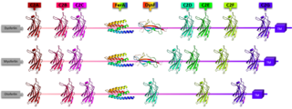
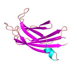
Ferlins are an ancient protein family involved in vesicle fusion and membrane trafficking. Ferlins are distinguished by their multiple tandem C2 domains, and sometimes a FerA and a DysF domain. Mutations in ferlins can cause human diseases such as muscular dystrophy and deafness. Abnormalities in expression of myoferlin, a human ferlin protein, is also directly associated with higher mortality rate and tumor recurrence in several types of cancer, including pancreatic, colorectal, breast, cervical, stomach, ovarian, cervical, thyroid, endometrial, and oropharyngeal squamous cell carcinoma. In other animals, ferlin mutations can cause infertility.

References

  1. 1 2 3 GRCh38: Ensembl release 89: ENSG00000135636 - Ensembl, May 2017
  2. 1 2 3 GRCm38: Ensembl release 89: ENSMUSG00000033788 - Ensembl, May 2017
  3. "Human PubMed Reference:". National Center for Biotechnology Information, U.S. National Library of Medicine.
  4. "Mouse PubMed Reference:". National Center for Biotechnology Information, U.S. National Library of Medicine.
  5. Passos-Bueno MR, Richard I, Vainzof M, Fougerousse F, Weissenbach J, Broux O, Cohen D, Akiyama J, Marie SK, Carvalho AA (May 1993). "Evidence of genetic heterogeneity in the autosomal recessive adult forms of limb-girdle muscular dystrophy following linkage analysis with 15q probes in Brazilian families". Journal of Medical Genetics . 30 (5): 385–7. doi:10.1136/jmg.30.5.385. PMC   1016373 . PMID   8320700.
  6. "Entrez Gene: DYSF dysferlin, limb girdle muscular dystrophy 2B (autosomal recessive)".
  7. Kerr JP, Ziman AP, Mueller AL, et al. (December 2013). "Dysferlin Stabilizes Stress-induced Ca2+ Signaling in the Transverse Tubule Membrane". Proc. Natl. Acad. Sci. USA. 110 (51): 20831–20836. Bibcode:2013PNAS..11020831K. doi: 10.1073/pnas.1307960110 . PMC   3870721 . PMID   24302765.
  8. Kerr JP, Ward CW, Bloch RJ (March 2014). "Dysferlin at Transverse Tubules Regulates Ca2+ Homeostasis in Skeletal Muscle". Frontiers in Physiology. 5: 89. doi: 10.3389/fphys.2014.00089 . PMC   3944681 . PMID   24639655.
  9. Lukyanenko V, Muriel JM, Bloch RJ (August 2017). "Coupling of Excitation to Ca2+ Release is Modulated by Dysferlin". J. Physiol. 595 (15): 5191–5207. doi:10.1113/JP274515. PMC   5538227 . PMID   8568606.
  10. Hofhuis J, Bersch K, Wagner S, Molina C, Fakuade FE, Iyer LM, Streckfuss-Bömeke K, Toischer K, Zelarayán LC, Voigt N, Nikolaev VO, Maier LS, Klinge L, Thoms S (July 2020). "Dysferlin links excitation-contraction coupling to structure and maintenance of the cardiac transverse-axial tubule system". Europace. 22 (7): 1119–1131. doi: 10.1093/europace/euaa093 . PMID   32572487.
  11. Leiden University Medical Center, Center for Human and Clinical Genetics - Dysferlin Retrieved 21 June 2007.
  12. Kawahara G, Serafini PR, Myers JA, Alexander MS, Kunkel LM (September 2011). "Characterization of zebrafish dysferlin by morpholino knockdown". Biochemical and Biophysical Research Communications. 413 (2): 358–363. doi:10.1016/j.bbrc.2011.08.105. PMC   4526276 . PMID   21893049.
  13. 1 2 3 Fuson K, Rice A, Mahling R, Snow A, Nayak K, Shanbhogue P, et al. (January 2014). "Alternate splicing of dysferlin C2A confers Ca²⁺-dependent and Ca²⁺-independent binding for membrane repair". Structure. 22 (1): 104–115. doi:10.1016/j.str.2013.10.001. PMC   5993433 . PMID   24239457.
  14. Demonbreun AR, Allen MV, Warner JL, Barefield DY, Krishnan S, Swanson KE, et al. (June 2016). "Enhanced Muscular Dystrophy from Loss of Dysferlin Is Accompanied by Impaired Annexin A6 Translocation after Sarcolemmal Disruption". The American Journal of Pathology. 186 (6): 1610–1622. doi:10.1016/j.ajpath.2016.02.005. PMC   4901136 . PMID   27070822.
  15. Cacciottolo M, Numitone G, Aurino S, Caserta IR, Fanin M, Politano L, et al. (September 2011). "Muscular dystrophy with marked Dysferlin deficiency is consistently caused by primary dysferlin gene mutations". European Journal of Human Genetics. 19 (9): 974–980. doi:10.1038/ejhg.2011.70. PMC   3179367 . PMID   21522182.
  16. Matsuda C, Kiyosue K, Nishino I, Goto Y, Hayashi YK (October 2015). "Dysferlinopathy Fibroblasts Are Defective in Plasma Membrane Repair". PLOS Currents. 7. doi: 10.1371/currents.md.5865add2d766f39a0e0411d38a7ba09c . PMC   4639325 . PMID   26579332.
  17. Vafiadaki E, Reis A, Keers S, Harrison R, Anderson LV, Raffelsberger T, et al. (March 2001). "Cloning of the mouse dysferlin gene and genomic characterization of the SJL-Dysf mutation". NeuroReport. 12 (3): 625–629. doi:10.1097/00001756-200103050-00039. PMID   11234777. S2CID   22800606.
  18. Abdullah N, Padmanarayana M, Marty NJ, Johnson CP (January 2014). "Quantitation of the calcium and membrane binding properties of the C2 domains of dysferlin". Biophysical Journal. 106 (2): 382–389. Bibcode:2014BpJ...106..382A. doi:10.1016/j.bpj.2013.11.4492. PMC   3907215 . PMID   24461013.
  19. Muriel J, Lukyanenko V, Kwiatkowski T, Bhattacharya S, Garman D, Weisleder N, Bloch RJ (April 2022). "The C2 domains of dysferlin: roles in membrane localization, Ca2+ signalling and sarcolemmal repair". The Journal of Physiology. 600 (8): 1953–1968. doi:10.1113/JP282648. PMC   9285653 . PMID   35156706.
  20. Therrien C, Di Fulvio S, Pickles S, Sinnreich M (March 2009). "Characterization of lipid binding specificities of dysferlin C2 domains reveals novel interactions with phosphoinositides". Biochemistry. 48 (11): 2377–2384. doi:10.1021/bi802242r. PMID   19253956.
  21. Lukyanenko V, Muriel J, Garman D, Breydo L, Bloch RJ (November 2022). "Elevated Ca2+ at the triad junction underlies dysregulation of Ca2+ signaling in dysferlin-null skeletal muscle". Frontiers in Physiology. 13: 1032447. doi: 10.3389/fphys.2022.1032447 . PMC   9669649 . PMID   36406982.
  22. 1 2 Harsini FM, Chebrolu S, Fuson KL, White MA, Rice AM, Sutton RB (July 2018). "FerA is a Membrane-Associating Four-Helix Bundle Domain in the Ferlin Family of Membrane-Fusion Proteins". Scientific Reports. 8 (1): 10949. Bibcode:2018NatSR...810949H. doi:10.1038/s41598-018-29184-1. PMC   6053371 . PMID   30026467.
  23. 1 2 Sula A, Cole AR, Yeats C, Orengo C, Keep NH (January 2014). "Crystal structures of the human Dysferlin inner DysF domain". BMC Structural Biology. 14: 3. doi: 10.1186/1472-6807-14-3 . PMC   3898210 . PMID   24438169.
  24. Bashir R, Britton S, Strachan T, Keers S, Vafiadaki E, Lako M, Richard I, Marchand S, Bourg N, Argov Z, Sadeh M, Mahjneh I, Marconi G, Passos-Bueno MR, Moreira Ede S, Zatz M, Beckmann JS, Bushby K (1998). "A gene related to Caenorhabditis elegans spermatogenesis factor fer-1 is mutated in limb-girdle muscular dystrophy type 2B". Nat. Genet. 20 (1): 37–42. doi:10.1038/1689. PMID   9731527. S2CID   24234676.
  25. Bansal D, Miyake K, Vogel SS, Groh S, Chen CC, Williamson R, McNeil PL, Campbell KP (2003). "Defective membrane repair in dysferlin-deficient muscular dystrophy". Nature. 423 (6936): 168–72. Bibcode:2003Natur.423..168B. doi:10.1038/nature01573. PMID   12736685. S2CID   4402938.
  26. Roostalu U, Strähle U (2012). "In vivo imaging of molecular interactions at damaged sarcolemma". Dev. Cell. 22 (3): 515–29. doi: 10.1016/j.devcel.2011.12.008 . PMID   22421042.
  27. Abdullah N, Padmanarayana M, Marty NJ, Johnson CP (2014). "Quantitation of the calcium and membrane binding properties of the C2 domains of dysferlin". Biophys. J. 106 (2): 382–9. Bibcode:2014BpJ...106..382A. doi:10.1016/j.bpj.2013.11.4492. PMC   3907215 . PMID   24461013.
  28. McDade JR, Michele DE (2014). "Membrane damage-induced vesicle-vesicle fusion of dysferlin-containing vesicles in muscle cells requires microtubules and kinesin". Hum. Mol. Genet. 23 (7): 1677–86. doi:10.1093/hmg/ddt557. PMC   3943514 . PMID   24203699.
  29. Chen JA, Wang Q, Davis-Turak J, et al. (April 2015). "A multiancestral genome-wide exome array study of Alzheimer disease, frontotemporal dementia, and progressive supranuclear palsy". JAMA Neurology. 72 (4): 414–22. doi:10.1001/jamaneurol.2014.4040. PMC   4397175 . PMID   25706306.
  30. Roche JA, Lovering RM, Vanapalli R, et al. (Feb 2010). "Extensive Mononuclear Infiltration and Myogenesis Characterize the Recovery of Dysferlin-Null Skeletal Muscle from Contraction-Induced Injuries". Am. J. Physiol. Cell Physiol. 298 (2): C298–C312. doi:10.1152/ajpcell.00122.2009. PMC   2822489 . PMID   9923419.
  31. Kerr JP, Ziman AP, Mueller AL, et al. (December 2013). "Dysferlin Stabilizes Stress-induced Ca2+ Signaling in the Transverse Tubule Membrane". Proc. Natl. Acad. Sci. USA. 110 (51): 20831–20836. Bibcode:2013PNAS..11020831K. doi: 10.1073/pnas.1307960110 . PMC   3870721 . PMID   24302765.
  32. Roche JA, Lovering RM, Vanapalli R, et al. (Feb 2010). "Extensive Mononuclear Infiltration and Myogenesis Characterize the Recovery of Dysferlin-Null Skeletal Muscle from Contraction-Induced Injuries". Am. J. Physiol. Cell Physiol. 298 (2): C298–C312. doi:10.1152/ajpcell.00122.2009. PMC   2822489 . PMID   9923419.
  33. Lukyanenko V, Muriel J, Bloch RJ (August 2017). "Coupling of Excitation to Ca2+ Release is Modulated by Dysferlin". J. Physiol. 595 (15): 5191–5207. doi:10.1113/JP274515. PMC   5538227 . PMID   8568606.
  34. Muriel J, Lukyanenko V, Kwiatkowski T, et al. (Apr 2022). "The C2 Domains of Dysferlin: Roles in Membrane Localization, Stabilization of Ca2+ Signaling, and Membrane Repair". J. Physiol. 600 (8): 1953–1968. doi:10.1113/JP282648. PMC   9285653 . PMID   35156706.
  35. Muriel J, Lukyanenko V, Kwiatkowski T, et al. (Apr 2022). "The C2 Domains of Dysferlin: Roles in Membrane Localization, Stabilization of Ca2+ Signaling, and Membrane Repair". J. Physiol. 600 (8): 1953–1968. doi:10.1113/JP282648. PMC   9285653 . PMID   35156706.
  36. Roche JA, Ru L, ONeill A, et al. (Nov 2011). "Unmasking intracellular dysferlin". J. Histochem. 59 (11): 964–975. doi:10.1369/0022155411423274. PMC   3261626 . PMID   22043020.
  37. Matsuda C, Hayashi YK, Ogawa M, Aoki M, Murayama K, Nishino I, Nonaka I, Arahata K, Brown RH (August 2001). "The sarcolemmal proteins dysferlin and caveolin-3 interact in skeletal muscle". Hum. Mol. Genet. 10 (17): 1761–6. doi: 10.1093/hmg/10.17.1761 . PMID   11532985.
  38. Hernández-Deviez DJ, Howes MT, Laval SH, Bushby K, Hancock JF, Parton RG (2008). "Caveolin regulates endocytosis of the muscle repair protein, dysferlin" (PDF). J. Biol. Chem. 283 (10): 6476–88. doi: 10.1074/jbc.M708776200 . PMID   18096699. S2CID   8337318.
  39. Cai C, Weisleder N, Ko JK, Komazaki S, Sunada Y, Nishi M, Takeshima H, Ma J (2009). "Membrane repair defects in muscular dystrophy are linked to altered interaction between MG53, caveolin-3, and dysferlin". J. Biol. Chem. 284 (23): 15894–902. doi: 10.1074/jbc.M109.009589 . PMC   2708885 . PMID   19380584.

Further reading