Myosatellite cell

Last updated
Myosatellite cell
Details
Identifiers
Latin myosatellitocytus
TH H2.00.05.2.01020
Anatomical terms of microanatomy

Myosatellite cells, also known as satellite cells, muscle stem cells or MuSCs, are small multipotent cells with very little cytoplasm found in mature muscle. [1] Satellite cells are precursors to skeletal muscle cells, able to give rise to satellite cells or differentiated skeletal muscle cells. [2] They have the potential to provide additional myonuclei to their parent muscle fiber, or return to a quiescent state. [3] More specifically, upon activation, satellite cells can re-enter the cell cycle to proliferate and differentiate into myoblasts. [4]

Contents

Myosatellite cells are located between the basement membrane and the sarcolemma of muscle fibers, [5] and can lie in grooves either parallel or transversely to the longitudinal axis of the fibre. Their distribution across the fibre can vary significantly. Non-proliferative, quiescent myosatellite cells, which adjoin resting skeletal muscles, can be identified by their distinct location between sarcolemma and basal lamina, a high nuclear-to-cytoplasmic volume ratio, few organelles (e.g. ribosomes, endoplasmic reticulum, mitochondria, golgi complexes), small nuclear size, and a large quantity of nuclear heterochromatin relative to myonuclei. On the other hand, activated satellite cells have an increased number of caveolae, cytoplasmic organelles, and decreased levels of heterochromatin. [2] Satellite cells are able to differentiate and fuse to augment existing muscle fibers and to form new fibers. These cells represent the oldest known adult stem cell niche, and are involved in the normal growth of muscle, as well as regeneration following injury or disease.

In undamaged muscle, the majority of satellite cells are quiescent; they neither differentiate nor undergo cell division. In response to mechanical strain, satellite cells become activated. Activated satellite cells initially proliferate as skeletal myoblasts before undergoing myogenic differentiation. [1]

Structure

Genetic markers

Satellite cells express a number of distinctive genetic markers. Current thinking is that most satellite cells express PAX7 and PAX3. [6] Satellite cells in the head musculature have a unique developmental program, [7] and are Pax3-negative. Moreover, both quiescent and activated human satellite cells can be identified by the membrane-bound neural cell adhesion molecule (N-CAM/CD56/Leu-19), a cell-surface glycoprotein. Myocyte nuclear factor (MNF), and c-met proto-oncogene (receptor for hepatocyte growth factor (HGF)) are less commonly used markers. [2]

CD34 and Myf5 markers specifically define the majority of quiescent satellite cells. [8] Activated satellite cells prove problematic to identify, especially as their markers change with the degree of activation; for example, greater activation results in the progressive loss of Pax7 expression as they enter the proliferative stage. However, Pax7 is expressed prominently after satellite cell differentiation. [9] Greater activation also results in increased expression of myogenic basic helix-loop-helix transcription factors MyoD, myogenin, and MRF4 – all responsible for the induction of myocyte-specific genes. [10] HGF testing is also used to identify active satellite cells. [2] Activated satellite cells also begin expressing muscle-specific filament proteins such as desmin as they differentiate.

The field of satellite cell biology suffers from the same technical difficulties as other stem cell fields. Studies rely almost exclusively on Flow cytometry and fluorescence activated cell sorting (FACS) analysis, which gives no information about cell lineage or behaviour. As such, the satellite cell niche is relatively ill-defined and it is likely that it consists of multiple sub-populations.

Function

Muscle repair

When muscle cells undergo injury, quiescent satellite cells are released from beneath the basement membrane. They become activated and re-enter the cell cycle. These dividing cells are known as the "transit amplifying pool" before undergoing myogenic differentiation to form new (post-mitotic) myotubes. There is also evidence suggesting that these cells are capable of fusing with existing myofibers to facilitate growth and repair. [1]

The process of muscle regeneration involves considerable remodeling of extracellular matrix and, where extensive damage occurs, is incomplete. Fibroblasts within the muscle deposit scar tissue, which can impair muscle function, and is a significant part of the pathology of muscular dystrophies.

Satellite cells proliferate following muscle trauma [11] and form new myofibers through a process similar to fetal muscle development. [12] After several cell divisions, the satellite cells begin to fuse with the damaged myotubes and undergo further differentiations and maturation, with peripheral nuclei as in hallmark. [12] One of the first roles described for IGF-1 was its involvement in the proliferation and differentiation of satellite cells. In addition, IGF-1 expression in skeletal muscle extends the capacity to activate satellite cell proliferation (Charkravarthy, et al., 2000), increasing and prolonging the beneficial effects to the aging muscle. [13] [14]

Effects of exercise

Satellite cell activation is measured by the extent of proliferation and differentiation. Typically, satellite cell content is expressed per muscle fiber or as a percentage of total nuclear content, the sum of satellite cell nuclei and myonuclei. While the adaptive response to exercise largely varies on an individual basis on factors such as genetics, age, diet, acclimatization to exercise, and exercise volume, human studies have demonstrated general trends. [2]

It is suggested that exercise triggers the release of signaling molecules including inflammatory substances, cytokines and growth factors from surrounding connective tissues and active skeletal muscles. [2] Notably, HGF, a cytokine, is transferred from the extracellular matrix into muscles through the nitric-oxide dependent pathway. It is thought that HGF activates satellite cells, while insulin-like growth factor-I (IGF-1) and fibroblast growth factor (FGF) enhance satellite cell proliferation rate following activation. [15] Studies have demonstrated that intense exercise generally increases IGF-1 production, though individual responses vary significantly. [16] [17] More specifically, IGF-1 exists in two isoforms: mechano growth factor (MGF) and IGF-IEa. [18] While the former induces activation and proliferation, the latter causes differentiation of proliferating satellite cells. [18]

Human studies have shown that both high resistance training and endurance training have yielded an increased number of satellite cells. [9] [19] These results suggest that a light, endurance training regimen may be useful to counteract the age-correlated satellite cell decrease. [2] In high-resistance training, activation and proliferation of satellite cells are evidenced by increased cyclin D1 mRNA, and p21 mRNA levels. This is consistent with the fact that cyclin D1 and p21 upregulation correlates to division and differentiation of cells. [3]

Satellite cell activation has also been demonstrated on an ultrastructural level following exercise. Aerobic exercise has been shown to significantly increase granular endoplasmic reticulum, free ribosomes, and mitochondria of the stimulated muscle groups. Additionally, satellite cells have been shown to fuse with muscle fibers, developing new muscle fibers. [20] Other ultrastructural evidence for activated satellite cells include increased concentration of Golgi apparatus and pinocytotic vesicles. [21]

Schematic of myosatellite cell transition into myofiber. Schematic of satellite cell myogenesis and markers typical of each stage.jpg
Schematic of myosatellite cell transition into myofiber.

Satellite cell activation and muscle regeneration

Satellite cells have a crucial role in muscle regeneration due to their ability to proliferate, differentiate, and self-renew. Prior to a severe injury to the muscle, satellite cells are in a dormant state. Slight proliferation can occur in times of light injuries but major injuries require greater numbers of satellite cells to activate. The activation of satellite cells from their dormant state is controlled through signals from the muscle niche. This signaling induces an inflammatory response in the muscle tissue. The behavior of satellite cells is a highly regulated process to accommodate the balance between dormant and active states. [22] In times of injury, satellite cells in myofibers receive signals to proliferate from proteins in the crushed skeletal muscle. Myofibers are fundamental elements in muscle made up of actin and myosin myofibrils. The proteins responsible for signaling the activation of satellite cells are called mitogens. A mitogen is a small protein that induces a cell to enter the cell cycle. When the cells receive signals from the neurons, it causes the myofibers to depolarize and release calcium from the sarcoplasmic reticulum. The release of calcium induces the actin and myosin filaments to move and contract the muscle. Studies found that transplanted satellite cells onto myofibers supported multiple regenerations of new muscle tissue. These findings support the hypothesis that satellite cells are the stem cells in muscles. Dependent on their relative position to daughter cells on myofibers, satellite cells undergo asymmetric and symmetric division. The niche and location determines the behavior of satellite cells in their proliferation and differentiation. In general, mammalian skeletal muscle is relatively stable with little myonuclei turnover. Minor injuries from daily activities can be repaired without inflammation or cell death. Major injuries contribute to myofiber necrosis, inflammation, and cause satellite cells to activate and proliferate. The process of myofiber necrosis to myofiber formation results in muscle regeneration. [23]

Muscle regeneration occurs in three overlapping stages. The inflammatory response, activation and differentiation of satellite cells, and maturation of the new myofibers are essential for muscle regeneration. This process begins with the death of damaged muscle fibers where dissolution of myofiber sarcolemma leads to an increase in myofiber permeability. The disruption in myofiber integrity is seen in increased plasma levels in muscle proteins. The death of myofibers drives a calcium influx from the sarcoplasmic reticulum to induce tissue degradation. An inflammatory response follows the necrosis of myofibers. During times of muscle growth and regeneration, satellite cells can travel over between myofibers and muscle and over connective tissue barriers. Signals from the damaged environment induce these behavioral changes in satellite cells. [23]

Research

Upon minimal stimulation, satellite cells in vitro or in vivo will undergo a myogenic differentiation program.

Unfortunately, it seems that transplanted satellite cells have a limited capacity for migration, and are only able to regenerate muscle in the region of the delivery site. As such, systemic treatments or even the treatment of an entire muscle in this way is not possible. However, other cells in the body such as pericytes and hematopoietic stem cells have all been shown to be able to contribute to muscle repair in a similar manner to the endogenous satellite cell. The advantage of using these cell types for therapy in muscle diseases is that they can be systemically delivered, autonomously migrating to the site of injury. Particularly successful recently has been the delivery of mesoangioblast cells into the Golden Retriever dog model of Duchenne muscular dystrophy, which effectively cured the disease. [24] However, the sample size used was relatively small and the study has since been criticized for a lack of appropriate controls for the use of immunosuppressive drugs. Recently, it has been reported that Pax7 expressing cells contribute to dermal wound repair by adopting a fibrotic phenotype through a Wnt/β-catenin mediated process. [25]

Regulation

Little is known of the regulation of satellite cells. Whilst together PAX3 and PAX7 currently form the definitive satellite markers, Pax genes are notoriously poor transcriptional activators. The dynamics of activation and quiesence and the induction of the myogenic program through the myogenic regulatory factors, Myf5, MyoD, myogenin, and MRF4 remains to be determined. [26]

There is some research indicating that satellite cells are negatively regulated by a protein called myostatin. Increased levels of myostatin up-regulate a cyclin-dependent kinase inhibitor called p21 and thereby inhibit the differentiation of satellite cells. [27]

Myosatellite cells and cultured meat

Myosatellite cells contribute the most to muscle regeneration and repair. [23] This makes them a prime target for the meat culturing field. These satellite cells are the main source of most muscle cell formation postnatally, with embryonic myoblasts being responsible for prenatal muscle generation. A single satellite cell can proliferate and become a larger amount of muscle cells. [28]

With the understanding that myosatellite cells are the progenitor of most skeletal muscle cells, it was theorized that if these cells could be grown in a lab and placed on scaffolds to make fibers, the muscle cells could then be used for food production. [29] This theory has been proven true with many companies sprouting around the globe in the field of cultured meat including Mosa Meat in the Netherlands, and Upside Foods in the USA. [30] [31]

An overview of the culturing process first involves the selection of a cell source. This initial stage is where the selection of a meat type happens, for example if the desired product is beef then cells are taken from a cow. The next part involves isolating and sorting out the myosatellite cells from whatever the selected cell source was. After being separated into the cellular components, the myosatellite cells need to be proliferated through the use of a bioreactor, a device used to grow microorganisms or cells in a media that can be easily controlled. [32] Whatever media chosen will simulate the cells being in prime condition to proliferate within an organism. After proliferation the cells are shaped using a scaffold. These scaffolds can be an organic structure like decellularized plant or animal tissues, inorganic such as polyacrylamide, or a mix of both. [33] Once the cells have attached themselves to the scaffold and fully matured, they have become a raw meat product. The final step will include any necessary food processes needed for the desired final product. [34]

See also

Related Research Articles

<span class="mw-page-title-main">Skeletal muscle</span> One of three major skeletal system types that connect to bones

Skeletal muscles are organs of the vertebrate muscular system and typically are attached by tendons to bones of a skeleton. The muscle cells of skeletal muscles are much longer than in the other types of muscle tissue, and are often known as muscle fibers. The muscle tissue of a skeletal muscle is striated – having a striped appearance due to the arrangement of the sarcomeres.

<span class="mw-page-title-main">Muscle cell</span> Type of cell found in muscle tissue

A muscle cell, also known as a myocyte, is a mature contractile cell in the muscle of an animal. In humans and other vertebrates there are three types: skeletal, smooth, and cardiac (cardiomyocytes). A skeletal muscle cell is long and threadlike with many nuclei and is called a muscle fiber. Muscle cells develop from embryonic precursor cells called myoblasts.

<span class="mw-page-title-main">Striated muscle tissue</span> Muscle tissue with repeating functional units called sarcomeres

Striated muscle tissue is a muscle tissue that features repeating functional units called sarcomeres. The presence of sarcomeres manifests as a series of bands visible along the muscle fibers, which is responsible for the striated appearance observed in microscopic images of this tissue. There are two types of striated muscle:

G<sub>0</sub> phase Quiescent stage of the cell cycle in which the cell does not divide

The G0 phase describes a cellular state outside of the replicative cell cycle. Classically, cells were thought to enter G0 primarily due to environmental factors, like nutrient deprivation, that limited the resources necessary for proliferation. Thus it was thought of as a resting phase. G0 is now known to take different forms and occur for multiple reasons. For example, most adult neuronal cells, among the most metabolically active cells in the body, are fully differentiated and reside in a terminal G0 phase. Neurons reside in this state, not because of stochastic or limited nutrient supply, but as a part of their developmental program.

<span class="mw-page-title-main">MyoD</span> Mammalian protein found in Homo sapiens

MyoD, also known as myoblast determination protein 1, is a protein in animals that plays a major role in regulating muscle differentiation. MyoD, which was discovered in the laboratory of Harold M. Weintraub, belongs to a family of proteins known as myogenic regulatory factors (MRFs). These bHLH transcription factors act sequentially in myogenic differentiation. Vertebrate MRF family members include MyoD1, Myf5, myogenin, and MRF4 (Myf6). In non-vertebrate animals, a single MyoD protein is typically found.

<span class="mw-page-title-main">Sarcopenia</span> Muscle loss due to ageing or immobility

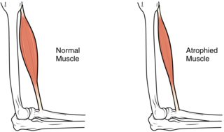
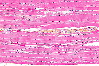
Sarcopenia is a type of muscle loss that occurs with aging and/or immobility. It is characterized by the degenerative loss of skeletal muscle mass, quality, and strength. The rate of muscle loss is dependent on exercise level, co-morbidities, nutrition and other factors. The muscle loss is related to changes in muscle synthesis signalling pathways. It is distinct from cachexia, in which muscle is degraded through cytokine-mediated degradation, although the two conditions may co-exist. Sarcopenia is considered a component of frailty syndrome. Sarcopenia can lead to reduced quality of life, falls, fracture, and disability.

<span class="mw-page-title-main">Pax genes</span> Family of transcription factors

In evolutionary developmental biology, Paired box (Pax) genes are a family of genes coding for tissue specific transcription factors containing an N-terminal paired domain and usually a partial, or in the case of four family members, a complete homeodomain to the C-terminus. An octapeptide as well as a Pro-Ser-Thr-rich C terminus may also be present. Pax proteins are important in early animal development for the specification of specific tissues, as well as during epimorphic limb regeneration in animals capable of such.

<span class="mw-page-title-main">PAX3</span> Paired box gene 3

The PAX3 gene encodes a member of the paired box or PAX family of transcription factors. The PAX family consists of nine human (PAX1-PAX9) and nine mouse (Pax1-Pax9) members arranged into four subfamilies. Human PAX3 and mouse Pax3 are present in a subfamily along with the highly homologous human PAX7 and mouse Pax7 genes. The human PAX3 gene is located in the 2q36.1 chromosomal region, and contains 10 exons within a 100 kb region.

<span class="mw-page-title-main">Myogenesis</span> Formation of muscular tissue, particularly during embryonic development

Myogenesis is the formation of skeletal muscular tissue, particularly during embryonic development.

<span class="mw-page-title-main">Mesoangioblast</span>

A mesoangioblast is a type of progenitor cell that is associated with vasculature walls. Mesoangioblasts exhibit many similarities to pericytes, which are found in the small vessels. Mesoangioblasts are multipotent stem cells with the potential to progress down the endothelial or mesodermal lineages. Mesoangioblasts express the critical marker of angiopoietic progenitors, KDR (FLK1). Because of these properties, mesoangioblasts are a precursor of skeletal, smooth, and cardiac muscle cells along with endothelial cells. Research has suggested their application for stem cell therapies for muscular dystrophy and cardiovascular disease.

<span class="mw-page-title-main">GDF11</span> Protein-coding gene in humans

Growth differentiation factor 11 (GDF11), also known as bone morphogenetic protein 11 (BMP-11), is a protein that in humans is encoded by the growth differentiation factor 11 gene. GDF11 is a member of the Transforming growth factor beta family.

Alveolar rhabdomyosarcoma (ARMS) is a subtype of the rhabdomyosarcoma soft tissue cancer family whose lineage is from mesenchymal cells and are related to skeletal muscle cells. ARMS tumors resemble the alveolar tissue in the lungs. Tumor location varies from patient to patient, but is commonly found in the head and neck region, male and female urogenital tracts, the torso, and extremities. Two fusion proteins can be associated with ARMS, but are not necessary, PAX3-FKHR. and PAX7-FKHR. In children and adolescents ARMS accounts for about 1 percent of all malignancies, has an incidence rate of 1 per million, and most cases occur sporadically with no genetic predisposition. PAX3-FOXO1 is now known to drive cancer-promoting gene expression programs through creation of distant genetic elements called super enhancers.

<span class="mw-page-title-main">PAX7</span> Paired box transcription factor protein

Paired box protein Pax-7 is a protein that in humans is encoded by the PAX7 gene.

<span class="mw-page-title-main">Denervation</span> Loss of nerve supply

Denervation is any loss of nerve supply regardless of the cause. If the nerves lost to denervation are part of the neuronal communication to a specific function in the body then altered or a loss of physiological functioning can occur. Denervation can be caused by injury or be a symptom of a disorder like ALS, post-polio syndrome, or POTS. Additionally, it can be a useful surgical technique to alleviate major negative symptoms, such as in renal denervation. Denervation can have many harmful side effects such as increased risk of infection and tissue dysfunction.

<span class="mw-page-title-main">C2C12</span> Mouse myoblast cell line

C2C12 is an immortalized mouse myoblast cell line. The C2C12 cell line is a subclone of myoblasts that were originally obtained by Yaffe and Saxel at the Weizmann Institute of Science in Israel in 1977. Developed for in vitro studies of myoblasts isolated from the complex interactions of in vivo conditions, C2C12 cells are useful in biomedical research. These cells are capable of rapid proliferation under high serum conditions and differentiation into myotubes under low serum conditions. Mononucleated myoblasts can later fuse to form multinucleated myotubes under low serum conditions or starvation, leading to the precursors of contractile skeletal muscle cells in the process of myogenesis. C2C12 cells are used to study the differentiation of myoblasts, osteoblasts, and myogenesis, to express various target proteins, and to explore mechanistic biochemical pathways.

A myokine is one of several hundred cytokines or other small proteins and proteoglycan peptides that are produced and released by skeletal muscle cells in response to muscular contractions. They have autocrine, paracrine and/or endocrine effects; their systemic effects occur at picomolar concentrations.

<span class="mw-page-title-main">MYF5</span> Protein-coding gene in the species Homo sapiens

Myogenic factor 5 is a protein that in humans is encoded by the MYF5 gene. It is a protein with a key role in regulating muscle differentiation or myogenesis, specifically the development of skeletal muscle. Myf5 belongs to a family of proteins known as myogenic regulatory factors (MRFs). These basic helix loop helix transcription factors act sequentially in myogenic differentiation. MRF family members include Myf5, MyoD (Myf3), myogenin, and MRF4 (Myf6). This transcription factor is the earliest of all MRFs to be expressed in the embryo, where it is only markedly expressed for a few days. It functions during that time to commit myogenic precursor cells to become skeletal muscle. In fact, its expression in proliferating myoblasts has led to its classification as a determination factor. Furthermore, Myf5 is a master regulator of muscle development, possessing the ability to induce a muscle phenotype upon its forced expression in fibroblastic cells.

<span class="mw-page-title-main">MYF6</span> Protein-coding gene in the species Homo sapiens

Myogenic factor 6 is a protein that in humans is encoded by the MYF6 gene. This gene is also known in the biomedical literature as MRF4 and herculin. MYF6 is a myogenic regulatory factor (MRF) involved in the process known as myogenesis.

Margaret Buckingham, is a British developmental biologist working in the fields of myogenesis and cardiogenesis. She is an honorary professor at the Pasteur Institute in Paris and emeritus director in the Centre national de la recherche scientifique (CNRS). She is a member of the European Molecular Biology Organization, the Academia Europaea and the French Academy of Sciences.

Immune system contribution to regeneration of tissues generally involves specific cellular components, transcription of a wide variety of genes, morphogenesis, epithelia renewal and proliferation of damaged cell types. However, current knowledge reveals more and more studies about immune system influence that cannot be omitted. As the immune system exhibits inhibitory or inflammatory functions during regeneration, the therapies are focused on either stopping these processes or control the immune cells setting in a regenerative way, suggesting that interplay between damaged tissue and immune system response must be well-balanced. Recent studies provide evidence that immune components are required not only after body injury but also in homeostasis or senescent cells replacement.

References

  1. 1 2 3 Birbrair A, Delbono O (August 2015). "Pericytes are Essential for Skeletal Muscle Formation". Stem Cell Reviews and Reports. 11 (4): 547–548. doi:10.1007/s12015-015-9588-6. PMID   25896402. S2CID   12812499.
  2. 1 2 3 4 5 6 7 Kadi F, Charifi N, Denis C, Lexell J, Andersen JL, Schjerling P, et al. (November 2005). "The behaviour of satellite cells in response to exercise: what have we learned from human studies?". Pflügers Archiv. 451 (2): 319–327. doi:10.1007/s00424-005-1406-6. PMID   16091958. S2CID   21822010.
  3. 1 2 Kadi F, Schjerling P, Andersen LL, Charifi N, Madsen JL, Christensen LR, Andersen JL (August 2004). "The effects of heavy resistance training and detraining on satellite cells in human skeletal muscles". The Journal of Physiology. 558 (Pt 3): 1005–1012. doi:10.1113/jphysiol.2004.065904. PMC   1665027 . PMID   15218062.
  4. Siegel AL, Kuhlmann PK, Cornelison DD (February 2011). "Muscle satellite cell proliferation and association: new insights from myofiber time-lapse imaging". Skeletal Muscle. 1 (1): 7. doi: 10.1186/2044-5040-1-7 . PMC   3157006 . PMID   21798086.
  5. Zammit PS, Partridge TA, Yablonka-Reuveni Z (November 2006). "The skeletal muscle satellite cell: the stem cell that came in from the cold". The Journal of Histochemistry and Cytochemistry. 54 (11): 1177–1191. doi: 10.1369/jhc.6r6995.2006 . PMID   16899758.
  6. Relaix F, Rocancourt D, Mansouri A, Buckingham M (June 2005). "A Pax3/Pax7-dependent population of skeletal muscle progenitor cells". Nature. 435 (7044): 948–953. Bibcode:2005Natur.435..948R. doi:10.1038/nature03594. hdl: 11858/00-001M-0000-0012-E8E0-9 . PMID   15843801. S2CID   4415583.
  7. Harel I, Nathan E, Tirosh-Finkel L, Zigdon H, Guimarães-Camboa N, Evans SM, Tzahor E (June 2009). "Distinct origins and genetic programs of head muscle satellite cells". Developmental Cell. 16 (6): 822–832. doi:10.1016/j.devcel.2009.05.007. PMC   3684422 . PMID   19531353.
  8. Beauchamp JR, Heslop L, Yu DS, Tajbakhsh S, Kelly RG, Wernig A, et al. (December 2000). "Expression of CD34 and Myf5 defines the majority of quiescent adult skeletal muscle satellite cells". The Journal of Cell Biology. 151 (6): 1221–1234. doi:10.1083/jcb.151.6.1221. PMC   2190588 . PMID   11121437.
  9. 1 2 Crameri R, Aagaard P, Qvortrup K, Kjaer M (2004). "N-CAM and Pax7 immunoreactive cells are expressed differently in the human vastus lateralis after a single bout of exhaustive eccentric exercise". J Physiol. 565: 165.
  10. Marchildon F, Lala N, Li G, St-Louis C, Lamothe D, Keller C, Wiper-Bergeron N (December 2012). "CCAAT/enhancer binding protein beta is expressed in satellite cells and controls myogenesis". Stem Cells. 30 (12): 2619–2630. doi: 10.1002/stem.1248 . PMID   23034923. S2CID   1219256.
  11. Seale P, Polesskaya A, Rudnicki MA (2003). "Adult stem cell specification by Wnt signaling in muscle regeneration". Cell Cycle. 2 (5): 418–419. doi: 10.4161/cc.2.5.498 . PMID   12963830.
  12. 1 2 Parker MH, Seale P, Rudnicki MA (July 2003). "Looking back to the embryo: defining transcriptional networks in adult myogenesis". Nature Reviews. Genetics. 4 (7): 497–507. doi:10.1038/nrg1109. PMID   12838342. S2CID   1800309.
  13. Mourkioti F, Rosenthal N (October 2005). "IGF-1, inflammation and stem cells: interactions during muscle regeneration". Trends in Immunology. 26 (10): 535–542. doi:10.1016/j.it.2005.08.002. PMID   16109502.
  14. Hawke TJ, Garry DJ (August 2001). "Myogenic satellite cells: physiology to molecular biology". Journal of Applied Physiology. 91 (2): 534–551. doi:10.1152/jappl.2001.91.2.534. PMID   11457764.
  15. Anderson JE, Wozniak AC (May 2004). "Satellite cell activation on fibers: modeling events in vivo--an invited review". Canadian Journal of Physiology and Pharmacology. 82 (5): 300–310. doi:10.1139/y04-020. PMID   15213729.
  16. Bamman MM, Shipp JR, Jiang J, Gower BA, Hunter GR, Goodman A, et al. (March 2001). "Mechanical load increases muscle IGF-I and androgen receptor mRNA concentrations in humans". American Journal of Physiology. Endocrinology and Metabolism. 280 (3): E383–E390. doi:10.1152/ajpendo.2001.280.3.E383. PMID   11171591.
  17. Hellsten Y, Hansson HA, Johnson L, Frandsen U, Sjödin B (June 1996). "Increased expression of xanthine oxidase and insulin-like growth factor I (IGF-I) immunoreactivity in skeletal muscle after strenuous exercise in humans". Acta Physiologica Scandinavica. 157 (2): 191–197. doi:10.1046/j.1365-201X.1996.492235000.x. PMID   8800359.
  18. 1 2 Yang SY, Goldspink G (July 2002). "Different roles of the IGF-I Ec peptide (MGF) and mature IGF-I in myoblast proliferation and differentiation". FEBS Letters. 522 (1–3): 156–160. doi:10.1016/s0014-5793(02)02918-6. PMID   12095637. S2CID   46646257.
  19. Charifi N, Kadi F, Féasson L, Denis C (July 2003). "Effects of endurance training on satellite cell frequency in skeletal muscle of old men". Muscle & Nerve. 28 (1): 87–92. doi:10.1002/mus.10394. PMID   12811778. S2CID   20002383.
  20. Appell HJ, Forsberg S, Hollmann W (August 1988). "Satellite cell activation in human skeletal muscle after training: evidence for muscle fiber neoformation". International Journal of Sports Medicine. 9 (4): 297–299. doi:10.1055/s-2007-1025026. PMID   3182162.
  21. Roth SM, Martel GF, Ivey FM, Lemmer JT, Tracy BL, Metter EJ, et al. (June 2001). "Skeletal muscle satellite cell characteristics in young and older men and women after heavy resistance strength training". The Journals of Gerontology. Series A, Biological Sciences and Medical Sciences. 56 (6): B240–B247. doi: 10.1093/gerona/56.6.B240 . PMID   11382785.
  22. Yablonka-Reuveni Z (December 2011). "The skeletal muscle satellite cell: still young and fascinating at 50". The Journal of Histochemistry and Cytochemistry. 59 (12): 1041–1059. doi:10.1369/0022155411426780. PMC   3283088 . PMID   22147605.
  23. 1 2 3 Yin H, Price F, Rudnicki MA (January 2013). "Satellite cells and the muscle stem cell niche". Physiological Reviews. 93 (1): 23–67. doi:10.1152/physrev.00043.2011. PMC   4073943 . PMID   23303905.
  24. Sampaolesi M, Blot S, D'Antona G, Granger N, Tonlorenzi R, Innocenzi A, et al. (November 2006). "Mesoangioblast stem cells ameliorate muscle function in dystrophic dogs" (PDF). Nature. 444 (7119): 574–579. Bibcode:2006Natur.444..574S. doi:10.1038/nature05282. PMID   17108972. S2CID   62808421.
  25. Amini-Nik S, Glancy D, Boimer C, Whetstone H, Keller C, Alman BA (September 2011). "Pax7 expressing cells contribute to dermal wound repair, regulating scar size through a β-catenin mediated process". Stem Cells. 29 (9): 1371–1379. doi: 10.1002/stem.688 . PMID   21739529. S2CID   206518139.
  26. McCroskery S, Thomas M, Maxwell L, Sharma M, Kambadur R (September 2003). "Myostatin negatively regulates satellite cell activation and self-renewal". The Journal of Cell Biology. 162 (6): 1135–1147. doi:10.1083/jcb.200207056. PMC   2172861 . PMID   12963705.
  27. McCroskery S, Thomas M, Maxwell L, Sharma M, Kambadur R (September 2003). "Myostatin negatively regulates satellite cell activation and self-renewal". The Journal of Cell Biology. 162 (6): 1135–1147. doi:10.1083/jcb.200207056. PMC   2172861 . PMID   12963705.
  28. Oh S, Park S, Park Y, Kim YA, Park G, Cui X, et al. (May 2023). "Culturing characteristics of Hanwoo myosatellite cells and C2C12 cells incubated at 37°C and 39°C for cultured meat". Journal of Animal Science and Technology. 65 (3): 664–678. doi:10.5187/jast.2023.e10. PMC   10271921 . PMID   37332290.
  29. Bhat ZF, Fayaz H (2011-04-01). "Prospectus of cultured meat—advancing meat alternatives". Journal of Food Science and Technology. 48 (2): 125–140. doi:10.1007/s13197-010-0198-7. ISSN   0975-8402. PMC   3551074 .
  30. "Mosa Meat". Mosa Meat. Retrieved 2023-11-17.
  31. "UPSIDE Foods". UPSIDE Foods. Retrieved 2023-11-17.
  32. "Bioreactors — Introduction to Chemical and Biological Engineering". www.engr.colostate.edu. Retrieved 2023-11-17.
  33. "Cultivated meat scaffolding | Deep dive | GFI". gfi.org. 2021-01-29. Retrieved 2023-11-17.
  34. Reiss J, Robertson S, Suzuki M (July 2021). "Cell Sources for Cultivated Meat: Applications and Considerations throughout the Production Workflow". International Journal of Molecular Sciences. 22 (14): 7513. doi: 10.3390/ijms22147513 . PMC   8307620 . PMID   34299132.