T-tubule

Last updated

T-tubule
Blausen 0801 SkeletalMuscle.png
Skeletal muscle fiber, with T-tubule labelled in zoomed in image.
1023 T-tubule.jpg
T-tubule structure and relationship to the sarcoplasmic reticulum in skeletal muscle
Details
Part of Cell membrane of skeletal and cardiac muscle cells
Identifiers
Latin tubulus transversus
TH H2.00.05.2.01018, H2.00.05.2.02013
Anatomical terminology

T-tubules (transverse tubules) are extensions of the cell membrane that penetrate into the center of skeletal and cardiac muscle cells. With membranes that contain large concentrations of ion channels, transporters, and pumps, T-tubules permit rapid transmission of the action potential into the cell, and also play an important role in regulating cellular calcium concentration.

Contents

Through these mechanisms, T-tubules allow heart muscle cells to contract more forcefully by synchronising calcium release from the sarcoplasmic reticulum throughout the cell. [1] T-tubule structure and function are affected beat-by-beat by cardiomyocyte contraction, [2] as well as by diseases, potentially contributing to heart failure and arrhythmias. Although these structures were first seen in 1897, research into T-tubule biology is ongoing.

Structure

T-tubules are tubules formed from the same phospholipid bilayer as the surface membrane or sarcolemma of skeletal or cardiac muscle cells. [1] They connect directly with the sarcolemma at one end before travelling deep within the cell, forming a network of tubules with sections running both perpendicular (transverse) to and parallel (axially) to the sarcolemma. [1] Due to this complex orientation, some refer to T-tubules as the transverse-axial tubular system. [3] The inside or lumen of the T-tubule is open at the cell surface, meaning that the T-tubule is filled with fluid containing the same constituents as the solution that surrounds the cell (the extracellular fluid). Rather than being just a passive connecting tube, the membrane that forms T-tubules is highly active, being studded with proteins including L-type calcium channels, sodium-calcium exchangers, calcium ATPases and Beta adrenoceptors. [1]

T-tubules are found in both atrial and ventricular cardiac muscle cells (cardiomyocytes), in which they develop in the first few weeks of life. [4] They are found in ventricular muscle cells in most species, and in atrial muscle cells from large mammals. [5] In cardiac muscle cells, across different species, T-tubules are between 20 and 450 nanometers in diameter and are usually located in regions called Z-discs where the actin myofilaments anchor within the cell. [1] T-tubules within the heart are closely associated with the intracellular calcium store known as the sarcoplasmic reticulum in specific regions referred to as terminal cisternae. The association of the T-tubule with a terminal cistern is known as a diad. [6]

In skeletal muscle cells, T-tubules are three to four times narrower than those in cardiac muscle cells, and are between 20 and 40 nm in diameter. [1] They are typically located at either side of the myosin strip, at the junction of overlap (A-I junction) between the A and I bands. [7] T-tubules in skeletal muscle are associated with two terminal cisternae, known as a triad. [1] [8]

Regulators

The shape of the T-tubule system is produced and maintained by a variety of proteins. The protein amphiphysin-2 is encoded by the gene BIN1 and is responsible for forming the structure of the T-tubule and ensuring that the appropriate proteins (in particular L-type calcium channels) are located within the T-tubule membrane. [9] Junctophilin-2 is encoded by the gene JPH2 and helps to form a junction between the T-tubule membrane and the sarcoplasmic reticulum, vital for excitation-contraction coupling. [6] Titin capping protein known as telethonin is encoded by the TCAP gene and helps with T-tubule development and is potentially responsible for the increasing number of T-tubules seen as muscles grow. [6]

Function

Excitation-contraction coupling

T-tubules are an important link in the chain from electrical excitation of a cell to its subsequent contraction (excitation-contraction coupling). When contraction of a muscle is needed, stimulation from a nerve or an adjacent muscle cell causes a characteristic flow of charged particles across the cell membrane known as an action potential. At rest, there are fewer positively charged particles on the inner side of the membrane compared to the outer side, and the membrane is described as being polarised. During an action potential, positively charged particles (predominantly sodium and calcium ions) flow across the membrane from the outside to the inside. This reverses the normal imbalance of charged particles and is referred to as depolarization. One region of membrane depolarizes adjacent regions, and the resulting wave of depolarization then spreads along the cell membrane. [10] The polarization of the membrane is restored as potassium ions flow back across the membrane from the inside to the outside of the cell.

In cardiac muscle cells, as the action potential passes down the T-tubules it activates L-type calcium channels in the T-tubular membrane. Activation of the L-type calcium channel allows calcium to pass into the cell. T-tubules contain a higher concentration of L-type calcium channels than the rest of the sarcolemma and therefore the majority of the calcium that enters the cell occurs via T-tubules. [11] This calcium binds to and activates a receptor, known as a ryanodine receptor, located on the cell's own internal calcium store, the sarcoplasmic reticulum. Activation of the ryanodine receptor causes calcium to be released from the sarcoplasmic reticulum, causing the muscle cell to contract. [12] In skeletal muscle cells, however, the L-type calcium channel is directly attached to the ryanodine receptor on the sarcoplasmic reticulum allowing activation of the ryanodine receptor directly without the need for an influx of calcium. [13]

The importance of T-tubules is not solely due to their concentration of L-type calcium channels, but lies also within their ability to synchronise calcium release within the cell. The rapid spread of the action potential along the T-tubule network activates all of the L-type calcium channels near-simultaneously. As T-tubules bring the sarcolemma very close to the sarcoplasmic reticulum at all regions throughout the cell, calcium can then be released from the sarcoplasmic reticulum across the whole cell at the same time. This synchronisation of calcium release allows muscle cells to contract more forcefully. [14] In cells lacking T-tubules such as smooth muscle cells, diseased cardiomyocytes, or muscle cells in which T-tubules have been artificially removed, the calcium that enters at the sarcolemma has to diffuse gradually throughout the cell, activating the ryanodine receptors much more slowly as a wave of calcium leading to less forceful contraction. [14]

As the T-tubules are the primary location for excitation-contraction coupling, the ion channels and proteins involved in this process are concentrated here - there are 3 times as many L-type calcium channels located within the T-tubule membrane compared to the rest of the sarcolemma. Furthermore, beta adrenoceptors are also highly concentrated in the T-tubular membrane, [15] and their stimulation increases calcium release from the sarcoplasmic reticulum. [16]

Calcium control

As the space within the lumen of the T-tubule is continuous with the space that surrounds the cell (the extracellular space), ion concentrations between the two are very similar. However, due to the importance of the ions within the T-tubules (particularly calcium in cardiac muscle), it is very important that these concentrations remain relatively constant. As the T-tubules are very thin, they essentially trap the ions. This is important as, regardless of the ion concentrations elsewhere in the cell, T-tubules still have enough calcium ions to permit muscle contraction. Therefore, even if the concentration of calcium outside the cell falls (hypocalcaemia), the concentration of calcium within the T-tubule remains relatively constant, allowing cardiac contraction to continue. [6]

As well as T-tubules being a site for calcium entry into the cell, they are also a site for calcium removal. This is important as it means that calcium levels within the cell can be tightly controlled in a small area (i.e. between the T-tubule and sarcoplasmic reticulum, known as local control). [17] Proteins such as the sodium-calcium exchanger and the sarcolemmal ATPase are located mainly in the T-tubule membrane. [6] The sodium-calcium exchanger passively removes one calcium ion from the cell in exchange for three sodium ions. As a passive process it can therefore allow calcium to flow into or out of the cell depending on the combination of the relative concentrations of these ions and the voltage across the cell membrane (the electrochemical gradient). [10] The calcium ATPase removes calcium from the cell actively, using energy derived from adenosine triphosphate (ATP). [10]

Detubulation

In order to study T-tubule function, T-tubules can be artificially uncoupled from the surface membrane using a technique known as detubulation. Chemicals such as glycerol [18] or formamide [14] (for skeletal and cardiac muscle respectively) can be added to the extracellular solution that surrounds the cells. These agents increase the osmolarity of the extracellular solution, causing the cells to shrink. When these agents are withdrawn, the cells rapidly expand and return to their normal size. This shrinkage and re-expansion of the cell causes T-tubules to detach from the surface membrane. [19] Alternatively, the osmolarity of the extracellular solution can be decreased, using for example hypotonic saline, causing a transient cell swelling. Returning the extracellular solution to a normal osmolarity allows the cells to return to their previous size, again leading to detubulation. [20]

History

The idea of a cellular structure that later became known as a T-tubule was first proposed in 1881. The very brief time lag between stimulating a striated muscle cell and its subsequent contraction was too short to have been caused by a signalling chemical travelling the distance between the sarcolemma and the sarcoplasmic reticulum. It was therefore suggested that pouches of membrane reaching into the cell might explain the very rapid onset of contraction that had been observed. [21] [22]  It took until 1897 before the first T-tubules were seen, using light microscopy to study cardiac muscle injected with India ink.  Imaging technology advanced, and with the advent of transmission electron microscopy the structure of T-tubules became more apparent [23] leading to the description of the longitudinal component of the T-tubule network in 1971. [24] In the 1990s and 2000s confocal microscopy enabled three-dimensional reconstruction of the T-tubule network and quantification of T-tubule size and distribution, [25] and the important relationships between T-tubules and calcium release began to be unravelled with the discovery of calcium sparks. [26] While early work focussed on ventricular cardiac muscle and skeletal muscle, in 2009 an extensive T-tubule network in atrial cardiac muscle cells was observed. [27] Ongoing research focusses on the regulation of T-tubule structure and how T-tubules are affected by and contribute to cardiovascular diseases. [28]

Clinical significance

The structure of T-tubules can be altered by disease, which in the heart may contribute to weakness of the heart muscle or abnormal heart rhythms. The alterations seen in disease range from a complete loss of T-tubules to more subtle changes in their orientation or branching patterns. [29] T-tubules may be lost or disrupted following a myocardial infarction, [29] and are also disrupted in the ventricles of patients with heart failure, contributing to reduced force of contraction and potentially decreasing the chances of recovery. [30] Heart failure can also cause the near-complete loss of T-tubules from atrial cardiomyocytes, reducing atrial contractility and potentially contributing to atrial fibrillation. [27]

Structural changes in T-tubules can lead to the L-type calcium channels moving away from the ryanodine receptors. This can increase the time taken for calcium levels within the cell to rise leading to weaker contractions and arrhythmias. [6] [27] However, disordered T-tubule structure may not be permanent, as some suggest that T-tubule remodelling might be reversed through the use of interval training. [6]

See also

Related Research Articles

<span class="mw-page-title-main">Sarcomere</span> Repeating unit of a myofibril in a muscle cell

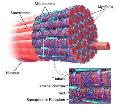
A sarcomere is the smallest functional unit of striated muscle tissue. It is the repeating unit between two Z-lines. Skeletal muscles are composed of tubular muscle cells which are formed during embryonic myogenesis. Muscle fibers contain numerous tubular myofibrils. Myofibrils are composed of repeating sections of sarcomeres, which appear under the microscope as alternating dark and light bands. Sarcomeres are composed of long, fibrous proteins as filaments that slide past each other when a muscle contracts or relaxes. The costamere is a different component that connects the sarcomere to the sarcolemma.

<span class="mw-page-title-main">Sarcoplasmic reticulum</span> Menbrane-bound structure in muscle cells for storing calcium

The sarcoplasmic reticulum (SR) is a membrane-bound structure found within muscle cells that is similar to the smooth endoplasmic reticulum in other cells. The main function of the SR is to store calcium ions (Ca2+). Calcium ion levels are kept relatively constant, with the concentration of calcium ions within a cell being 10,000 times smaller than the concentration of calcium ions outside the cell. This means that small increases in calcium ions within the cell are easily detected and can bring about important cellular changes (the calcium is said to be a second messenger). Calcium is used to make calcium carbonate (found in chalk) and calcium phosphate, two compounds that the body uses to make teeth and bones. This means that too much calcium within the cells can lead to hardening (calcification) of certain intracellular structures, including the mitochondria, leading to cell death. Therefore, it is vital that calcium ion levels are controlled tightly, and can be released into the cell when necessary and then removed from the cell.

<span class="mw-page-title-main">Cardiac muscle</span> Muscular tissue of heart in vertebrates

Cardiac muscle is one of three types of vertebrate muscle tissues, with the other two being skeletal muscle and smooth muscle. It is an involuntary, striated muscle that constitutes the main tissue of the wall of the heart. The cardiac muscle (myocardium) forms a thick middle layer between the outer layer of the heart wall and the inner layer, with blood supplied via the coronary circulation. It is composed of individual cardiac muscle cells joined by intercalated discs, and encased by collagen fibers and other substances that form the extracellular matrix.

<span class="mw-page-title-main">Muscle cell</span> Type of cell found in muscle tissue

A muscle cell, also known as a myocyte, is a mature contractile cell in the muscle of an animal. In humans and other vertebrates there are three types: skeletal, smooth, and cardiac (cardiomyocytes). A skeletal muscle cell is long and threadlike with many nuclei and is called a muscle fiber. Muscle cells develop from embryonic precursor cells called myoblasts.

<span class="mw-page-title-main">Striated muscle tissue</span> Muscle tissue with repeating functional units called sarcomeres

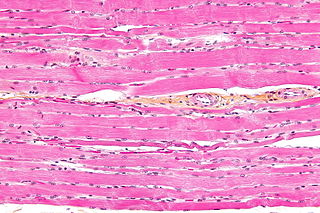
Striated muscle tissue is a muscle tissue that features repeating functional units called sarcomeres. The presence of sarcomeres manifests as a series of bands visible along the muscle fibers, which is responsible for the striated appearance observed in microscopic images of this tissue. There are two types of striated muscle:

<span class="mw-page-title-main">Cardiac action potential</span> Biological process in the heart

Unlike the action potential in skeletal muscle cells, the cardiac action potential is not initiated by nervous activity. Instead, it arises from a group of specialized cells known as pacemaker cells, that have automatic action potential generation capability. In healthy hearts, these cells form the cardiac pacemaker and are found in the sinoatrial node in the right atrium. They produce roughly 60–100 action potentials every minute. The action potential passes along the cell membrane causing the cell to contract, therefore the activity of the sinoatrial node results in a resting heart rate of roughly 60–100 beats per minute. All cardiac muscle cells are electrically linked to one another, by intercalated discs which allow the action potential to pass from one cell to the next. This means that all atrial cells can contract together, and then all ventricular cells.

<span class="mw-page-title-main">Muscle contraction</span> Activation of tension-generating sites in muscle

Muscle contraction is the activation of tension-generating sites within muscle cells. In physiology, muscle contraction does not necessarily mean muscle shortening because muscle tension can be produced without changes in muscle length, such as when holding something heavy in the same position. The termination of muscle contraction is followed by muscle relaxation, which is a return of the muscle fibers to their low tension-generating state.

<span class="mw-page-title-main">Sarcolemma</span> Cell membrane of a muscle fibre

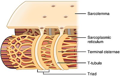
The sarcolemma, also called the myolemma, is the cell membrane surrounding a skeletal muscle fibre or a cardiomyocyte. It consists of a lipid bilayer and a thin outer coat of polysaccharide material (glycocalyx) that contacts the basement membrane. The basement membrane contains numerous thin collagen fibrils and specialized proteins such as laminin that provide a scaffold to which the muscle fibre can adhere. Through transmembrane proteins in the plasma membrane, the actin skeleton inside the cell is connected to the basement membrane and the cell's exterior. At each end of the muscle fibre, the surface layer of the sarcolemma fuses with a tendon fibre, and the tendon fibres, in turn, collect into bundles to form the muscle tendons that adhere to bones.

Ryanodine receptors form a class of intracellular calcium channels in various forms of excitable animal tissue like muscles and neurons. There are three major isoforms of the ryanodine receptor, which are found in different tissues and participate in different signaling pathways involving calcium release from intracellular organelles. The RYR2 ryanodine receptor isoform is the major cellular mediator of calcium-induced calcium release (CICR) in animal cells.

Calcium-induced calcium release (CICR) describes a biological process whereby calcium is able to activate calcium release from intracellular Ca2+ stores (e.g., endoplasmic reticulum or sarcoplasmic reticulum). Although CICR was first proposed for skeletal muscle in the 1970s, it is now known that CICR is unlikely to be the primary mechanism for activating SR calcium release. Instead, CICR is thought to be crucial for excitation-contraction coupling in cardiac muscle. It is now obvious that CICR is a widely occurring cellular signaling process present even in many non-muscle cells, such as in the insulin-secreting pancreatic beta cells, epithelium, and many other cells. Since CICR is a positive-feedback system, it has been of great interest to elucidate the mechanism(s) responsible for its termination.

<span class="mw-page-title-main">Calsequestrin</span> Calcium-binding protein

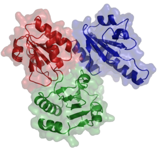
Calsequestrin is a calcium-binding protein that acts as a calcium buffer within the sarcoplasmic reticulum. The protein helps hold calcium in the cisterna of the sarcoplasmic reticulum after a muscle contraction, even though the concentration of calcium in the sarcoplasmic reticulum is much higher than in the cytosol. It also helps the sarcoplasmic reticulum store an extraordinarily high amount of calcium ions. Each molecule of calsequestrin can bind 18 to 50 Ca2+ ions. Sequence analysis has suggested that calcium is not bound in distinct pockets via EF-hand motifs, but rather via presentation of a charged protein surface. Two forms of calsequestrin have been identified. The cardiac form Calsequestrin-2 (CASQ2) is present in cardiac and slow skeletal muscle and the fast skeletal form Calsequestrin-1(CASQ1) is found in fast skeletal muscle. The release of calsequestrin-bound calcium (through a calcium release channel) triggers muscle contraction. The active protein is not highly structured, more than 50% of it adopting a random coil conformation. When calcium binds there is a structural change whereby the alpha-helical content of the protein increases from 3 to 11%. Both forms of calsequestrin are phosphorylated by casein kinase 2, but the cardiac form is phosphorylated more rapidly and to a higher degree. Calsequestrin is also secreted in the gut where it deprives bacteria of calcium ions..

<span class="mw-page-title-main">Catecholaminergic polymorphic ventricular tachycardia</span> Medical condition

Catecholaminergic polymorphic ventricular tachycardia (CPVT) is an inherited genetic disorder that predisposes those affected to potentially life-threatening abnormal heart rhythms or arrhythmias. The arrhythmias seen in CPVT typically occur during exercise or at times of emotional stress, and classically take the form of bidirectional ventricular tachycardia or ventricular fibrillation. Those affected may be asymptomatic, but they may also experience blackouts or even sudden cardiac death.

A calcium spark is the microscopic release of calcium (Ca2+) from a store known as the sarcoplasmic reticulum (SR), located within muscle cells. This release occurs through an ion channel within the membrane of the SR, known as a ryanodine receptor (RyR), which opens upon activation. This process is important as it helps to maintain Ca2+ concentration within the cell. It also initiates muscle contraction in skeletal and cardiac muscles and muscle relaxation in smooth muscles. Ca2+ sparks are important in physiology as they show how Ca2+ can be used at a subcellular level, to signal both local changes, known as local control, as well as whole cell changes.

Within the muscle tissue of animals and humans, contraction and relaxation of the muscle cells (myocytes) is a highly regulated and rhythmic process. In cardiomyocytes, or cardiac muscle cells, muscular contraction takes place due to movement at a structure referred to as the diad, sometimes spelled "dyad." The dyad is the connection of transverse- tubules (t-tubules) and the junctional sarcoplasmic reticulum (jSR). Like skeletal muscle contractions, Calcium (Ca2+) ions are required for polarization and depolarization through a voltage-gated calcium channel. The rapid influx of calcium into the cell signals for the cells to contract. When the calcium intake travels through an entire muscle, it will trigger a united muscular contraction. This process is known as excitation-contraction coupling. This contraction pushes blood inside the heart and from the heart to other regions of the body.

<span class="mw-page-title-main">Calmodulin 1</span> Protein-coding gene in the species Homo sapiens

Calmodulin 1 is a protein in humans that is encoded by the CALM1 gene.

<span class="mw-page-title-main">Ryanodine receptor 2</span> Transport protein and coding gene in humans

Ryanodine receptor 2 (RYR2) is one of a class of ryanodine receptors and a protein found primarily in cardiac muscle. In humans, it is encoded by the RYR2 gene. In the process of cardiac calcium-induced calcium release, RYR2 is the major mediator for sarcoplasmic release of stored calcium ions.

<span class="mw-page-title-main">Terminal cisternae</span> Enlarged areas of the sarcoplasmic reticulum surrounding the transverse tubules

Terminal cisternae are enlarged areas of the sarcoplasmic reticulum surrounding the transverse tubules.

Calcium buffering describes the processes which help stabilise the concentration of free calcium ions within cells, in a similar manner to how pH buffers maintain a stable concentration of hydrogen ions. The majority of calcium ions within the cell are bound to intracellular proteins, leaving a minority freely dissociated. When calcium is added to or removed from the cytoplasm by transport across the cell membrane or sarcoplasmic reticulum, calcium buffers minimise the effect on changes in cytoplasmic free calcium concentration by binding calcium to or releasing calcium from intracellular proteins. As a result, 99% of the calcium added to the cytosol of a cardiomyocyte during each cardiac cycle becomes bound to calcium buffers, creating a relatively small change in free calcium.

The dyadic space is the name for the volume of cytoplasm between pairs (dyads) of areas where the cell membrane and an organelle such as the endoplasmic reticulum come into close contact of each other, creating what are known as dyadic clefts.

Cardiac excitation-contraction coupling (CardiacEC coupling) describes the series of events, from the production of an electrical impulse (action potential) to the contraction of muscles in the heart. This process is of vital importance as it allows for the heart to beat in a controlled manner, without the need for conscious input. EC coupling results in the sequential contraction of the heart muscles that allows blood to be pumped, first to the lungs (pulmonary circulation) and then around the rest of the body (systemic circulation) at a rate between 60 and 100 beats every minute, when the body is at rest. This rate can be altered, however, by nerves that work to either increase heart rate (sympathetic nerves) or decrease it (parasympathetic nerves), as the body's oxygen demands change. Ultimately, muscle contraction revolves around a charged atom (ion), calcium (Ca2+), which is responsible for converting the electrical energy of the action potential into mechanical energy (contraction) of the muscle. This is achieved in a region of the muscle cell, called the transverse tubule during a process known as calcium induced calcium release.

References

  1. 1 2 3 4 5 6 7 Hong, TingTing; Shaw, Robin M. (2017-01-01). "Cardiac T-Tubule Microanatomy and Function". Physiological Reviews. 97 (1): 227–252. doi:10.1152/physrev.00037.2015. ISSN   0031-9333. PMC   6151489 . PMID   27881552.
  2. Rog-Zielinska EA, et al. (2021). "Beat-by-Beat Cardiomyocyte T-Tubule Deformation Drives Tubular Content Exchange". Circ. Res. 128 (2): 203–215. doi: 10.1161/CIRCRESAHA.120.317266 . PMC   7834912 . PMID   33228470.
  3. Ferrantini, Cecilia; Coppini, Raffaele; Sacconi, Leonardo; Tosi, Benedetta; Zhang, Mei Luo; Wang, Guo Liang; Vries, Ewout de; Hoppenbrouwers, Ernst; Pavone, Francesco (2014-06-01). "Impact of detubulation on force and kinetics of cardiac muscle contraction". The Journal of General Physiology. 143 (6): 783–797. doi:10.1085/jgp.201311125. PMC   4035744 . PMID   24863933.
  4. Haddock, Peter S.; Coetzee, William A.; Cho, Emily; Porter, Lisa; Katoh, Hideki; Bers, Donald M.; Jafri, M. Saleet; Artman, Michael (1999-09-03). "Subcellular [Ca2+]i Gradients During Excitation-Contraction Coupling in Newborn Rabbit Ventricular Myocytes". Circulation Research. 85 (5): 415–427. doi: 10.1161/01.RES.85.5.415 . ISSN   0009-7330. PMID   10473671.
  5. Richards, M. A.; Clarke, J. D.; Saravanan, P.; Voigt, N.; Dobrev, D.; Eisner, D. A.; Trafford, A. W.; Dibb, K. M. (November 2011). "Transverse tubules are a common feature in large mammalian atrial myocytes including human". American Journal of Physiology. Heart and Circulatory Physiology. 301 (5): H1996–2005. doi:10.1152/ajpheart.00284.2011. ISSN   1522-1539. PMC   3213978 . PMID   21841013.
  6. 1 2 3 4 5 6 7 Ibrahim, M.; Gorelik, J.; Yacoub, M. H.; Terracciano, C. M. (2011-09-22). "The structure and function of cardiac t-tubules in health and disease". Proceedings of the Royal Society B: Biological Sciences. 278 (1719): 2714–2723. doi:10.1098/rspb.2011.0624. PMC   3145195 . PMID   21697171.
  7. di Fiore, Mariano SH; Eroschenko, Victor P (2008). Di Fiore's Atlas of histology: with functional correlations. Philadelphia: Wolters Kluwer Health/Lippincott Williams & Wilkins. p. 124. ISBN   978-0-7817-7057-6.
  8. "4. Calcium reuptake and relaxation". www.bristol.ac.uk. Retrieved 2017-02-21.
  9. Caldwell, Jessica L.; Smith, Charlotte E. R.; Taylor, Rebecca F.; Kitmitto, Ashraf; Eisner, David A.; Dibb, Katharine M.; Trafford, Andrew W. (2014-12-05). "Dependence of cardiac transverse tubules on the BAR domain protein amphiphysin II (BIN-1)". Circulation Research. 115 (12): 986–996. doi:10.1161/CIRCRESAHA.116.303448. ISSN   1524-4571. PMC   4274343 . PMID   25332206.
  10. 1 2 3 M., Bers, D. (2001). Excitation-contraction coupling and cardiac contractile force (2nd ed.). Dordrecht: Kluwer Academic Publishers. ISBN   9780792371588. OCLC   47659382.{{cite book}}: CS1 maint: multiple names: authors list (link)
  11. Scriven, D. R.; Dan, P.; Moore, E. D. (November 2000). "Distribution of proteins implicated in excitation-contraction coupling in rat ventricular myocytes". Biophysical Journal. 79 (5): 2682–2691. Bibcode:2000BpJ....79.2682S. doi:10.1016/S0006-3495(00)76506-4. ISSN   0006-3495. PMC   1301148 . PMID   11053140.
  12. Bers, Donald M. (2002-01-10). "Cardiac excitation-contraction coupling". Nature. 415 (6868): 198–205. Bibcode:2002Natur.415..198B. doi:10.1038/415198a. ISSN   0028-0836. PMID   11805843. S2CID   4337201.
  13. Rebbeck, Robyn T.; Karunasekara, Yamuna; Board, Philip G.; Beard, Nicole A.; Casarotto, Marco G.; Dulhunty, Angela F. (2014-03-01). "Skeletal muscle excitation-contraction coupling: who are the dancing partners?". The International Journal of Biochemistry & Cell Biology. 48: 28–38. doi:10.1016/j.biocel.2013.12.001. ISSN   1878-5875. PMID   24374102.
  14. 1 2 3 Ferrantini, Cecilia; Coppini, Raffaele; Sacconi, Leonardo; Tosi, Benedetta; Zhang, Mei Luo; Wang, Guo Liang; de Vries, Ewout; Hoppenbrouwers, Ernst; Pavone, Francesco (2014-06-01). "Impact of detubulation on force and kinetics of cardiac muscle contraction". The Journal of General Physiology. 143 (6): 783–797. doi:10.1085/jgp.201311125. ISSN   1540-7748. PMC   4035744 . PMID   24863933.
  15. Laflamme, M. A.; Becker, P. L. (1999-11-01). "G(s) and adenylyl cyclase in transverse tubules of heart: implications for cAMP-dependent signaling". The American Journal of Physiology. 277 (5 Pt 2): H1841–1848. doi:10.1152/ajpheart.1999.277.5.H1841. ISSN   0002-9513. PMID   10564138.
  16. Bers, Donald M. (2006-05-15). "Cardiac ryanodine receptor phosphorylation: target sites and functional consequences". Biochemical Journal. 396 (Pt 1): e1–3. doi:10.1042/BJ20060377. ISSN   0264-6021. PMC   1450001 . PMID   16626281.
  17. Hinch, R., Greenstein, J.L., Tanskanen, A.J., Xu, L. and Winslow, R.L. (2004) ‘A simplified local control model of calcium-induced calcium release in cardiac ventricular Myocytes’, 87(6).
  18. Fraser, James A.; Skepper, Jeremy N.; Hockaday, Austin R.; Huang1, Christopher L.-H. (1998-08-01). "The tubular vacuolation process in amphibian skeletal muscle". Journal of Muscle Research & Cell Motility. 19 (6): 613–629. doi:10.1023/A:1005325013355. ISSN   0142-4319. PMID   9742446. S2CID   12312117.{{cite journal}}: CS1 maint: numeric names: authors list (link)
  19. Kawai M, Hussain M, Orchard CH (1999). "Excitation-contraction coupling in rat ventricular myocytes after formamide-induced detubulation". Am J Physiol. 277 (2): H603-9. doi:10.1152/ajpheart.1999.277.2.H603. PMID   10444485.
  20. Moench, I.; Meekhof, K. E.; Cheng, L. F.; Lopatin, A. N. (July 2013). "Resolution of hyposmotic stress in isolated mouse ventricular myocytes causes sealing of t-tubules". Experimental Physiology. 98 (7): 1164–1177. doi:10.1113/expphysiol.2013.072470. ISSN   1469-445X. PMC   3746342 . PMID   23585327.
  21. Huxley, A. F. (1971-06-15). "The activation of striated muscle and its mechanical response". Proceedings of the Royal Society of London. Series B, Biological Sciences. 178 (1050): 1–27. doi:10.1098/rspb.1971.0049. ISSN   0950-1193. PMID   4397265. S2CID   30218942.
  22. Hill, A. V. (October 1949). "The abrupt transition from rest to activity in muscle". Proceedings of the Royal Society of London. Series B, Biological Sciences. 136 (884): 399–420. Bibcode:1949RSPSB.136..399H. doi:10.1098/rspb.1949.0033. ISSN   0950-1193. PMID   18143369. S2CID   11863605.
  23. Lindner, E. (1957). "[Submicroscopic morphology of the cardiac muscle]". Zeitschrift für Zellforschung und Mikroskopische Anatomie. 45 (6): 702–746. ISSN   0340-0336. PMID   13456982.
  24. Sperelakis, N.; Rubio, R. (August 1971). "An orderly lattice of axial tubules which interconnect adjacent transverse tubules in guinea-pig ventricular myocardium". Journal of Molecular and Cellular Cardiology. 2 (3): 211–220. doi:10.1016/0022-2828(71)90054-x. ISSN   0022-2828. PMID   5117216.
  25. Savio-Galimberti, Eleonora; Frank, Joy; Inoue, Masashi; Goldhaber, Joshua I.; Cannell, Mark B.; Bridge, John H. B.; Sachse, Frank B. (August 2008). "Novel features of the rabbit transverse tubular system revealed by quantitative analysis of three-dimensional reconstructions from confocal images". Biophysical Journal. 95 (4): 2053–2062. Bibcode:2008BpJ....95.2053S. doi:10.1529/biophysj.108.130617. ISSN   1542-0086. PMC   2483780 . PMID   18487298.
  26. Cheng, H.; Lederer, W. J.; Cannell, M. B. (1993-10-29). "Calcium sparks: elementary events underlying excitation-contraction coupling in heart muscle". Science. 262 (5134): 740–744. Bibcode:1993Sci...262..740C. doi:10.1126/science.8235594. ISSN   0036-8075. PMID   8235594.
  27. 1 2 3 Dibb, Katharine M.; Clarke, Jessica D.; Horn, Margaux A.; Richards, Mark A.; Graham, Helen K.; Eisner, David A.; Trafford, Andrew W. (September 2009). "Characterization of an extensive transverse tubular network in sheep atrial myocytes and its depletion in heart failure". Circulation: Heart Failure. 2 (5): 482–489. doi: 10.1161/CIRCHEARTFAILURE.109.852228 . ISSN   1941-3297. PMID   19808379.
  28. Eisner, David A.; Caldwell, Jessica L.; Kistamás, Kornél; Trafford, Andrew W. (2017-07-07). "Calcium and Excitation-Contraction Coupling in the Heart". Circulation Research. 121 (2): 181–195. doi:10.1161/CIRCRESAHA.117.310230. ISSN   1524-4571. PMC   5497788 . PMID   28684623.
  29. 1 2 Pinali, Christian; Malik, Nadim; Davenport, J. Bernard; Allan, Laurence J.; Murfitt, Lucy; Iqbal, Mohammad M.; Boyett, Mark R.; Wright, Elizabeth J.; Walker, Rachel (2017-05-04). "Post-Myocardial Infarction T-tubules Form Enlarged Branched Structures With Dysregulation of Junctophilin-2 and Bridging Integrator 1 (BIN-1)". Journal of the American Heart Association. 6 (5). doi:10.1161/JAHA.116.004834. ISSN   2047-9980. PMC   5524063 . PMID   28473402.
  30. Seidel, Thomas; Navankasattusas, Sutip; Ahmad, Azmi; Diakos, Nikolaos A.; Xu, Weining David; Tristani-Firouzi, Martin; Bonios, Michael J.; Taleb, Iosif; Li, Dean Y. (2017-04-25). "Sheet-Like Remodeling of the Transverse Tubular System in Human Heart Failure Impairs Excitation-Contraction Coupling and Functional Recovery by Mechanical Unloading". Circulation. 135 (17): 1632–1645. doi:10.1161/CIRCULATIONAHA.116.024470. ISSN   1524-4539. PMC   5404964 . PMID   28073805.