Related Research Articles

A motor neuron is a neuron whose cell body is located in the motor cortex, brainstem or the spinal cord, and whose axon (fiber) projects to the spinal cord or outside of the spinal cord to directly or indirectly control effector organs, mainly muscles and glands. There are two types of motor neuron – upper motor neurons and lower motor neurons. Axons from upper motor neurons synapse onto interneurons in the spinal cord and occasionally directly onto lower motor neurons. The axons from the lower motor neurons are efferent nerve fibers that carry signals from the spinal cord to the effectors. Types of lower motor neurons are alpha motor neurons, beta motor neurons, and gamma motor neurons.
The muscular system is an organ system consisting of skeletal, smooth, and cardiac muscle. It permits movement of the body, maintains posture, and circulates blood throughout the body. The muscular systems in vertebrates are controlled through the nervous system although some muscles can be completely autonomous. Together with the skeletal system in the human, it forms the musculoskeletal system, which is responsible for the movement of the body.

Smooth muscle is an involuntary non-striated muscle, so-called because it has no sarcomeres and therefore no striations. It is divided into two subgroups, single-unit and multiunit smooth muscle. Within single-unit muscle, the whole bundle or sheet of smooth muscle cells contracts as a syncytium.

Skeletal muscles are organs of the vertebrate muscular system and typically are attached by tendons to bones of a skeleton. The muscle cells of skeletal muscles are much longer than in the other types of muscle tissue, and are often known as muscle fibers. The muscle tissue of a skeletal muscle is striated – having a striped appearance due to the arrangement of the sarcomeres.
Weakness is a symptom of many different medical conditions. The causes are many and can be divided into conditions that have true or perceived muscle weakness. True muscle weakness is a primary symptom of a variety of skeletal muscle diseases, including muscular dystrophy and inflammatory myopathy. It occurs in neuromuscular junction disorders, such as myasthenia gravis.

Muscle spindles are stretch receptors within the body of a skeletal muscle that primarily detect changes in the length of the muscle. They convey length information to the central nervous system via afferent nerve fibers. This information can be processed by the brain as proprioception. The responses of muscle spindles to changes in length also play an important role in regulating the contraction of muscles, for example, by activating motor neurons via the stretch reflex to resist muscle stretch.
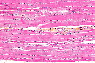
Striated muscle tissue is a muscle tissue that features repeating functional units called sarcomeres. The presence of sarcomeres manifests as a series of bands visible along the muscle fibers, which is responsible for the striated appearance observed in microscopic images of this tissue. There are two types of striated muscle:
Muscle fatigue is when muscles that were initially generating a normal amount of force, then experience a declining ability to generate force. It can be a result of vigorous exercise, but abnormal fatigue may be caused by barriers to or interference with the different stages of muscle contraction. There are two main causes of muscle fatigue: the limitations of a nerve’s ability to generate a sustained signal ; and the reduced ability of the muscle fiber to contract.
Muscle contraction is the activation of tension-generating sites within muscle cells. In physiology, muscle contraction does not necessarily mean muscle shortening because muscle tension can be produced without changes in muscle length, such as when holding something heavy in the same position. The termination of muscle contraction is followed by muscle relaxation, which is a return of the muscle fibers to their low tension-generating state.

Motor unit recruitment is the activation of additional motor units to accomplish an increase in contractile strength in a muscle. A motor unit consists of one motor neuron and all of the muscle fibers it stimulates. All muscles consist of a number of motor units and the fibers belonging to a motor unit are dispersed and intermingle amongst fibers of other units. The muscle fibers belonging to one motor unit can be spread throughout part, or most of the entire muscle, depending on the number of fibers and size of the muscle. When a motor neuron is activated, all of the muscle fibers innervated by the motor neuron are stimulated and contract. The activation of one motor neuron will result in a weak but distributed muscle contraction. The activation of more motor neurons will result in more muscle fibers being activated, and therefore a stronger muscle contraction. Motor unit recruitment is a measure of how many motor neurons are activated in a particular muscle, and therefore is a measure of how many muscle fibers of that muscle are activated. The higher the recruitment the stronger the muscle contraction will be. Motor units are generally recruited in order of smallest to largest as contraction increases. This is known as Henneman's size principle.
Muscle weakness is a lack of muscle strength. Its causes are many and can be divided into conditions that have either true or perceived muscle weakness. True muscle weakness is a primary symptom of a variety of skeletal muscle diseases, including muscular dystrophy and inflammatory myopathy. It occurs in neuromuscular junction disorders, such as myasthenia gravis. Muscle weakness can also be caused by low levels of potassium and other electrolytes within muscle cells. It can be temporary or long-lasting. The term myasthenia is from my- from Greek μυο meaning "muscle" + -asthenia ἀσθένεια meaning "weakness".

Muscle is a soft tissue, one of the four basic types of animal tissue. Muscle tissue gives skeletal muscles the ability to contract. Muscle is formed during embryonic development, in a process known as myogenesis. Muscle tissue contains special contractile proteins called actin and myosin which interact to cause movement. Among many other muscle proteins present are two regulatory proteins, troponin and tropomyosin.
Myomeres are blocks of skeletal muscle tissue arranged in sequence, commonly found in aquatic chordates. Myomeres are separated from adjacent myomeres by connective fascia (myosepta) and most easily seen in larval fishes or in the olm. Myomere counts are sometimes used for identifying specimens, since their number corresponds to the number of vertebrae in the adults. Location varies, with some species containing these only near the tails, while some have them located near the scapular or pelvic girdles. Depending on the species, myomeres could be arranged in an epaxial or hypaxial manner. Hypaxial refers to ventral muscles and related structures while epaxial refers to more dorsal muscles. The horizontal septum divides these two regions in vertebrates from cyclostomes to gnathostomes. In terrestrial chordates, the myomeres become fused as well as indistinct, due to the disappearance of myosepta.

Myosin-2 is a protein that in humans is encoded by the MYH2 gene.
A motor pool consists of all individual motor neurons that innervate a single muscle. Each individual muscle fiber is innervated by only one motor neuron, but one motor neuron may innervate several muscle fibers. This distinction is physiologically significant because the size of a given motor pool determines the activity of the muscle it innervates: for example, muscles responsible for finer movements are innervated by motor pools consisting of higher numbers of individual motor neurons. Motor pools are also distinguished by the different classes of motor neurons that they contain. The size, composition, and anatomical location of each motor pool is tightly controlled by complex developmental pathways.
The motor unit consists of a voluntary alpha motoneuron and all of the collective muscle fibers that it controls, known as the effector muscle. The alpha motoneuron communicates with acetylcholine receptors on the motor end plate of the effector muscle. Reception of acetylcholine neurotransmitters on the motor end plate causes contraction of that effector muscle.
Normal aging movement control in humans is about the changes in the muscles, motor neurons, nerves, sensory functions, gait, fatigue, visual and manual responses, in men and women as they get older but who do not have neurological, muscular or neuromuscular disorder. With aging, neuromuscular movements are impaired, though with training or practice, some aspects may be prevented.
Muscle architecture is the physical arrangement of muscle fibers at the macroscopic level that determines a muscle’s mechanical function. There are several different muscle architecture types including: parallel, pennate and hydrostats. Force production and gearing vary depending on the different muscle parameters such as muscle length, fiber length, pennation angle, and the physiological cross-sectional area (PCSA).
Henneman’s size principle describes relationships between properties of motor neurons and the muscle fibers they innervate and thus control, which together are called motor units. Motor neurons with large cell bodies tend to innervate fast-twitch, high-force, less fatigue-resistant muscle fibers, whereas motor neurons with small cell bodies tend to innervate slow-twitch, low-force, fatigue-resistant muscle fibers. In order to contract a particular muscle, motor neurons with small cell bodies are recruited before motor neurons with large cell bodies. It was proposed by Elwood Henneman.
Even before the very beginning of human space exploration, serious and reasonable concerns were expressed about exposure of humans to the microgravity of space due to the potential systemic effects on terrestrially-evolved life forms adapted to Earth gravity. Unloading of skeletal muscle, both on Earth via bed-rest experiments and during spaceflight, result in remodeling of muscle. As a result, decrements occur in skeletal muscle strength, fatigue resistance, motor performance, and connective tissue integrity. In addition, there are cardiopulmonary and vascular changes, including a significant decrease in red blood cell mass, that affect skeletal muscle function. This normal adaptive response to the microgravity environment may become a liability resulting in increased risk of an inability or decreased efficiency in crewmember performance of physically demanding tasks during extravehicular activity (EVA) or upon return to Earth.
References
- Altshuler, Douglas; K. Welch; B. Cho; D. Welch; A. Lin; W. Dickinson; M. Dickinson (April 2010). "Neuromuscular control of wingbeat kinematics in Annas hummingbirds". The Journal of Experimental Biology. 213 (Pt 14): 2507–2514. doi:10.1242/jeb.043497. PMC 2892424 . PMID 20581280.
- 1 2 Buchtal, F; H. Schmalbruch (1 January 1980). "Motor Unit of Mammalian Muscle". Physiological Reviews. 60 (1): 90–142. doi:10.1152/physrev.1980.60.1.90. PMID 6766557.
- ↑ Kandel, Eric (2013). Principles of Neural Science, 5th ed. McGraw-Hill, New York. p. 768. ISBN 978-0-07-139011-8.
- ↑ Milner-Brown HS, Stein RB, Yemm R (September 1973). "The orderly recruitment of human motor units during voluntary isometric contractions". J. Physiol. 230 (2): 359–70. doi:10.1113/jphysiol.1973.sp010192. PMC 1350367 . PMID 4350770.
- ↑ Robinson R (February 2009). "In mammalian muscle, axonal wiring takes surprising paths". PLOS Biol. 7 (2): e1000050. doi: 10.1371/journal.pbio.1000050 . PMC 2637923 . PMID 20076726.
- ↑ Farina, Dario; Merletti R; Enoka R.M. (2004). "The extraction of neural strategies from the surface EMG". Journal of Applied Physiology. 96 (4): 1486–1495. doi:10.1152/japplphysiol.01070.2003. PMID 15016793.
- ↑ Spiegel KM.; Stratton J.; Burke JR.; Glendinning DS; Enoka RM (November 2012). "The influence of age on the assessment of motor unit activation in a human hand muscle". Experimental Physiology. 81 (5): 805–819. doi: 10.1113/expphysiol.1996.sp003978 . PMID 8889479. S2CID 29034955.
- ↑
 This article incorporates text available under the CC BY 4.0 license.Betts, J Gordon; Desaix, Peter; Johnson, Eddie; Johnson, Jody E; Korol, Oksana; Kruse, Dean; Poe, Brandon; Wise, James; Womble, Mark D; Young, Kelly A (May 14, 2023). Anatomy & Physiology. Houston: OpenStax CNX. 10.3 Muscle Fiber Contraction and Relaxation. ISBN 978-1-947172-04-3.
 - ↑ De Luca, Carlo; William J. Forrest (December 1972). "Some Properties of Motor Unit Action Potential Trains Recorded during Constant Force Isometric Contractions in Man". Kybernetik. 12 (3): 160–168. doi:10.1007/bf00289169. PMID 4712973. S2CID 11373497.
- 1 2 Burke RE, Levine DN, Tsairis P, Zajac FE (November 1973). "Physiological types and histochemical profiles in motor units of the cat gastrocnemius". J. Physiol. 234 (3): 723–48. doi:10.1113/jphysiol.1973.sp010369. PMC 1350696 . PMID 4148752.
- ↑ Collatos TC, Edgerton VR, Smith JL, Botterman BR (November 1977). "Contractile properties and fiber type compositions of flexors and extensors of elbow joint in cat: implications for motor control". J. Neurophysiol. 40 (6): 1292–300. doi:10.1152/jn.1977.40.6.1292. PMID 925731.
- ↑ Altshuler D.; Welch K.; Cho B.; Welch D.; Lin A.; Dickinson W.; Dickinson M. (April 2010). "Neuromuscular control of wingbeat kinematics in Annas hummingbirds". The Journal of Experimental Biology. 213 (Pt 14): 2507–2514. doi:10.1242/jeb.043497. PMC 2892424 . PMID 20581280.
- 1 2 Schiaffino S, Reggiani C (August 1994). "Myosin isoforms in mammalian skeletal muscle". J. Appl. Physiol. 77 (2): 493–501. doi:10.1152/jappl.1994.77.2.493. PMID 8002492.
- 1 2 Caiozzo VJ, Baker MJ, Huang K, Chou H, Wu YZ, Baldwin KM (September 2003). "Single-fiber myosin heavy chain polymorphism: how many patterns and what proportions?". Am. J. Physiol. Regul. Integr. Comp. Physiol. 285 (3): R570–80. doi:10.1152/ajpregu.00646.2002. PMID 12738613. S2CID 860317.
- ↑ Baldwin KM, Haddad F (January 2001). "Effects of different activity and inactivity paradigms on myosin heavy chain gene expression in striated muscle". J. Appl. Physiol. 90 (1): 345–57. doi:10.1152/jappl.2001.90.1.345. PMID 11133928. S2CID 9677583.
- 1 2 3 4 Feinstein, Bertram; Lindegård, Bengt; Nyman, Eberhard; Wohlfart, Gunnar (2008-06-18). "Morphologic Studies of Motor Units in Normal Human Muscles". Acta Anatomica. 23 (2): 127–142. doi:10.1159/000140989. ISSN 0001-5180. PMID 14349537.
- ↑ Karpati, George (2010). Disorders of Voluntary Muscle (PDF). Cambridge University Press. p. 7. ISBN 9780521876292. referenced Feinstein, B; Lindegard, B; Nyman, E; Wohlfart, G (1955). "Morphologic studies of motor units in normal human muscles". Acta Anat (Basel). 23 (2): 127–142. doi:10.1159/000140989. PMID 14349537.