
A motor neuron is a neuron whose cell body is located in the motor cortex, brainstem or the spinal cord, and whose axon (fiber) projects to the spinal cord or outside of the spinal cord to directly or indirectly control effector organs, mainly muscles and glands. There are two types of motor neuron – upper motor neurons and lower motor neurons. Axons from upper motor neurons synapse onto interneurons in the spinal cord and occasionally directly onto lower motor neurons. The axons from the lower motor neurons are efferent nerve fibers that carry signals from the spinal cord to the effectors. Types of lower motor neurons are alpha motor neurons, beta motor neurons, and gamma motor neurons.
A motor nerve is a nerve that transmits motor signals from the central nervous system (CNS) to the muscles of the body. This is different from the motor neuron, which includes a cell body and branching of dendrites, while the nerve is made up of a bundle of axons. Motor nerves act as efferent nerves which carry information out from the CNS to muscles, as opposed to afferent nerves, which transfer signals from sensory receptors in the periphery to the CNS. Efferent nerves can also connect to glands or other organs/issues instead of muscles. The vast majority of nerves contain both sensory and motor fibers and are therefore called mixed nerves.

The somatic nervous system (SNS) is made up of nerves that link the brain and spinal cord to voluntary or skeletal muscles that are under conscious control as well as to skin sensory receptors. Specialized nerve fiber ends called sensory receptors are responsible for detecting information within and outside of the body.

Muscle spindles are stretch receptors within the body of a skeletal muscle that primarily detect changes in the length of the muscle. They convey length information to the central nervous system via afferent nerve fibers. This information can be processed by the brain as proprioception. The responses of muscle spindles to changes in length also play an important role in regulating the contraction of muscles, for example, by activating motor neurons via the stretch reflex to resist muscle stretch.

Afferent nerve fibers are axons of sensory neurons that carry sensory information from sensory receptors to the central nervous system. Many afferent projections arrive at a particular brain region.
Efferent nerve fibers refer to axonal projections that exit a particular region; as opposed to afferent projections that arrive at the region. These terms have a slightly different meaning in the context of the peripheral nervous system (PNS) and central nervous system (CNS). The efferent fiber is a long process projecting far from the neuron's body that carries nerve impulses away from the central nervous system toward the peripheral effector organs. A bundle of these fibers is called an efferent nerve. The opposite direction of neural activity is afferent conduction, which carries impulses by way of the afferent nerve fibers of sensory neurons.

The grey column refers to a somewhat ridge-shaped mass of grey matter in the spinal cord. This presents as three columns: the anterior grey column, the posterior grey column, and the lateral grey column, all of which are visible in cross-section of the spinal cord.
A nuclear bag fiber is a type of intrafusal muscle fiber that lies in the center of a muscle spindle. Each has many nuclei concentrated in bags and they cause excitation of the primary sensory fibers.
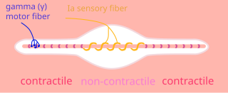
A nuclear chain fiber is a specialized sensory organ contained within a muscle. Nuclear chain fibers are intrafusal fibers that, along with nuclear bag fibers, make up the muscle spindle responsible for the detection of changes in muscle length.
A type Ia sensory fiber, or a primary afferent fiber is a type of afferent nerve fiber. It is the sensory fiber of a stretch receptor called the muscle spindle found in muscles, which constantly monitors the rate at which a muscle stretch changes. The information carried by type Ia fibers contributes to the sense of proprioception.

The patellar reflex, also called the knee reflex or knee-jerk, is a stretch reflex which tests the L2, L3, and L4 segments of the spinal cord. Many animals, most significantly humans, have been seen to have the patellar reflex, including dogs, cats, horses, and other mammalian species.
Lower motor neurons (LMNs) are motor neurons located in either the anterior grey column, anterior nerve roots or the cranial nerve nuclei of the brainstem and cranial nerves with motor function. Many voluntary movements rely on spinal lower motor neurons, which innervate skeletal muscle fibers and act as a link between upper motor neurons and muscles. Cranial nerve lower motor neurons also control some voluntary movements of the eyes, face and tongue, and contribute to chewing, swallowing and vocalization. Damage to the lower motor neurons can lead to flaccid paralysis, absent deep tendon reflexes and muscle atrophy.
A gamma motor neuron, also called gamma motoneuron, or fusimotor neuron, is a type of lower motor neuron that takes part in the process of muscle contraction, and represents about 30% of (Aγ) fibers going to the muscle. Like alpha motor neurons, their cell bodies are located in the anterior grey column of the spinal cord. They receive input from the reticular formation of the pons in the brainstem. Their axons are smaller than those of the alpha motor neurons, with a diameter of only 5 μm. Unlike the alpha motor neurons, gamma motor neurons do not directly adjust the lengthening or shortening of muscles. However, their role is important in keeping muscle spindles taut, thereby allowing the continued firing of alpha neurons, leading to muscle contraction. These neurons also play a role in adjusting the sensitivity of muscle spindles.
Extrafusal muscle fibers are the standard skeletal muscle fibers that are innervated by alpha motor neurons and generate tension by contracting, thereby allowing for skeletal movement. They make up the large mass of skeletal striated muscle tissue and are attached to bone by fibrous tissue extensions (tendons).

The stretch reflex, or more accurately "muscle stretch reflex", is a muscle contraction in response to stretching a muscle. The function of the reflex is generally thought to be maintaining the muscle at a constant length but the response is often coordinated across multiple muscles and even joints. The older term deep tendon reflex is now criticized as misleading. Tendons have little to do with the response, and some muscles with stretch reflexes have no tendons. Rather, muscle spindles detect a stretch and convey the information to the central nervous system.

Alpha (α) motor neurons (also called alpha motoneurons), are large, multipolar lower motor neurons of the brainstem and spinal cord. They innervate extrafusal muscle fibers of skeletal muscle and are directly responsible for initiating their contraction. Alpha motor neurons are distinct from gamma motor neurons, which innervate intrafusal muscle fibers of muscle spindles.

The general (spinal) somatic efferent neurons arise from motor neuron cell bodies in the ventral horns of the gray matter within the spinal cord. They exit the spinal cord through the ventral roots, carrying motor impulses to skeletal muscle through a neuromuscular junction.
The Golgi tendon reflex (also called inverse stretch reflex, autogenic inhibition, tendon reflex) is an inhibitory effect on the muscle resulting from the muscle tension stimulating Golgi tendon organs (GTO) of the muscle, and hence it is self-induced. The reflex arc is a negative feedback mechanism preventing too much tension on the muscle and tendon. When the tension is extreme, the inhibition can be so great it overcomes the excitatory effects on the muscle's alpha motoneurons causing the muscle to suddenly relax. This reflex is also called the inverse myotatic reflex, because it is the inverse of the stretch reflex.
A motor pool consists of all individual motor neurons that innervate a single muscle. Each individual muscle fiber is innervated by only one motor neuron, but one motor neuron may innervate several muscle fibers. This distinction is physiologically significant because the size of a given motor pool determines the activity of the muscle it innervates: for example, muscles responsible for finer movements are innervated by motor pools consisting of higher numbers of individual motor neurons. Motor pools are also distinguished by the different classes of motor neurons that they contain. The size, composition, and anatomical location of each motor pool is tightly controlled by complex developmental pathways.
Beta motor neurons, also called beta motoneurons, are a kind of lower motor neuron, along with alpha motor neurons and gamma motor neurons. Beta motor neurons innervate intrafusal fibers of muscle spindles with collaterals to extrafusal fibers - a type of slow twitch fiber. Also, axons of alpha, beta, and gamma motor neurons become myelinated. Moreover, these efferent neurons originate from the anterior grey column of the spinal cord and travel to skeletal muscles. However, the larger diameter alpha motor fibers require higher conduction velocity than beta and gamma.
