MHC class I

Last updated
MHC class I
MHC Class 1.svg
Schematic representation of MHC class I
Identifiers
SymbolMHC class I
Membranome 63

MHC class I molecules are one of two primary classes of major histocompatibility complex (MHC) molecules (the other being MHC class II) and are found on the cell surface of all nucleated cells in the bodies of vertebrates. [1] [2] They also occur on platelets, but not on red blood cells. Their function is to display peptide fragments of proteins from within the cell to cytotoxic T cells; this will trigger an immediate response from the immune system against a particular non-self antigen displayed with the help of an MHC class I protein. Because MHC class I molecules present peptides derived from cytosolic proteins, the pathway of MHC class I presentation is often called cytosolic or endogenous pathway. [3]

Contents

In humans, the HLAs corresponding to MHC class I are HLA-A, HLA-B, and HLA-C.

Function

Class I MHC molecules bind peptides generated mainly from degradation of cytosolic proteins by the proteasome. The MHC I:peptide complex is then inserted via endoplasmic reticulum into the external plasma membrane of the cell. The epitope peptide is bound on extracellular parts of the class I MHC molecule. Thus, the function of the class I MHC is to display intracellular proteins to cytotoxic T cells (CTLs). However, class I MHC can also present peptides generated from exogenous proteins, in a process known as cross-presentation.

A normal cell will display peptides from normal cellular protein turnover on its class I MHC, and CTLs will not be activated in response to them due to central and peripheral tolerance mechanisms. When a cell expresses foreign proteins, such as after viral infection, a fraction of the class I MHC will display these peptides on the cell surface. Consequently, CTLs specific for the MHC:peptide complex will recognize and kill presenting cells.

Alternatively, class I MHC itself can serve as an inhibitory ligand for natural killer cells (NKs). Reduction in the normal levels of surface class I MHC, a mechanism employed by some viruses [4] and certain tumors to evade CTL responses, activates NK cell killing.

PirB and visual plasticity

Paired-immunoglobulin-like receptor B (PirB), an MHCI-binding receptor, is involved in the regulation of visual plasticity. [5] PirB is expressed in the central nervous system and diminishes ocular dominance plasticity in the developmental critical period and adulthood. [5] When the function of PirB was abolished in mutant mice, ocular dominance plasticity became more pronounced at all ages. [5] PirB loss of function mutant mice also exhibited enhanced plasticity after monocular deprivation during the critical period. [5] These results suggest PirB may be involved in modulation of synaptic plasticity in the visual cortex.

Structure

MHC class I molecules are heterodimers that consist of two polypeptide chains, α and β2-microglobulin (B2M). The two chains are linked noncovalently via interaction of B2M and the α3 domain. Only the α chain is polymorphic and encoded by a HLA gene, while the B2M subunit is not polymorphic and encoded by the Beta-2 microglobulin gene. The α3 domain is plasma membrane-spanning and interacts with the CD8 co-receptor of T-cells. The α3-CD8 interaction holds the MHC I molecule in place while the T cell receptor (TCR) on the surface of the cytotoxic T cell binds its α12 heterodimer ligand, and checks the coupled peptide for antigenicity. The α1 and α2 domains fold to make up a groove for peptides to bind. MHC class I molecules bind peptides that are predominantly 8-10 amino acid in length (Parham 87), but the binding of longer peptides have also been reported. [6]

While a high-affinity peptide and the B2M subunit are normally required to maintain a stable ternary complex between the peptide, MHC I, and B2M, under subphysiological temperatures, stable, peptide-deficient MHC I/B2M heterodimers have been observed. [7] [8] Synthetic stable, peptide-receptive MHC I molecules have been generated using a disulfide bond between the MHC I and B2M, named "open MHC-I". [9]

Synthesis

Simplified diagram of cytoplasmic protein degradation by the proteasome, transport into endoplasmic reticulum by TAP complex, loading on MHC class I, and transport to the surface for presentation MHC Class I processing.svg
Simplified diagram of cytoplasmic protein degradation by the proteasome, transport into endoplasmic reticulum by TAP complex, loading on MHC class I, and transport to the surface for presentation

The peptides are generated mainly in the cytosol by the proteasome. The proteasome is a macromolecule that consists of 28 subunits, of which half affect proteolytic activity. The proteasome degrades intracellular proteins into small peptides that are then released into the cytosol. Proteasomes can also ligate distinct peptide fragments (termed spliced peptides), producing sequences that are noncontiguous and therefore not linearly templated in the genome. The origin of spliced peptide segments can be from the same protein (cis-splicing) or different proteins (trans-splicing). [10] [11] The peptides have to be translocated from the cytosol into the endoplasmic reticulum (ER) to meet the MHC class I molecule, whose peptide-binding site is in the lumen of the ER. They have membrane proximal Ig fold

Translocation and peptide loading

The peptide translocation from the cytosol into the lumen of the ER is accomplished by the transporter associated with antigen processing (TAP). TAP is a member of the ABC transporter family and is a heterodimeric multimembrane-spanning polypeptide consisting of TAP1 and TAP2. The two subunits form a peptide binding site and two ATP binding sites that face the cytosol. TAP binds peptides on the cytoplasmic side and translocates them under ATP consumption into the lumen of the ER. The MHC class I molecule is then, in turn, loaded with peptides in the lumen of the ER.

The peptide-loading process involves several other molecules that form a large multimeric complex called the Peptide loading complex [12] consisting of TAP, tapasin, calreticulin, calnexin, and Erp57 (PDIA3). Calnexin acts to stabilize the class I MHC α chains prior to β2m binding. Following complete assembly of the MHC molecule, calnexin dissociates. The MHC molecule lacking a bound peptide is inherently unstable and requires the binding of the chaperones calreticulin and Erp57. Additionally, tapasin binds to the MHC molecule and serves to link it to the TAP proteins and facilitates the selection of peptide in an iterative process called peptide editing, [13] [14] [15] thus facilitating enhanced peptide loading and colocalization.

Once the peptide is loaded onto the MHC class I molecule, the complex dissociates and it leaves the ER through the secretory pathway to reach the cell surface. The transport of the MHC class I molecules through the secretory pathway involves several posttranslational modifications of the MHC molecule. Some of the posttranslational modifications occur in the ER and involve change to the N-glycan regions of the protein, followed by extensive changes to the N-glycans in the Golgi apparatus. The N-glycans mature fully before they reach the cell surface.

Peptide removal

Peptides that fail to bind MHC class I molecules in the lumen of the endoplasmic reticulum (ER) are removed from the ER via the sec61 channel into the cytosol, [16] [17] where they might undergo further trimming in size, and might be translocated by TAP back into ER for binding to a MHC class I molecule.

For example, an interaction of sec61 with bovine albumin has been observed. [18]

Effect of viruses

MHC class I molecules are loaded with peptides generated from the degradation of ubiquitinated cytosolic proteins in proteasomes. As viruses induce cellular expression of viral proteins, some of these products are tagged for degradation, with the resulting peptide fragments entering the endoplasmic reticulum and binding to MHC I molecules. It is in this way, the MHC class I-dependent pathway of antigen presentation, that the virus infected cells signal T-cells that abnormal proteins are being produced as a result of infection.

The fate of the virus-infected cell is almost always induction of apoptosis through cell-mediated immunity, reducing the risk of infecting neighboring cells. As an evolutionary response to this method of immune surveillance, many viruses are able to down-regulate or otherwise prevent the presentation of MHC class I molecules on the cell surface. In contrast to cytotoxic T lymphocytes, natural killer (NK) cells are normally inactivated upon recognizing MHC I molecules on the surface of cells. Therefore, in the absence of MHC I molecules, NK cells are activated and recognize the cell as aberrant, suggesting that it may be infected by viruses attempting to evade immune destruction. Several human cancers also show down-regulation of MHC I, giving transformed cells the same survival advantage of being able to avoid normal immune surveillance designed to destroy any infected or transformed cells. [19]

Genes and isotypes

Evolutionary history

The MHC class I genes originated in the most recent common ancestor of all jawed vertebrates, and have been found in all living jawed vertebrates that have been studied thus far. [2] Since their emergence in jawed vertebrates, this gene family has been subjected to many divergent evolutionary paths as speciation events have taken place. There are, however, documented cases of trans-species polymorphisms in MHC class I genes, where a particular allele in an evolutionary related MHC class I gene remains in two species, likely due to strong pathogen-mediated balancing selection by pathogens that can infect both species. [20] Birth-and-death evolution is one of the mechanistic explanations for the size of the MHC class I gene family.

Birth-and-death of MHC class I genes

Birth-and-death evolution asserts that gene duplication events cause the genome to contain multiple copies of a gene which can then undergo separate evolutionary processes. Sometimes these processes result in pseudogenization (death) of one copy of the gene, though sometimes this process results in two new genes with divergent function. [21] It is likely that human MHC class Ib loci (HLA-E, -F, and -G) as well as MHC class I pseudogenes arose from MHC class Ia loci (HLA-A, -B, and -C) in this birth-and-death process. [22]

Related Research Articles

<span class="mw-page-title-main">Major histocompatibility complex</span> Cell surface proteins, part of the acquired immune system

The major histocompatibility complex (MHC) is a large locus on vertebrate DNA containing a set of closely linked polymorphic genes that code for cell surface proteins essential for the adaptive immune system. These cell surface proteins are called MHC molecules.

Antigen processing, or the cytosolic pathway, is an immunological process that prepares antigens for presentation to special cells of the immune system called T lymphocytes. It is considered to be a stage of antigen presentation pathways. This process involves two distinct pathways for processing of antigens from an organism's own (self) proteins or intracellular pathogens, or from phagocytosed pathogens ; subsequent presentation of these antigens on class I or class II major histocompatibility complex (MHC) molecules is dependent on which pathway is used. Both MHC class I and II are required to bind antigens before they are stably expressed on a cell surface. MHC I antigen presentation typically involves the endogenous pathway of antigen processing, and MHC II antigen presentation involves the exogenous pathway of antigen processing. Cross-presentation involves parts of the exogenous and the endogenous pathways but ultimately involves the latter portion of the endogenous pathway.

<span class="mw-page-title-main">Bare lymphocyte syndrome</span> Medical condition

Bare lymphocyte syndrome is a condition caused by mutations in certain genes of the major histocompatibility complex or involved with the processing and presentation of MHC molecules. It is a form of severe combined immunodeficiency.

<span class="mw-page-title-main">Antigen presentation</span> Vital immune process that is essential for T cell immune response triggering

Antigen presentation is a vital immune process that is essential for T cell immune response triggering. Because T cells recognize only fragmented antigens displayed on cell surfaces, antigen processing must occur before the antigen fragment can be recognized by a T-cell receptor. Specifically, the fragment, bound to the major histocompatibility complex (MHC), is transported to the surface of the cell, a process known as presentation. If there has been an infection with viruses or bacteria, the cell will present an endogenous or exogenous peptide fragment derived from the antigen by MHC molecules. There are two types of MHC molecules which differ in the behaviour of the antigens: MHC class I molecules (MHC-I) bind peptides from the cell cytosol, while peptides generated in the endocytic vesicles after internalisation are bound to MHC class II (MHC-II). Cellular membranes separate these two cellular environments - intracellular and extracellular. Each T cell can only recognize tens to hundreds of copies of a unique sequence of a single peptide among thousands of other peptides presented on the same cell, because an MHC molecule in one cell can bind to quite a large range of peptides. Predicting which antigens will be presented to the immune system by a certain MHC/HLA type is difficult, but the technology involved is improving.

<span class="mw-page-title-main">MHC class II</span> Protein of the immune system

MHC Class II molecules are a class of major histocompatibility complex (MHC) molecules normally found only on professional antigen-presenting cells such as dendritic cells, mononuclear phagocytes, some endothelial cells, thymic epithelial cells, and B cells. These cells are important in initiating immune responses.

<span class="mw-page-title-main">HLA-A</span> Protein-coding gene in the species Homo sapiens

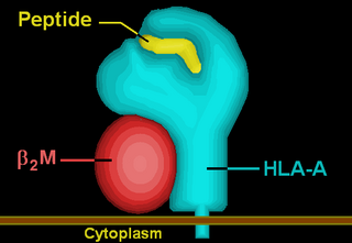
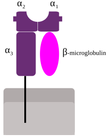
HLA-A is a group of human leukocyte antigens (HLA) that are encoded by the HLA-A locus, which is located at human chromosome 6p21.3. HLA is a major histocompatibility complex (MHC) antigen specific to humans. HLA-A is one of three major types of human MHC class I transmembrane proteins. The others are HLA-B and HLA-C. The protein is a heterodimer, and is composed of a heavy α chain and smaller β chain. The α chain is encoded by a variant HLA-A gene, and the β chain (β2-microglobulin) is an invariant β2 microglobulin molecule. The β2 microglobulin protein is encoded by the B2M gene, which is located at chromosome 15q21.1 in humans.

<span class="mw-page-title-main">CLIP (protein)</span>

CLIP or Class II-associated invariant chain peptide is the part of the invariant chain (Ii) that binds to the peptide binding groove of MHC class II and remains there until the MHC receptor is fully assembled. CLIP is one of the most prevalent self peptides found in the thymic cortex of most antigen-presenting cells. The purpose of CLIP is to prevent the degradation of MHC II dimers before antigenic peptides bind, and to prevent autoimmunity.

<span class="mw-page-title-main">HLA-E</span> Protein-coding gene in the species Homo sapiens

HLA class I histocompatibility antigen, alpha chain E (HLA-E) also known as MHC class I antigen E is a protein that in humans is encoded by the HLA-E gene. The human HLA-E is a non-classical MHC class I molecule that is characterized by a limited polymorphism and a lower cell surface expression than its classical paralogues. The functional homolog in mice is called Qa-1b, officially known as H2-T23.

<span class="mw-page-title-main">Minor histocompatibility antigen</span>

Minor histocompatibility antigen are peptides presented on the cellular surface of donated organs that are known to give an immunological response in some organ transplants. They cause problems of rejection less frequently than those of the major histocompatibility complex (MHC). Minor histocompatibility antigens (MiHAs) are diverse, short segments of proteins and are referred to as peptides. These peptides are normally around 9-12 amino acids in length and are bound to both the major histocompatibility complex (MHC) class I and class II proteins. Peptide sequences can differ among individuals and these differences arise from SNPs in the coding region of genes, gene deletions, frameshift mutations, or insertions. About a third of the characterized MiHAs come from the Y chromosome. Prior to becoming a short peptide sequence, the proteins expressed by these polymorphic or diverse genes need to be digested in the proteasome into shorter peptides. These endogenous or self peptides are then transported into the endoplasmic reticulum with a peptide transporter pump called TAP where they encounter and bind to the MHC class I molecule. This contrasts with MHC class II molecules's antigens which are peptides derived from phagocytosis/endocytosis and molecular degradation of non-self entities' proteins, usually by antigen-presenting cells. MiHA antigens are either ubiquitously expressed in most tissue like skin and intestines or restrictively expressed in the immune cells.

<span class="mw-page-title-main">Tapasin</span> Type of protein

TAP-associated glycoprotein, also known as tapasin or TAPBP, is a protein that in humans is encoded by the TAPBP gene.

<span class="mw-page-title-main">PDIA3</span> Protein-coding gene in the species Homo sapiens

Protein disulfide-isomerase A3 (PDIA3), also known as glucose-regulated protein, 58-kD (GRP58), is an isomerase enzyme encoded by the autosomal gene PDIA3 in humans. This protein localizes to the endoplasmic reticulum (ER) and interacts with lectin chaperones calreticulin and calnexin (CNX) to modulate folding of newly synthesized glycoproteins. It is thought that complexes of lectins and this protein mediate protein folding by promoting formation of disulfide bonds in their glycoprotein substrates.

<span class="mw-page-title-main">HLA-DM</span>

HLA-DM is an intracellular protein involved in the mechanism of antigen presentation on antigen presenting cells (APCs) of the immune system. It does this by assisting in peptide loading of major histocompatibility complex (MHC) class II membrane-bound proteins. HLA-DM is encoded by the genes HLA-DMA and HLA-DMB.

<span class="mw-page-title-main">TAP1</span> Protein-coding gene in the species Homo sapiens

Transporter associated with antigen processing 1 (TAP1) is a protein that in humans is encoded by the TAP1 gene. A member of the ATP-binding cassette transporter family, it is also known as ABCB2.

<span class="mw-page-title-main">CD74</span> Mammalian protein found in Homo sapiens

HLA class II histocompatibility antigen gamma chain also known as HLA-DR antigens-associated invariant chain or CD74, is a protein that in humans is encoded by the CD74 gene. The invariant chain is a polypeptide which plays a critical role in antigen presentation. It is involved in the formation and transport of MHC class II peptide complexes for the generation of CD4+ T cell responses. The cell surface form of the invariant chain is known as CD74. CD74 is a cell surface receptor for the cytokine macrophage migration inhibitory factor (MIF).

<span class="mw-page-title-main">HLA-F</span> Protein-coding gene in the species Homo sapiens

HLA class I histocompatibility antigen, alpha chain F is a protein that in humans is encoded by the HLA-F gene. It is an empty intracellular molecule that encodes a non-classical heavy chain anchored to the membrane and forming a heterodimer with a β-2 microglobulin light chain. It belongs to the HLA class I heavy chain paralogues that separate from most of the HLA heavy chains. HLA-F is localized in the endoplasmic reticulum and Golgi apparatus, and is also unique in the sense that it exhibits few polymorphisms in the human population relative to the other HLA genes; however, there have been found different isoforms from numerous transcript variants found for the HLA-F gene. Its pathways include IFN-gamma signaling and CDK-mediated phosphorylation and removal of the Saccharomycescerevisiae Cdc6 protein, which is crucial for functional DNA replication.

<span class="mw-page-title-main">ERAP1</span> Protein-coding gene in the species Homo sapiens

Type 1 tumor necrosis factor receptor shedding aminopeptidase regulator, also known as endoplasmic reticulum aminopeptidase 1 (ARTS-1), is a protein which in humans is encoded by the ARTS-1 gene.

<span class="mw-page-title-main">HM13</span> Protein-coding gene in the species Homo sapiens

Minor histocompatibility antigen H13 is a protein that in humans is encoded by the HM13 gene.

<span class="mw-page-title-main">TAP2</span> Protein-coding gene in the species Homo sapiens

TAP2 is a gene in humans that encodes the protein Antigen peptide transporter 2.

Immunoevasins are proteins expressed by some viruses that enable the virus to evade immune recognition by interfering with MHC I complexes in the infected cell, therefore blocking the recognition of viral protein fragments by CD8+ cytotoxic T lymphocytes. Less frequently, MHC II antigen presentation and induced-self molecules may also be targeted. Some viral immunoevasins block peptide entry into the endoplasmic reticulum (ER) by targeting the TAP transporters. Immunoevasins are particularly abundant in viruses that are capable of establishing long-term infections of the host, such as herpesviruses.

<span class="mw-page-title-main">Peptide loading complex</span>

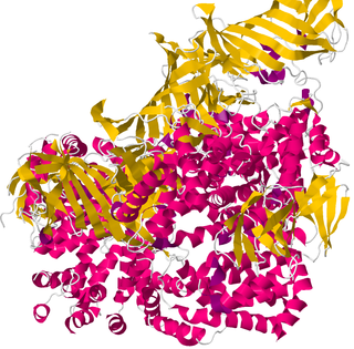
The peptide-loading complex (PLC) is a short-lived, multisubunit membrane protein complex that is located in the endoplasmic reticulum (ER). It orchestrates peptide translocation and selection by major histocompatibility complex class I (MHC-I) molecules. Stable peptide-MHC I complexes are released to the cell surface to promote T-cell response against malignant or infected cells. In turn, T-cells recognize the activated peptides, which could be immunogenic or non-immunogenic.

References

  1. Hewitt EW (October 2003). "The MHC class I antigen presentation pathway: strategies for viral immune evasion". Immunology. 110 (2): 163–9. doi:10.1046/j.1365-2567.2003.01738.x. PMC   1783040 . PMID   14511229.
  2. 1 2 Kulski JK, Shiina T, Anzai T, Kohara S, Inoko H (December 2002). "Comparative genomic analysis of the MHC: the evolution of class I duplication blocks, diversity and complexity from shark to man". Immunological Reviews. 190: 95–122. doi:10.1034/j.1600-065x.2002.19008.x. PMID   12493009. S2CID   41765680.
  3. http://users.rcn.com/jkimball.ma.ultranet/BiologyPages/H/HLA.html#Class_I_Histocompatibility_Molecules Archived 2016-02-04 at the Wayback Machine Kimball's Biology Pages, Histocompatibility Molecules
  4. Hansen TH, Bouvier M (July 2009). "MHC class I antigen presentation: learning from viral evasion strategies". Nature Reviews. Immunology. 9 (7): 503–13. doi:10.1038/nri2575. PMID   19498380. S2CID   9278263.
  5. 1 2 3 4 Syken J, Grandpre T, Kanold PO, Shatz CJ (September 2006). "PirB restricts ocular-dominance plasticity in visual cortex". Science. 313 (5794): 1795–800. Bibcode:2006Sci...313.1795S. doi:10.1126/science.1128232. PMID   16917027. S2CID   1860730.
  6. Burrows SR, Rossjohn J, McCluskey J (January 2006). "Have we cut ourselves too short in mapping CTL epitopes?". Trends in Immunology. 27 (1): 11–6. doi:10.1016/j.it.2005.11.001. PMID   16297661.
  7. Ljunggren HG, Stam NJ, Öhlén C, Neefjes JJ, Höglund P, Heemels MT, Bastin J, Schumacher TN, Townsend A, Kärre K, Ploegh HL (1990-08-02). "Empty MHC class I molecules come out in the cold". Nature. 346 (6283): 476–480. Bibcode:1990Natur.346..476L. doi:10.1038/346476a0. ISSN   0028-0836. PMID   2198471.
  8. Schumacher TN, Heemels MT, Neefjes JJ, Kast W, Melief CJ, Ploegh HL (August 1990). "Direct binding of peptide to empty MHC class I molecules on intact cells and in vitro". Cell. 62 (3): 563–567. doi:10.1016/0092-8674(90)90020-F. PMID   2199065.
  9. Sun Y, Young MC, Woodward CH, Danon JN, Truong HV, Gupta S, Winters TJ, Font-Burgada J, Burslem GM, Sgourakis NG (2023-06-20). "Universal open MHC-I molecules for rapid peptide loading and enhanced complex stability across HLA allotypes". Proceedings of the National Academy of Sciences. 120 (25): e2304055120. Bibcode:2023PNAS..12004055S. doi:10.1073/pnas.2304055120. ISSN   0027-8424. PMC   10288639 . PMID   37310998.
  10. Faridi P, Li C, Ramarathinam SH, Vivian JP, Illing PT, Mifsud NA, Ayala R, Song J, Gearing LJ, Hertzog PJ, Ternette N, Rossjohn J, Croft NP, Purcell AW (12 October 2018). "A subset of HLA-I peptides are not genomically templated: Evidence for cis- and trans-spliced peptide ligands" (PDF). Science Immunology. 3 (28): eaar3947. doi: 10.1126/sciimmunol.aar3947 . PMID   30315122.
  11. Liepe J, Marino F, Sidney J, Jeko A, Bunting DE, Sette A, Kloetzel PM, Stumpf MP, Heck AJ, Mishto M (21 October 2016). "A large fraction of HLA class I ligands are proteasome-generated spliced peptides" (PDF). Science. 354 (6310): 354–358. Bibcode:2016Sci...354..354L. doi:10.1126/science.aaf4384. hdl: 10044/1/42330 . PMID   27846572. S2CID   41095551.
  12. Blees A, Januliene D, Hofmann T, Koller N, Schmidt C, Trowitzsch S, Moeller A, Tampé R (November 2017). "Structure of the human MHC-I peptide-loading complex". Nature. 551 (7681): 525–528. Bibcode:2017Natur.551..525B. doi:10.1038/nature24627. PMID   29107940. S2CID   4447406.
  13. Howarth M, Williams A, Tolstrup AB, Elliott T (August 2004). "Tapasin enhances MHC class I peptide presentation according to peptide half-life". Proceedings of the National Academy of Sciences of the United States of America. 101 (32): 11737–42. Bibcode:2004PNAS..10111737H. doi: 10.1073/pnas.0306294101 . PMC   511045 . PMID   15286279.
  14. Wearsch PA, Cresswell P (August 2007). "Selective loading of high-affinity peptides onto major histocompatibility complex class I molecules by the tapasin-ERp57 heterodimer". Nature Immunology. 8 (8): 873–81. doi:10.1038/ni1485. PMID   17603487. S2CID   29762957.
  15. Thirdborough SM, Roddick JS, Radcliffe JN, Howarth M, Stevenson FK, Elliott T (February 2008). "Tapasin shapes immunodominance hierarchies according to the kinetic stability of peptide-MHC class I complexes". European Journal of Immunology. 38 (2): 364–9. doi: 10.1002/eji.200737832 . PMID   18196518. S2CID   28659293.
  16. Koopmann JO, Albring J, Hüter E, Bulbuc N, Spee P, Neefjes J, Hämmerling GJ, Momburg F, et al. (July 2000). "Export of antigenic peptides from the endoplasmic reticulum intersects with retrograde protein translocation through the Sec61p channel". Immunity. 13 (1): 117–27. doi: 10.1016/S1074-7613(00)00013-3 . PMID   10933400.
  17. Albring J, Koopmann JO, Hämmerling GJ, Momburg F (January 2004). "Retrotranslocation of MHC class I heavy chain from the endoplasmic reticulum to the cytosol is dependent on ATP supply to the ER lumen". Molecular Immunology. 40 (10): 733–41. doi:10.1016/j.molimm.2003.08.008. PMID   14644099.
  18. Imai J, Hasegawa H, Maruya M, Koyasu S, Yahara I (January 2005). "Exogenous antigens are processed through the endoplasmic reticulum-associated degradation (ERAD) in cross-presentation by dendritic cells". International Immunology. 17 (1): 45–53. doi: 10.1093/intimm/dxh184 . PMID   15546887.
  19. Wang Z, Zhang L, Qiao A, Watson K, Zhang J, Fan GH (February 2008). "Activation of CXCR4 triggers ubiquitination and down-regulation of major histocompatibility complex class I (MHC-I) on epithelioid carcinoma HeLa cells". The Journal of Biological Chemistry. 283 (7): 3951–9. doi: 10.1074/jbc.m706848200 . PMID   18083706.
  20. Azevedo L, Serrano C, Amorim A, Cooper DN (September 2015). "Trans-species polymorphism in humans and the great apes is generally maintained by balancing selection that modulates the host immune response". Human Genomics. 9 (1): 21. doi: 10.1186/s40246-015-0043-1 . PMC   4559023 . PMID   26337052.
  21. Nei M, Rooney AP (2005-11-14). "Concerted and birth-and-death evolution of multigene families". Annual Review of Genetics. 39 (1): 121–52. doi:10.1146/annurev.genet.39.073003.112240. PMC   1464479 . PMID   16285855.
  22. Hughes AL (March 1995). "Origin and evolution of HLA class I pseudogenes". Molecular Biology and Evolution. 12 (2): 247–58. doi: 10.1093/oxfordjournals.molbev.a040201 . PMID   7700152.