Histocompatibility

Last updated

Histocompatibility, or tissue compatibility, is the property of having the same, or sufficiently similar, alleles of a set of genes called human leukocyte antigens (HLA), or major histocompatibility complex (MHC). [1] Each individual expresses many unique HLA proteins on the surface of their cells, which signal to the immune system whether a cell is part of the self or an invading organism. [2] T cells recognize foreign HLA molecules and trigger an immune response to destroy the foreign cells. [3] Histocompatibility testing is most relevant for topics related to whole organ, tissue, or stem cell transplants, where the similarity or difference between the donor's HLA alleles and the recipient's triggers the immune system to reject the transplant. [4] The wide variety of potential HLA alleles lead to unique combinations in individuals and make matching difficult.

Contents

Discovery

The discovery of the MHC and role of histocompatibility in transplantation was a combined effort of many scientists in the 20th century. A genetic basis for transplantation rejection was proposed in a 1914 Nature paper by C.C. Little and Ernest Tyyzer, which showed that tumors transplanted between genetically identical mice grew normally, but tumors transplanted between non-identical mice were rejected and failed to grow. [5] The role of the immune system in transplant reject was proposed by Peter Medawar, whose skin graft transplants in world war two victims showed that skin transplants between individuals had much higher rejection rates than self-transplants within an individual, and that suppressing the immune system delayed skin transplant rejection. [6] Medawar shared the 1960 Nobel Prize in part for this work. [7]

In the 1930s and 1940s, George Snell and Peter Gorer individually isolated the genetic factors that when similar allowed transplantation between mouse strains, naming them H and antigen II respectively. These factors were in fact one and the same, and the locus was named H-2. Snell coined the term "histocompatibility" to describe the relationship between the H-2 cell-surface proteins and transplant acceptance. [8] The human version of the histocompatibility complex was found by Jean Dausset in the 1950s, when he noticed that recipients of blood transfusions were producing antibodies directed against only the donor cells. [9] The target of these antibodies, or the human leukocyte antigens (HLA), were discovered to be the human homologue of Snell and Gorer's mouse MHC. Snell, Dausset and Baruj Benacerraf shared the 1980 Nobel Prize for the discovery of the MHC and HLA. [10]

Major histocompatibility complex (MHC)

HLA, the human form of the major histocompatibility complex (MHC), is located on chromosome 6 at 6p21.3. [11] Individuals inherit two different HLA haplotypes, one from each parent, each containing more than 200 genes relevant to helping the immune system recognize foreign invaders. These genes include MHC class I and class II cell-surface proteins. [12] MHC Class I moleculesHLA-A, HLA-B, and HLA-C—are present on all nucleated cells and are responsible for signaling to an immune cell that an antigen is inside the cell. [2] MHC Class II molecules—HLA-DR, and HLA-DQ and HLA-DP—are only present on antigen presenting cells and are responsible for presenting molecules from invading organisms to cells of the immune system. [13]

The MHC genes are highly polymorphic, with thousands of versions of the MHC receptors in the population, though any one individual can have no more than two versions for any one locus. [14] MHC receptors are codominantly expressed, meaning all inherited alleles are expressed by the individual. [15] The wide variety of potential alleles and multiple loci in the HLA allow for many unique combinations in individuals.

Role in transplantation

HLA genes and their location on chromosome 6 HLA.svg
HLA genes and their location on chromosome 6

After receiving a transplant, the recipient's T cells will become activated by foreign MHC molecules on the donor tissue and trigger the immune system to attack the donated tissue [3] The more similar HLA alleles are between donor and recipient, the fewer foreign targets exist on the donor tissue for the host immune system to recognize and attack. [16] The number and selection of MHC molecules to be considered when determining whether two individuals are histocompatible fluctuates based on application, however matching HLA-A, HLA-B, and HLA-DR has been shown to improve patient outcomes. [17] Histocompatibility has a measurable effect on whole organ transplantation, increasing life expectancy of both the patient and organ. [3] HLA similarity is therefore a relevant factor when choosing donors for tissue or organ transplant. This is especially important for pancreas and kidney transplants.

Due to the inherited nature of HLA genes, family members are more likely to be histocompatible. The odds of a sibling having received the same haplotypes from both parents is 25%, while there is a 50% chance that the sibling would share just one haplotype and a 25% chance they would share neither. However, variability due to crossing over, haplotypes may rearrange between generations and siblings may be intermediate matches. [18]

The degree of histocompatibility required is dependent on individual factors, including the type of tissue or organ and the medical condition of the recipient. While whole organ transplants can be successful between unmatched individuals, increased histocompatibility lowers rates of rejection, result in longer lifespans, and overall lower associated hospital costs. [19] The impact of HLA matching differs even among whole organ transplants, with some studies reporting less importance in liver transplants as compared to heart, lung, and other organs. [17] In comparison, hematopoietic stem cell transplants are often require higher degrees of matching due to the increased risk of graft-versus-host disease, in which the donor's immune system recognizes the recipient's MHC molecules as foreign and mounts an immune response. [20] Some transplanted tissue is not exposed to T cells that could detect foreign MHC molecules, such as corneas, and thus histocompatibility is not a factor in transplantation. [21] Individual factors such as age sometimes factors into matching protocol, as the immune response of older transplant patients towards MHC proteins is slower and therefore less compatibility is necessary for positive results. [22] Post-operative immunosuppressant therapy is often used to lessen the immune response and prevent tissue rejection by dampening the immune system's response to the foreign HLA molecules, [23] and can increase the likelihood of successful transplantation in non-identical transplant recipients. [24]

Testing

Because of the clinical significance of histocompatibility in tissue transplants, several methods of typing are used to check for HLA allele expression.

Serological Typing

Serological typing involves incubating lymphocytes from the recipient with serum containing known antibodies against the varying HLA alleles. If the serum contains an antibody specific for a HLA allele that is present on the recipient's lymphocyte, the antibodies will bind to the cell and activate a complement signaling cascade resulting in cell lysis. A lysed cell will take up an added dye such as trypan blue allowing for identification. Comparing which serums triggers cell lysis allows identification of HLA alleles present on the cell surface of the recipients cells. [25]

Serological typing has the benefit of quickly identifying expressed HLA alleles, and ignores any non-expressed alleles that could be of little immunological significance. However, it does not recognize subclasses of alleles, which are sometimes necessary for matching. [25]

Molecular Typing

HLA alleles can be determined by directly analyzing the HLA loci on chromosome 6. Sequence specific oligonucleotide probes, sequence specific primer PCR amplification, and direct sequencing can all be used to identify HLA alleles, often providing amino acid level resolution. Molecular methods can more accurately identify rare and unique alleles, but do not provide information about expression levels. [25]

See also

Related Research Articles

<span class="mw-page-title-main">Major histocompatibility complex</span> Cell surface proteins, part of the acquired immune system

The major histocompatibility complex (MHC) is a large locus on vertebrate DNA containing a set of closely linked polymorphic genes that code for cell surface proteins essential for the adaptive immune system. These cell surface proteins are called MHC molecules.

<span class="mw-page-title-main">Human leukocyte antigen</span> Genes on human chromosome 6

The human leukocyte antigen (HLA) system or complex is a complex of genes on chromosome 6 in humans which encode cell-surface proteins responsible for regulation of the immune system. The HLA system is also known as the human version of the major histocompatibility complex (MHC) found in many animals.

<span class="mw-page-title-main">Transplant rejection</span> Rejection of transplanted tissue by the recipients immune system

Transplant rejection occurs when transplanted tissue is rejected by the recipient's immune system, which destroys the transplanted tissue. Transplant rejection can be lessened by determining the molecular similitude between donor and recipient and by use of immunosuppressant drugs after transplant.

<span class="mw-page-title-main">Graft-versus-host disease</span> Medical condition

Graft-versus-host disease (GvHD) is a syndrome, characterized by inflammation in different organs. GvHD is commonly associated with bone marrow transplants and stem cell transplants.

Alloimmunity is an immune response to nonself antigens from members of the same species, which are called alloantigens or isoantigens. Two major types of alloantigens are blood group antigens and histocompatibility antigens. In alloimmunity, the body creates antibodies against the alloantigens, attacking transfused blood, allotransplanted tissue, and even the fetus in some cases. Alloimmune (isoimmune) response results in graft rejection, which is manifested as deterioration or complete loss of graft function. In contrast, autoimmunity is an immune response to the self's own antigens. Alloimmunization (isoimmunization) is the process of becoming alloimmune, that is, developing the relevant antibodies for the first time.

<span class="mw-page-title-main">Serotype</span> Distinct variation within a species of bacteria or virus or among immune cells

A serotype or serovar is a distinct variation within a species of bacteria or virus or among immune cells of different individuals. These microorganisms, viruses, or cells are classified together based on their surface antigens, allowing the epidemiologic classification of organisms to the subspecies level. A group of serovars with common antigens is called a serogroup or sometimes serocomplex.

<span class="mw-page-title-main">Jan Klein</span> Czech American immunologist


Jan Klein | Czech - American immunologist | January 18, 1936 - May 7, 2023

<span class="mw-page-title-main">HLA-DR</span> Subclass of HLA-D antigens that consist of alpha and beta chains

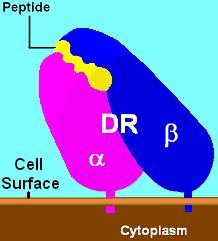
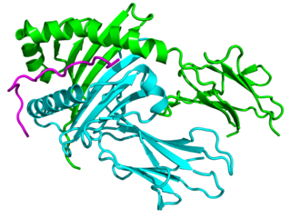
HLA-DR is an MHC class II cell surface receptor encoded by the human leukocyte antigen complex on chromosome 6 region 6p21.31. The complex of HLA-DR and peptide, generally between 9 and 30 amino acids in length, constitutes a ligand for the T-cell receptor (TCR). HLA were originally defined as cell surface antigens that mediate graft-versus-host disease. Identification of these antigens has led to greater success and longevity in organ transplant.

<span class="mw-page-title-main">MHC class II</span> Protein of the immune system

MHC Class II molecules are a class of major histocompatibility complex (MHC) molecules normally found only on professional antigen-presenting cells such as dendritic cells, mononuclear phagocytes, some endothelial cells, thymic epithelial cells, and B cells. These cells are important in initiating immune responses.

<span class="mw-page-title-main">HLA-DQ</span> Cell surface receptor protein found on antigen-presenting cells.

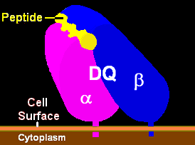
HLA-DQ (DQ) is a cell surface receptor protein found on antigen-presenting cells. It is an αβ heterodimer of type MHC class II. The α and β chains are encoded by two loci, HLA-DQA1 and HLA-DQB1, that are adjacent to each other on chromosome band 6p21.3. Both α-chain and β-chain vary greatly. A person often produces two α-chain and two β-chain variants and thus 4 isoforms of DQ. The DQ loci are in close genetic linkage to HLA-DR, and less closely linked to HLA-DP, HLA-A, HLA-B and HLA-C.

HLA-DP is a protein/peptide-antigen receptor and graft-versus-host disease antigen that is composed of 2 subunits, DPα and DPβ. DPα and DPβ are encoded by two loci, HLA-DPA1 and HLA-DPB1, that are found in the MHC Class II region in the Human Leukocyte Antigen complex on human chromosome 6 . Less is known about HLA-DP relative to HLA-DQ and HLA-DR but the sequencing of DP types and determination of more frequent haplotypes has progressed greatly within the last few years.

<span class="mw-page-title-main">HLA-A</span> Protein-coding gene in the species Homo sapiens

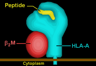
HLA-A is a group of human leukocyte antigens (HLA) that are encoded by the HLA-A locus, which is located at human chromosome 6p21.3. HLA is a major histocompatibility complex (MHC) antigen specific to humans. HLA-A is one of three major types of human MHC class I transmembrane proteins. The others are HLA-B and HLA-C. The protein is a heterodimer, and is composed of a heavy α chain and smaller β chain. The α chain is encoded by a variant HLA-A gene, and the β chain (β2-microglobulin) is an invariant β2 microglobulin molecule. The β2 microglobulin protein is encoded by the B2M gene, which is located at chromosome 15q21.1 in humans.

Tissue typing is a procedure in which the tissues of a prospective donor and recipient are tested for compatibility prior to transplantation. Mismatched donor and recipient tissues can lead to rejection of the tissues. There are multiple methods of tissue typing.

<span class="mw-page-title-main">Minor histocompatibility antigen</span>

Minor histocompatibility antigen are peptides presented on the cellular surface of donated organs that are known to give an immunological response in some organ transplants. They cause problems of rejection less frequently than those of the major histocompatibility complex (MHC). Minor histocompatibility antigens (MiHAs) are diverse, short segments of proteins and are referred to as peptides. These peptides are normally around 9-12 amino acids in length and are bound to both the major histocompatibility complex (MHC) class I and class II proteins. Peptide sequences can differ among individuals and these differences arise from SNPs in the coding region of genes, gene deletions, frameshift mutations, or insertions. About a third of the characterized MiHAs come from the Y chromosome. Prior to becoming a short peptide sequence, the proteins expressed by these polymorphic or diverse genes need to be digested in the proteasome into shorter peptides. These endogenous or self peptides are then transported into the endoplasmic reticulum with a peptide transporter pump called TAP where they encounter and bind to the MHC class I molecule. This contrasts with MHC class II molecules's antigens which are peptides derived from phagocytosis/endocytosis and molecular degradation of non-self entities' proteins, usually by antigen-presenting cells. MiHA antigens are either ubiquitously expressed in most tissue like skin and intestines or restrictively expressed in the immune cells.

<span class="mw-page-title-main">HLA-A*02</span>

HLA-A*02 (A*02) is a human leukocyte antigen serotype within the HLA-A serotype group. The serotype is determined by the antibody recognition of the α2 domain of the HLA-A α-chain. For A*02, the α chain is encoded by the HLA-A*02 gene and the β chain is encoded by the B2M locus. In 2010 the World Health Organization Naming Committee for Factors of the HLA System revised the nomenclature for HLAs. Before this revision, HLA-A*02 was also referred to as HLA-A2, HLA-A02, and HLA-A*2.

<span class="mw-page-title-main">HLA-G</span>

HLA-G histocompatibility antigen, class I, G, also known as human leukocyte antigen G (HLA-G), is a protein that in humans is encoded by the HLA-G gene.

<span class="mw-page-title-main">Major histocompatibility complex, class II, DQ alpha 1</span> Protein-coding gene in the species Homo sapiens

Major histocompatibility complex, class II, DQ alpha 1, also known as HLA-DQA1, is a human gene present on short arm of chromosome 6 (6p21.3) and also denotes the genetic locus which contains this gene. The protein encoded by this gene is one of two proteins that are required to form the DQ heterodimer, a cell surface receptor essential to the function of the immune system.

The following outline is provided as an overview of and topical guide to immunology:

Human leukocyte antigens (HLA) began as a list of antigens identified as a result of transplant rejection. The antigens were initially identified by categorizing and performing massive statistical analyses on interactions between blood types. This process is based upon the principle of serotypes. HLA are not typical antigens, like those found on surface of infectious agents. HLAs are alloantigens, they vary from individual to individual as a result of genetic differences. An organ called the thymus is responsible for ensuring that any T-cells that attack self proteins are not allowed to live. In essence, every individual's immune system is tuned to the specific set of HLA and self proteins produced by that individual; where this goes awry is when tissues are transferred to another person. Since individuals almost always have different "banks" of HLAs, the immune system of the recipient recognizes the transplanted tissue as non-self and destroys the foreign tissue, leading to transplant rejection. It was through the realization of this that HLAs were discovered.

Alloantigen recognition refers to immune system recognition of genetically encoded polymorphisms among the genetically distinguishable members of same species. Post-transplant recognition of alloantigens occurs in secondary lymphoid organs. Donor specific antigens are recognized by recipient’s T lymphocytes and triggers adaptive pro-inflammatory response which consequently leads to rejection of allogenic transplants. Allospecific T lymphocytes may be stimulated by three major pathways: direct recognition, indirect recognition or semidirect recognition. The pathway involved in specific cases is dictated by intrinsic and extrinsic factors of allograft and directly influence nature and magnitude of T lymphocytes mediated immune response. Furthermore, variant tissues and organs such as skin or cornea or solid organ transplants can be recognized in different pathways and therefore are rejected in different fashion.

References

  1. "Histocompatibility". Dorlands Illustrated Medical Dictionary. Philadelphia, PA: Elsevier. 2012.
  2. 1 2 "Human leukocyte antigens". Genetics Home Reference. Retrieved 2018-01-25.
  3. 1 2 3 Ingulli E (January 2010). "Mechanism of cellular rejection in transplantation". Pediatric Nephrology. 25 (1): 61–74. doi:10.1007/s00467-008-1020-x. PMC   2778785 . PMID   21476231.
  4. Kindt TJ, Goldsby RA, Osborne BA, Kuby J (2006). Kurby Immunology. W. H. Freeman & Company. ISBN   978-1-4292-0211-4.
  5. Auchincloss H, Winn HJ (February 2004). "Clarence Cook Little (1888-1971): the genetic basis of transplant immunology". American Journal of Transplantation. 4 (2): 155–9. doi: 10.1046/j.1600-6143.2003.00324.x . PMID   14974934. S2CID   39596314.
  6. Starzl TE (March 1995). "Peter Brian Medawar: father of transplantation". Journal of the American College of Surgeons. 180 (3): 332–6. PMC   2681237 . PMID   7874344.
  7. "The Nobel Prize in Physiology or Medicine 1960 - Speed Read". www.nobelprize.org. Retrieved 2018-02-26.
  8. Elgert KD (2009). Immunology : understanding the immune system (2nd ed.). Hoboken, N.J.: Wiley-Blackwell. ISBN   9780470081570. OCLC   320494512.
  9. "The Nobel Prize in Physiology or Medicine 1980 - Speed Read". www.nobelprize.org. Retrieved 2018-02-26.
  10. "The Nobel Prize in Physiology or Medicine 1980 - Speed Read". www.nobelprize.org. Retrieved 2018-02-26.
  11. Kasahara M (2000). Major Histocompatibility Complex: Evolution, Structure, and Function. New York: Springer. ISBN   978-4-431-70276-4.
  12. Delves PJ (January 2017). "Human Leukocyte Antigen (HLA) System". Merck Manual.
  13. Schwartz RH (1985-01-01). "T-lymphocyte recognition of antigen in association with gene products of the major histocompatibility complex". Annual Review of Immunology. 3 (1): 237–61. doi:10.1146/annurev.iy.03.040185.001321. PMID   2415139.
  14. Ayala García MA, González Yebra B, López Flores AL, Guaní Guerra E (2012). "The major histocompatibility complex in transplantation". Journal of Transplantation. 2012: 842141. doi: 10.1155/2012/842141 . PMC   3388305 . PMID   22778908.
  15. Janeway CA, Travers P, Walport M, Shlomchik M (2001). "The Major Histocompatibility Complex and Its Functions". Immunobiology: The Immune System in Health and Disease (5th ed.). Elsevier España. ISBN   978-0-8153-3642-6.
  16. Trowsdale J, Knight JC (2013). "Major histocompatibility complex genomics and human disease". Annual Review of Genomics and Human Genetics. 14: 301–23. doi:10.1146/annurev-genom-091212-153455. PMC   4426292 . PMID   23875801.
  17. 1 2 Zachary AA, Leffell MS (2016). "HLA Mismatching Strategies for Solid Organ Transplantation - A Balancing Act". Frontiers in Immunology. 7: 575. doi: 10.3389/fimmu.2016.00575 . PMC   5141243 . PMID   28003816.
  18. Cruz-Tapias P, Castiblanco J, Anaya J (2013-07-18). Major histocompatibility complex: Antigen processing and presentation. El Rosario University Press.
  19. Takemoto S, Port FK, Claas FH, Duquesnoy RJ (December 2004). "HLA matching for kidney transplantation". Human Immunology. 65 (12): 1489–505. doi:10.1016/j.humimm.2004.06.008. PMID   15603878.
  20. Apperley J, Niederwieser D, Huang XJ, Nagler A, Fuchs E, Szer J, Kodera Y (January 2016). "Haploidentical Hematopoietic Stem Cell Transplantation: A Global Overview Comparing Asia, the European Union, and the United States". Biology of Blood and Marrow Transplantation. 22 (1): 23–6. doi: 10.1016/j.bbmt.2015.11.001 . PMID   26551633.
  21. Immune response and the eye: in memoriam J. Wayne Streilein. Niederkorn, J. Y. (Jerry Y.), 1946-, Kaplan, Henry J., Streilein, J. Wayne. (2nd, rev. ed.). Basel: Karger. 2007. ISBN   9783805581875. OCLC   85243138.{{cite book}}: CS1 maint: others (link)
  22. Dreyer GJ, Hemke AC, Reinders ME, de Fijter JW (October 2015). "Transplanting the elderly: Balancing aging with histocompatibility". Transplantation Reviews. 29 (4): 205–11. doi:10.1016/j.trre.2015.08.003. PMID   26411382.
  23. van Sandwijk MS, Bemelman FJ, Ten Berge IJ (July 2013). "Immunosuppressive drugs after solid organ transplantation". The Netherlands Journal of Medicine. 71 (6): 281–9. PMID   23956308.
  24. Petersdorf EW (2017-05-03). "Role of major histocompatibility complex variation in graft-versus-host disease after hematopoietic cell transplantation". F1000Research. 6: 617. doi: 10.12688/f1000research.10990.1 . PMC   5419254 . PMID   28529723.
  25. 1 2 3 Chandraker A, Sayegh MH (2012). Core concepts in renal transplantation. New York: Springer Science+Business Media, LLC. p. 1960. ISBN   9781461400073. OCLC   768245800.